Rate of ventilatory disorders in patients with the idiopathic scoliosis after the corrective operation and influence of rehabilitation on ventilatory parameters
7
0
0
Pełen tekst
(2) Aleksander Barinow-Wojewódzki, Tadeusz Rychlewski, Maria Laurentowska. types of scoliosis and it controls the development of scoliosis. Disturbances in muscular balance contribute to the progression of the deformities. There are plenty arguments indicating on changes in the neuromuscular system as responsible for the idiopathic scoliosis [19, 24]. Regardless of the attitude towards pathogenesis of scoliosis, there is no doubt that etiologic factors cause disturbances in the static balance of the vertebral column. The following factors are necessary for a proper function of the respiratory system: the regular shape and compliance of the chest, regular lung elasticity, regular resistance of the respiratory tract, and proper function of respiratory muscles. During respiration, the major work is performed by the inspiratory muscles, while the expiratory muscles are recruited during forced respiration. However, both groups of muscles keep an active tension during the whole respiratory cycle. The main inspiratory muscle is the diaphragm. The internal intercostal muscles, and other muscles of the chest and abdomen assist during the expiration. The external intercostal muscles are inspiratory, but are also active during abdominal press and during fixation of the chest [15, 24]. Disturbances of statics and bone symmetry of the chest during the idiopathic scoliosis augment changes in mobility of costovertebral joints and as consequence lead to alterations of ribs’ position during respiration. At the same time intercostal spaces change together with the activity of intercostal muscles. This in turn causes a decrease of force of the remaining respiratory muscles and contributes to disorders in spatial relations among bronchi, bronchioles and pulmonary alveoli. These changes are of great importance for their expansion and ventilation [13, 23, 27]. In parallel with a child’s growth, development of the chest becomes irregular, leading in some cases to a lower mobility of its one half. This may be accompanied by the additional activity of the neck and arm muscles. The above mentioned changes are responsible for the restrictive ventilatory insufficiency what may result in development of the cor pulmonale in a proportion of patients [5, 14]. Conservative treatment is sometimes insufficient. The standard treatment of the idiopathic scoliosis is mainly posterior vertebral arthrodesis after previous correction and stabilization of a curvature with Harrington’s instruments. However, the Cotrel-Dubousset (CD) method was the first considering a possibility to correct scoliosis in three planes. The multisegmental fixation and better stabilization protect the majority of patients from the 40. postoperative immobilization [7, 8, 9, 12]. In available literature referring to the correction with the CD method, improvement of spirometric parameters immediately after the operation has been reported [20]. Motor rehabilitation and respiratory exercises, likewise surgical treatment, restrain general pathological changes coinciding with the idiopathic scoliosis and improve the exercise tolerance and respiratory capacity of patients. The aim of the study was to investigate ventilatory disorders and the functional efficiency of the respiratory system in patients with the idiopathic scoliosis by determination of: – ventilatory parameters – the influence of the four-weeks rehabilitation under conditions of hospital care.. METHODS The study was performed on thirty 13-19 years old girls with the idiopathic scoliosis, one and a half year after surgical treatment with the CD method. The extended spirometric measurements to determine the efficiency of the respiratory system were made in the Hospital of Lung Diseases and Tuberculosis in Ludwikowo, by the ABC PNEUMO PC apparatus with the Microsoft PNEUMO PC software compatible to Windows 95 TM. The investigations were made before the fourweeks rehabilitation camp and after its completion in the spirometric laboratory, by the same person, under identical conditions: temperature 25oC, humidity 65oC, pressure 985 HPa. The mean values were calculated from three successive measurements. The obtained results were analyzed by comparisons with values attributed to age, height and gender. The following parameters were determined: the forced ventilatory capacity (FVC), the inspiratory vital capacity (VC In), the expiratory vital capacity (VC Ex), the minute volume of ventilation (MV), the forced expiratory volume in one second (FEV1), the maximal expiratory flow (MEF), 25%, 50%, 75% of the forced expiratory capacity (FVC), the FEV1/FVC ratio, the peak expiratory flow (PEF), and the maximal voluntary volume (MVV). In the paper, however, only selected parameters were presented. On the basis of the obtained physiological parameters, a load to reach the ventilatory threshold was determined in the girls under the study. These values were used for realization of the rehabilitation.
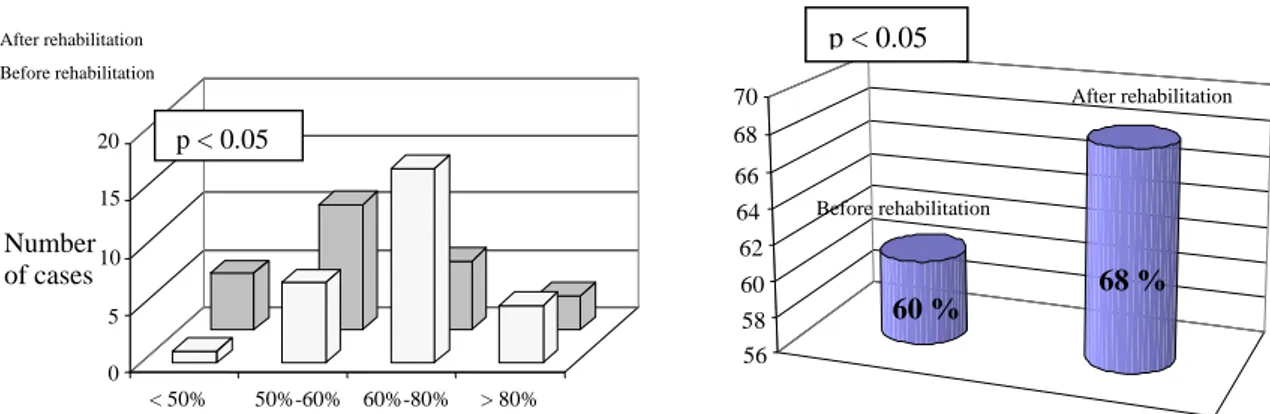
(3) Rate of ventilatory disorders in patients with the idiopathic scoliosis .... process. The endurance training on the cycloergometer was performed 3 times a week for 40 minutes, with a 5-minutes warm-up, 30 minutes of exercise with a previously determined threshold load, and a 5-minutes exercise without a load. Moreover, respiratory exercises directed towards the effective expiration were performed every day for 30 minutes. They consisted of exercises mobilizing the chest, increasing expiration, increasing respiratory movements of lower ribs, mobilizing upper parts of the chest, exercises of the diaphragmatic respiration, and of the abdominal respiration. Additionally, 45-minutes general exercises were performed every second day and girls were swimming in a pool for 45 minutes, once a week. The analyzed parameters were described with the arithmetic means, minimum and maximum values, and standard deviations. The Shapiro-Wilk test was used to assess the regular distribution. For comparisons of differences before and after the rehabilitation, Student’s t-test for combined variables was used when compliance with the regular distribution was confirmed. Otherwise, nonparametric Wilcoxon’s test was used. Values at p < 0.05 were considered as statistically significant. All calculations were made with the use of the STATISTICA for Windows 5.5 program.. RESULTS The positive influence of the rehabilitation process on the ventilatory parameters was observed in girls with the idopathic scoliosis after the correction. The vital capacity of the lungs increased from 2.869 to 3.036 l (p<0.05), though it was lower than regular, either before or after the rehabilitation camp. Analogical changes were revealed in values of the forced expiratory volume in one second. Before the rehabilitation period it amounted to 2.49, while after the rehabilitation 2.75 l (p<0.01). Both results were lower than suitable values amounting to 3.15 and 3.10 l, respectively. The FEV1/VC index, previously known as the Tiffeneau test, characterizing obturatory changes in girls with the idiopathic scoliosis, was not decreased neither before nor after the rehabilitation camp, what indicates absence of symptoms of the bronchial stricture in the studied patients. The peak expiratory flow slightly increased. Before the rehabilitation it amounted to the mean 5.66 l/s, and after the rehabilitation 5.82 l/s, what makes 82.3 and 85.2% of suitable values, respec-. tively (no statistically significant differences were found). Similar results were obtained for the maximal voluntary ventilation (MVV) in the girls investigated. Mean values of MVV before the rehabilitation were 60.7%, while after the rehabilitation 68% in relation to suitable values (p<0.05). Absolute values of the MVV, amounted to 75 l/min and 83.2 l/min, respectively (p<0.05). During the period of the study, no severe ventilatory disorders and no considerable decrease of the vital capacity were observed. As an effect of the rehabilitation program, number of girls with the medium and small impairments of ventilation decreased, while number of girls with regular values of the ventilatory parameters increased from 13 to 20. This positive tendency was also noted for the forced expiratory volume in one second. After the rehabilitation, number of girls with regular values of this parameter increased from 17 to 26. Moreover, no disorders of the high degree were observed after the rehabilitation. The rehabilitation exercises led to the increase of the peak expiratory flow in the girls investigated. As a result, no severe impairments were determined in the second term of the study, and number of patients with regular values of this parameter increased from 17 to 20. Values of the maximal minute voluntary ventilation were also positively changed. Number of girls with considerable impairments of this parameter decreased from five to one, and number with regular minute ventilation of the lungs increased from three to five.. After rehabilitation Before rehabilitation. 20. p < 0.05. 15. Number of cases. 10 5 0 < 50%. 50%-60% 60%-80% > 80% VC %. Figure 1. Structure of the vital capacity (VC) impairment in patients with the idiopathic scoliosis, before and after the rehabilitation, as a ratio of the measured to suitable values, in percents. 41.
(4) Aleksander Barinow-Wojewódzki, Tadeusz Rychlewski, Maria Laurentowska After rehabilitation Before rehabilitation. p < 0.05. After rehabilitation. p < 0.01. 30. 84. 25 20. 82 Before rehabilitation. Number 15 of cases. 80 78. 78 %. 84 %. 10. 76. 5. 74. 0 < 50% 50%-60% 60%-80%. > 80%. FEV 1 %. Figure 2. Mean values of the lung vital capacity (VC) in patients with the idiopathic scoliosis before and after the rehabilitation, % of suitable values. Figure 3. Structure of the forced expiratory volume in one second (FEV1) in girls with the idiopathic scoliosis before and after the rehabilitation, as a ratio of the measured to suitable values, in percents After rehabilitation Before rehabilitation. After rehabilitation. 90 85. Before rehabilitation. Number of cases. 80. 79%. 75. 89 %. 70. 35 30 25 20 15 10 5 0 < 50%. p < 0.01. 50%-60% 60%-80%. > 80%. FEV1 / VC. Figure 4. Mean values of the forced expiratory volume in one second (FEV1) in girls with the idiopathic scoliosis before and after the rehabilitation, % of suitable values. Figure 5. The percentage index of the forced expiration (FEV1/VC), in patients with the idiopathic scoliosis before and after the rehabilitation. After rehabilitation Before rehabilitation After rehabilitation. 86 20. 85 84. 15. Number of cases. Before rehabilitation. 83 10 82 5. 81. 82 %. 85 %. 80. 0 < 50%. 50%-60% 60%-80%. > 80%. PEF %. Figure 6. Structure of the peak expiratory flow (PEF) impairment in girls with the idiopathic scoliosis before and after the rehabilitation, as a ratio of the measured to suitable values in percents. 42. Figure 7. Mean values of the peak expiratory flow (PEF) in patients with the idiopathic scoliosis before and after the rehabilitation, % of suitable values.
(5) Rate of ventilatory disorders in patients with the idiopathic scoliosis .... p < 0.05. After rehabilitation Before rehabilitation. 70 20. After rehabilitation. 68. p < 0.05. 66 15. 64. Number 10 of cases. 62. 5. 58 56. Before rehabilitation. 68 %. 60. 0 < 50%. 50%-60% 60%-80%. 60 %. > 80%. MVV %. Figure 8. Structure of the maximal voluntary ventilation (MVV) impairment in girls with the idiopathic scoliosis before and after the rehabilitation, as a ratio of the measured to suitable values, in percents. DISCUSSION Structural scoliosis causes changes not only in individual vertebrae but also of the whole vertebral column and are always accompanied by its rotation along the long axis. This leads to the chest deformity with impairments of respiratory and circulatory functions [30, 32]. Depending on the angle of the scoliosis, pressure exerted on the lung may completely exclude it from respiration causing fibrosis. The lung on a concave side compensates loss by emphysematous changes. As a result, the vital capacity decreases and the image of the cor pulmonale is observed [1, 5, 32]. The displacement of the heart, mediastinum and large vessels exerts pressure on internal organs, and as a consequence algetic disturbances of ventilation, circulatory disorders and considerable decrease of the physical capacity [25, 28]. The Cotrel-Dubousset (CD) method has appeared to be the first that considered a possibility to correct scoliosis in three planes. The multisegmental fixation and a better stabilization have protected the majority of patients from the postoperative immobilization [7, 9]. There is no unequivocal answer to the fundamental question, whether reconstruction of regular vertebral curvatures influences on the functional improvement of either the respiratory or circulatory systems, and on the overall physical efficiency. It is also not known if it prevents circulatory-respiratory insufficiency in the future [17, 20]. In our research study, performed on children with the idiopathic scoliosis, 16 years old at the average, we have indicated that one and a half year. Figure 9. Mean values of maximal voluntary ventilation (MVV) in patients with the idiopathic scoliosis before and after the rehabilitation, % of suitable values. after the surgery ventilatory parameters may be improved by a four-weeks rehabilitation camp. In girls below 16 years old, parameters of physical capacity increase up to the top during the puberty period, and later interpersonal differences may be observed depending on the level of physical activity. We have revealed the profitable influence of physical rehabilitation on ventilatory parameters which, according to several authors, are generally worse in patients in comparison to healthy people at the same age. This is undoubtedly caused by a weakness of respiratory muscles, the displacement of internal organs in the chest, the mediastimum and the large vessels, as well as deformations of pulmonary tissue [1, 3, 11]. In the presented material, the impairment in the vital capacity (VC) was noticed in 59.4% of cases, however, after the rehabilitation it was observed only in 34.5%, while the vital capacity increased in subjects under the study by 5.69%, which appears to be a satisfactory result. It has been confirmed that in patients with the idiopathic scoliosis prevail ventilatory changes evoked by the decrease of the lung volume and the chest capacity as well as the decrease of the heart and chest mobility, either before or after surgical operation [2, 5, 22]. Contrary to few other authors, no cases of obturatory changes have been observed before or after the rehabilitation camp [17, 18, 21]. The peak expiratory flow would seem to be a more sensitive index, however, the present results have not confirmed this supposition. Mean values of PEF before and after the rehabilitation only slightly changed, from 82.35% to 85.23% of the suitable values, respectively. Values of the maximal 43.
(6) Aleksander Barinow-Wojewódzki, Tadeusz Rychlewski, Maria Laurentowska. voluntary ventilation (MVV) were impaired in 88.9% of the girls investigated before the rehabilitation and slightly increased (by 5.6%) after its completion. Studies concerning views on the respiratory capacity in patients with scoliosis most frequently stress on the decrease of this capacity, sometimes to a high degree, until the development of the cor pulmonale, though only over 40 years of life [1, 5, 6]. The vertebral deformity, in spite of unprofitable changes, not always leads to considerably high disorders of the respiratory functions and no correlations between the angle of a curvature and impairments of ventilation have been established [18, 34]. Numerous investigators indicate that the rehabilitation positively influences ventilatory parameters, however in a longer time perspective its significance decreases [20, 21]. On the basis of the described results of the study, one may conclude as follows: 1. The decrease of the ventilatory indices with respect to suitable values has been revealed as disturbances of a restrictive type in girls with idiopathic scoliosis, 1.5 years after the surgical treatment with the CD method. 2. No obturatory disorders have been revealed before or after the rehabilitation camp. 3. The rehabilitation program applied during the four-weeks camp, comprised of the physical training and respiratory exercises leads to: a. the increase of the vital capacity, b. the increase of the forced expiratory volume in one second, c. the increase of the maximal voluntary ventilation, what indicates the improvement of the ventilatory parameters. One may suppose that systematic rehabilitation in girls with the idiopathic scoliosis under conditions of a rehabilitation hospital or a sanatorium may prevent intensification of ventilatory disorders.. REFERENCES [1] [2]. [3]. 44. Aaro S., 0hlund C., Scoliosis and pulmonary fuction. Spine, 1984, 9: 220-222. Aaro S., Computer tomography in studies of scoliotic deformities. Thesis. Karolinska Institute Stokholm 1980, pp. 2-3. Bjure J., Grimby G., Kasalicki J., et al., Respiratory impairment and airway closure in patients with untreated idiopathic scoliosis, Thorax, 1970, 25: 451-456.. [4] [5] [6]. [7]. [8]. [9]. [10]. [11]. [12]. [13]. [14]. [15]. [16] [17]. [18]. Bunnell W.P., The natural history of idiopathic scoliosis, Clinical Orthopedics, 1988, 229: 20-23. Caro C.G., DuBois A.B., Pulmonary function in kyphoscoliosis, Thorax, 1961, 16: 282-290. Cook C.D., Barrie H., Dr Forest S.A., Helliesen P.J., Pulmonary physiology in children. Lung Volumes. Mechanics of respiration and respiratory muscle strength in scoliosis, Pediatrics, 1960, 25: 766-774. Cotrel Y. et al., New universal instrumentation in spinal surgery, Clinical Orthopedics, 1988, 227: 10-23. Cochran T., Irstam L., Nachemson A., Long term anatomic and fuctional changes in patients with adolescent idiopathic scoliosis treated by Harrington rod fusion, Spine, 1983, 8: 576-579. Dubousset J., Herring J.A., Sfufflebarger H., The Crankshaft phenomenon, Journal of Pediatric Orthopedics, 1989, 9: 541-543. Dubousset J., Scoliosis in infants and young children. European Instumentation Course Lectures, Vol 2, 1995: 121-127. Gagnon S., Jodoin A., Martin R., Pulmonary fuction test study and after spinal fusion in yong idiopathic scoliosis, Spine, 1989, 14: 486-490. Graf H., Hecquet. J., Dubousset J., 3-dimensional approaches to spinal deformities. Application to the study of the prognosis of pediatric scoliosis. Rev. Chir. Orthop., 1983, 69: 400-407. Grassi V., Tantucci C., Respiratory prognosis in chest wall diseases. Monaldi Archives of Chest Disorders, 1993, 48: 183-187. Haluszka J., d`Almeida P., Sadoul P., Testy określające stan obwodowych dróg oddechowych – niecałkowicie spełnione nadzieje diagnostyczne (Tests determining status of the peripheral respiratory tract – diagnostic hopes not fullfilled), Pneumologia Polska, 1999, 45: 397-402. Ivy J.L., Withers R., Van Handel P., Elger D., Costill D., Muscle respiratory capacity and fiber tzpe as determiants of the lactate thershold, Journal of Applied Physiology, 1980, 48: 523-527. James J.I.P., The etiology of scoliosis, 1970, 52: 410-419. Janiszewski M., Dobrowolski J., Bandyra A., Ocena zachowania się wskaźników wentylacyjnych płuc, wydolności fizycznej i tolerancji wysiłkowej w trakcie postępowania korekcyjnego u dzieci z idiopatycznymi skrzywieniami kręgosłupa (Estimation of the ventilatory parameters, physical capacity and exercise tolerance during corrective treatment in children with the idiopathic scoliosis), Kwartalnik Ortopedyczny, 1995, 1: 18-23. Kearon C., Viviani G., Kirkley A., Kilian, Factors determining pulmonary function in adolescent.
(7) Rate of ventilatory disorders in patients with the idiopathic scoliosis .... [19]. [20]. [21]. [22]. [23]. [24]. [25]. [26]. idiopatyhic thoracis scoliosis, American Review Of Respiratory Disorders, 1993, 148: 288-299. Kozłowski S., Nazar K., eds., Wprowadzenie do fizjologii klinicznej (Introduction to the clinical physiology), PZWL, Warszawa 1995, pp. 23-27. Laurentowska M., Rychlewski T., Głowacki M., Jastrzębski A., Michalak E., Szczepanowska E., Deskur-Śmielecka E., The spiroergometric assessment of physical performance in patients operated for lateral spinal curvature, Studies in Physical Culture and Tourism, 2000: Vol. VII: 87-92. Lenke G.L., Bridwell H.K., Blanke K., Baldus C., Analysis of pulmonary function and chest cage dimension changes after thoracoplastry in idiopathic scoliosis, Spine, 1994, 20: 1343-1350. Lonstein J.E., Carlson J.M., The prediction of curve progresion in untreated idiopathic scoliosis during growth, Journal of Bone and Joint Surgery (Am), 1984, 66: 1061-1063. Lonstein J.E., Winter R.B, Bradford D.S., Ogilvie J.W., Moe`s Textbook of scoliosis and other spinal deformities. W. B. Saunders Company, 1995, 97: 153-167. Metha M.H., Infantile idiopathic scoliosis, (in:) D.S. Bradford and R. Dicson, eds., Management of Spinal Deformities, Butterworths, Boston, 1984, pp. 114-116. Milanowska K., Dega W., Rehabilitacja medyczna (Medical rehabilitation), PZWL, Warszawa 1998, pp. 228-270. Nowakowski A., Postępy w diagnostyce i leczeniu skoliozy idiopatycznej u dzieci i młodzieży. [27]. [28]. [29] [30]. [31]. [32]. [33]. (Progress in diagnostics and treatment of the idiopathic scoliosis in children and youths), Chirurgia Narządu Ruchu i Ortopedia Polska, 1995, 6: 445-457. Sevastic J.A., Aaro S., Normelli H., Scoliosis. Experimental and clinical studies. Clinical Orthopedics, 1984, 191: 27-34. Smyth R.J., Chapman K.R., Wright T.A. et al., Pulmonary function in adolescents with mild idiopathic scoliosis, Thorax, 1984, 39: 901-904. Weinstein S.L., Idiopathis scoliosis. Natural history, Spine, 1986, 11: 780-786. Willner S., Etiology of adolescent idiopathic scoliosis, (in:) S.L. Weinstein, ed., The pediatric spine: Principles and practice, Raven Press, New York, 1984, pp. 445-462. Wierusz-Kozłowska M. et al., Ocena czynności układu oddechowego u dorosłych leczonych operacyjnie w dzieciństwie z powodu bocznego idiopatycznego skrzywienia kręgosłupa (Assessment of the respiratory system function in adults after surgical treatment of the idiopathic scoliosis during childhood), IV Sympozjum Sekcji Spondyloortopedii PTO i TV, Lądek Zdrój 1986, 35-38. Wierusz-Kozłowska M., Kubacki A., Wczesne wykrywanie i zapobieganie progresji bocznych skrzywień kręgosłupa (Early diagnosis and prevention of scoliosis progression), PZWL, Warszawa 1983, pp. 229-304. Wynne-Davies R., Familial idopathic scoliosis, Journal of Bone and Joint Surgery, 1968, 50: 24-30.. 45.
(8)
Obraz

Powiązane dokumenty
Evaluation of the relationship between the le- vels of chronic inflammation markers (fibrinogen, hs-CRP, IL-6 and TNF-a) and markers of obstruc- tion, hyperinflation, and blood
Wyniki: Stwierdzono znamienną poprawę jakości życia u chorych oraz wydłużenie czasu trwania testu wysiłkowego, zarówno bezpośrednio po zakończeniu rehabilitacji, jak i 3
The aim of the study was to establish whether patients with dyspepsia may potentially be a group at high risk of developing diabetes in which screening for glucose
The aim of the study was to assess the mental health status and the risk of mental disorders in adult patients with DSD and Y chromosome in the Polish population, as well as
The great- est improvement was observed regarding the reduction of cases with joint pain (61.3%) but we observed still a high percentage of patients with joint oedema (74.5%,
In the group of patients with left knee joint involvement (n = 11), the values of differences in active dorsal flexion of the left and right foot are nearly half the value of
The assessment of functional and clinical condition of patients with coxarthrosis was conducted on the basis of the WOMAC (Western Ontario and McMaster Universities
Self-feeding respondents scored higher on the Barthel scale as far as their functional capacity was concerned both at the beginning of rehabilitation and after its end (Table
