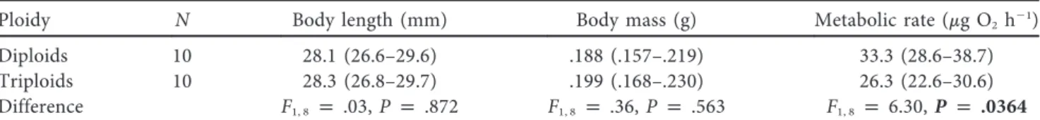
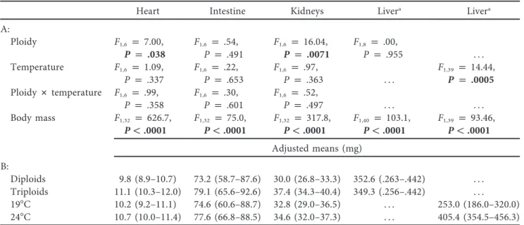
Wydział Biologiczno-Chemiczny
Adam Hermaniuk
The impact of cell size on metabolic rate, growth
rate and development in amphibians: A case
study on the diploid and triploid edible frogs
(Pelophylax esculentus)
Rozprawa doktorska
wykonana w Instytucie Biologii
Uniwersytetu w Białymstoku
pod kierunkiem
prof. dr hab. Jana R. E. Taylora
BIAŁYSTOK
2016
2
The selection forces working for the diminution of genome and cell size may be very
powerful and may change these cell characters in a comparatively short time...
3
I dedicate this dissertation to my mom, my wife, Katarzyna; and my son, Tymon.
Without your love, patience and support, this thesis would not have been possible.
Acknowledgments
I am most grateful to my supervisor Jan R.E. Taylor for giving me the opportunity to
conduct my thesis in his research group. I would like to thank him for generous support
and valuable advice and for training me to became a better scientist.
I am grateful to Włodzimierz Chętnicki for inspiring me with scientific passion and for
introducing me the fascinating world of amphibians.
Many thanks to the water frog researchers Mariusz Rybacki, Piotr Kierzkowski, Nicolas
Pruvost and Maria Ogielska, who showed me various laboratory techniques that were
applied in my study. It was a pleasure to work with you.
For their assistance with laboratory work and maintenance of amphibians, I thank
Anna Koszewnik, Marta Strzałka and a large group of students: Grzegorz Gutowski,
Marta Rurka, Aleksandra Świniarska, Izabela Kaliszuk, Anna Miruć, Natalia
Gawrysiuk, Emilia Murawska.
Many thanks to the technical staff at the Department of Animal Ecology, Małgorzata
Lewoc, Bogusław Lewończuk, and Stanisław Płonowski, for their technical support and
help in parts of the laboratory work.
I also thank Joanna Leśniewska for help with adapting the fluorescence microscope to
my specific needs. I am grateful to Barbara Łaszkiewicz-Tiszczenko and Elżbieta
Bonda-Ostaszewska for support in their lab.
4
Contents
Streszczenie ………... 5
Abstract ………. 8
Introduction ………. 10
Chapter I. Low temperature and polyploidy result in larger cell and body size in an
ectothermic vertebrate………... 14
Chapter II. Genetic and cytogenetic characteristics of pentaploidy in water frogs... 28
Chapter III. Metabolic rate of diploid and triploid edible frog, Pelophylax esculentus,
correlates inversely with cell size in tadpoles but not in frogs………... 40
General Discussion ……….. 52
References ………...
56
5
Wielkość komórek a tempo metabolizmu i rozwoju diploidalnych i
triploidalnych żab wodnych (Pelophylax esculentus)
Streszczenie
Wśród organizmów wielokomórkowych obserwuje się znaczne zróżnicowanie
wielkości komórek, co w dużej mierze związane jest z wielkością genomu. Wiadomo
też, że mniejsze komórki mają wyższe tempo metabolizmu w przeliczeniu na jednostkę
masy niż komórki większe. Według jednej z hipotez, tempo metabolizmu całego
organizmu powinno być odzwierciedleniem tempa metabolizmu pojedynczych
komórek, które składają się na organizm. Poprzez związek z tempem metabolizmu,
rozmiar komórek powinien również wpływać na tempo podziałów komórkowych, a co
za tym idzie – na tempo wzrostu i rozwoju. Porównania międzygatunkowe nie dają
jednak jednoznacznej odpowiedzi co do związku pomiędzy wielkością komórek,
tempem metabolizmu na poziomie całego organizmu i tempem wzrostu. U zwierząt
zmiennocieplnych, najsilniejszy wpływ na rozmiary komórek mają temperatura i
wielkość genomu (zwłaszcza poliploidalność). Powiększenie rozmiarów komórek
spowodowane przez niską temperaturę może być przyczyną geograficznej zmienności
wielkości ciała; w chłodniejszych środowiskach większość zwierząt zmiennocieplnych
rośnie wolniej, ale osiąga większe rozmiary ciała (reguła zależności wielkości ciała od
temperatury otoczenia; TSR). U organizmów poliploidalnych nieliczne przykłady
wskazują, że większe genomy i komórki mogą powodować wzrost wielkości ciała, jak
również mogą obniżać tempo metabolizmu przeliczone na jednostkę masy. Natomiast
wpływ temperatury na wielkość komórek somatycznych u zwierząt poliploidalnych
prawdopodobnie nigdy wcześniej nie był badany.
Celem mojej pracy doktorskiej było zbadanie związku pomiędzy wielkością
komórek, wielkością ciała, tempem wzrostu i tempem metabolizmu u płazów. Obiektem
moich badań były diploidalne i triploidalne żaby wodne, Pelophylax esculentus, które
różniły się wielkością genomu i wielkością erytrocytów, co wykazano w poprzednich
pracach. Badanie konsekwencji zróżnicowania wielkości komórek w obrębie jednego
gatunku omija problem różnej historii ewolucyjnej gatunków porównywanych w
analizach międzygatunkowych. W pierwszej części mojej pracy skupiłem się na
wpływie poliploidii i temperatury na wielkość komórek i wielkość ciała kijanek
hodowanych w 19 °C i 24 °C. Wykazałem, że ploidia i temperatura istotnie wpływa na
wielkość komórek kijanek (erytrocytów i komórek naskórka). Kijanki di- i triploidalne
6
miały większe komórki w 19 °C, a komórki triploidów były większe niż u diploidów w
obu temperaturach. Średnia wielkość erytrocytów pentaploidalnej żabki, która
nieoczekiwanie pojawiła się w jednej z krzyżówek, nie była jednak proporcjonalnie
większa od tej u triploidów, czego można było oczekiwać na podstawie wielkości
genomu. U diploidalnych i triploidalnych żabek temperatura wody, w której żabki
rozwijały się jako kijanki, nie wpływała na wielkość ich komórek. Triploidy nadal
jednak posiadały większe komórki (hepatocyty i erytrocyty). W dalszej części mojej
pracy testowałem hipotezę, że osobniki zbudowane z mniejszych komórek mają wyższe
tempo metabolizmu niż osobniki o porównywalnej masie ciała, ale zbudowane z
większych komórek. Zgodnie z oczekiwaniami, diploidalne kijanki miały wyższe
standardowe tempo metabolizmu (SMR) niż triploidy. Co ciekawe, ploidia nie
wpływała jednak na SMR u żabek. Na podstawie uzyskanych wyników, jak również w
oparciu o obszerny przegląd literatury, wnioskuję, że wielkość komórek może mieć
większe znaczenie dla SMR w wodzie niż na lądzie, ponieważ ilość tlenu zawartego w
wodzie jest mniejsza, a jego dostępność w stosunku do zapotrzebowania maleje wraz ze
wzrostem temperatury. W wodzie poliploidy zbudowane z większych komórek (o
mniejszej powierzchni w stosunku do objętości) mogą być niewystarczająco
zaopatrywane w tlen, wobec czego wykazują niższe tempo metabolizmu. Wykazałem
również, że temperatura wody, w której hodowane były kijanki, nie miała wpływu na
SMR żabek po metamorfozie, co było spójne z brakiem wpływu temperatury wody na
wielkość komórek u żabek.
Kijanki obu ploidii były większe i rozwijały się dłużej w niskiej temperaturze,
co miało związek z ich większymi komórkami. Nie było to dotychczas obserwowane u
płazów i wskazuje, że wielkość komórek może mieć istotne znaczenie w wyjaśnieniu
TSR.
Wykazałem też, że poliploidia/wielkość komórek wpływa na rozmiar ciała
kijanek, ale ich tempo wzrostu zależy w dużej mierze od temperatury. Triploidy rosły
szybciej niż diploidy w 19 °C i miały większą masę ciała, ale w 24 °C nie było
wyraźnej różnicy w tempie wzrostu pomiędzy ploidiami.
Odrzuciłem więc hipotezę, że
triploidalne kijanki, zbudowane z większych komórek, rosną wolniej i rozwijają się
dłużej w danej temperaturze. Wydaje się, że większe komórki triploidów nie są
czynnikiem ograniczającym ich wzrost w niskiej temperaturze.
Wyniki mojej pracy doktorskiej wyraźnie wskazują, że zróżnicowanie wielkości
komórek, wywołane przez czynniki środowiskowe i cytogenetyczne, może odgrywać
7
istotną rolę w fizjologii zmiennocieplnych kręgowców. Po raz pierwszy wykazałem, że
poliploidia/wielkość komórek może różny sposób wpływać na tempo metabolizmu
całego organizmu w obrębie jednego gatunku w zależności od środowiska rozwoju –
wodnego lub lądowego. Wykazałem, że niska temperatura w połączeniu z poliploidią
skutkuje większymi rozmiarami komórek, jak również większymi rozmiarami ciała, co
było pierwszą taką obserwacją u kręgowców. Największą masę ciała osiągnęły kijanki
triploidalne w niskiej temperaturze, co może być potencjalnie korzystne na wyższych
szerokościach geograficznych i może tłumaczyć częstsze występowanie osobników
triploidalnych w północnych częściach geograficznego zasięgu P. esculentus.
8
Abstract
Among multicellular organisms, substantial variation in cell size has been
demonstrated, largely related to the size of the genome. It has been shown that small
cells have higher mass-specific metabolic rates (MRs) than do larger cells. One
hypothesis predicts that the metabolic rate of a whole organism should reflect the
metabolic rates of the individual cells that ultimately constitute that organism. Via the
link with metabolic rate, cell size should also affect the rate of mitotic divisions and
therefore the organismal growth rate and development. However, the interspecific
comparisons do not always reveal the links between cell size, whole-body MR and
growth rate. Of all tested factors, temperature and genome size (especially polyploidy)
produce the strongest effects on cell size in ectotherms. The increase in cell size induced
by low temperature may explain the pattern of geographic variation of body size; in
colder environments, the majority of ectotherms grow more slowly but mature at larger
body sizes (the temperature-size rule, TSR). Regarding polyploids, there is some, albeit
scarce, evidence that their larger genomes and cells may increase the whole body size
and decrease mass-specific MR. The impact of temperature on the size of somatic cells
in polyploid animals has presumably never been studied.
The objective of my thesis was to investigate the links between cell size, body
size, growth rate and whole-body metabolic rate in amphibians. My research objects
were diploid and triploid edible frogs, Pelophylax esculentus, which had previously
been shown to differ in genome size and erythrocyte size. Studying the consequences of
cell size variation within one species overcomes the problem of inter-specific
comparisons associated with the different evolutionary histories of the compared
species. In the first part of the thesis, I focused on the effect of polyploidy and
temperature on the cell size and body size in tadpoles that were reared at 19 °C and 24
°C. I found that ploidy and temperature significantly affected cell size in tadpoles
(erythrocytes and epidermal cells). The cells were larger in both diploid and triploid
tadpoles at 19 °C than at 24 °C, and triploids had larger cells than diploids at both
temperatures. The mean erythrocyte size in a pentaploid froglet, which was
unexpectedly found in one of the crosses, was not proportionally larger than that in
triploids, as might be expected on the basis of genome size. In diploid and triploid
froglets, the temperature in which they developed as tadpoles did not affect the size of
their cells, but triploids still had larger cells (hepatocytes and erythrocytes). In the
9
second part of the thesis, I tested the hypothesis that individuals composed of smaller
cells have a higher MR than individuals of a comparable body size but composed of
larger cells. As I expected, diploid tadpoles had a higher standard metabolic rate (SMR)
than triploids. Interestingly, in froglets, ploidy did not affect the SMR. Based on this
result and an extensive review of the literature, I suggest that cell size may have more
consequences for whole-body metabolic rates in aquatic than in terrestrial habitats
because oxygen is less available in water, and its availability in relation to oxygen
demand decreases with increasing temperatures. In water, polyploids composed of
larger cells (with a less favorable surface-to-volume ratio) may be more vulnerable to
insufficient oxygen supply and display lower MRs. I also found that water temperatures
in which tadpoles were reared had no effect on the SMR of froglets after
metamorphosis, consistent with no effect of these temperatures on cell size.
Tadpoles of both ploidies were larger and developed over a longer time period at
the lower temperature, which was associated with larger cells. This result has not been
observed in amphibians before and indicates that variation in cell size may be important
in explaining the TSR. I was able to demonstrate that polyploidy/cell size affects body
size in tadpoles but that their growth rate largely depends on temperature. At 19 °C,
triploids grew faster than diploids and had larger body mass, whereas at 24 °C there was
no clear difference between ploidies in growth rate. Thus, I rejected the hypothesis that
triploid tadpoles, composed of larger cells, grow more slowly and develop over a longer
time period at a given temperature. It seems that the larger cells of triploids are not a
limiting factor for their growth at lower water temperatures.
My thesis clearly demonstrates that the variation in cell size induced by internal
and external factors may play a significant role in the physiology of ectothermic
vertebrates. I present the first report of two distinct effects of polyploidy/cell size on the
whole-body metabolic rate within a single species developing in two different habitats,
aquatic and terrestrial. I was also able to demonstrate, for the first time in vertebrates,
that the combination of low temperature and polyploidy results in larger cells and body
size. Considering the potential advantages of large body size at higher latitudes, the
highest body mass of triploid tadpoles at lower temperature found in my study may
explain the observation that triploid individuals of P. esculentus prevail in the in the
northern parts of its geographic range.
10
Introduction
The observed substantial variation in cell size among animal species, determined
to a large degree by genome size, may have crucial consequences for physiological
functions affecting various parameters of the organism, such as body size or the
whole-animal metabolic rate (MR) (Gregory 2001; Dufresne and Jeffery 2011; Kozłowski et
al. 2003). On the cellular level, cell size is important because small cells have higher
mass-specific metabolic rates than do larger cells (Goniakowska 1970; Monnickendam
and Balls 1973). One of the explanations for this phenomenon is that the relatively
higher ratio of membrane surface to cell volume in smaller cells requires more energy
for phospholipids turnover and to maintain the ionic gradient between the cytoplasm
and the cell’s surroundings (Rolfe and Brown 1997; Konarzewski and Książek 2013).
The complementary approach points to the importance of cell membranes in exchanging
important components between the cytoplasm and its surroundings, especially to the
oxygen supply required to maintain the metabolism of a cell. Hence, the relatively
larger area of exchange in smaller cells coupled with the shorter distance of diffusion
could enhance their metabolic rate (Szarski 1983; Czarnoleski et al. 2013, 2015).
According to the cell metabolism hypothesis, the whole-body metabolic rate can
be considered as the sum of the metabolic rates of the constituent cells. Thus, at the
organismal level, individuals built of smaller cells should have a higher MR than
individuals of a comparable body size but composed of larger cells (Davison 1955;
Kozłowski et al. 2003). This prediction, however, was not always upheld when
interspecific comparisons were performed within different vertebrate classes.
Significant negative correlations between mass-specific MR and cell size were found in
mammals, birds and one group of reptiles (Vinogradov 1995; Gregory, 2002; Starostová
et al. 2009). In amphibians, only among salamanders was there a significant correlation
between cell size and mass-specific MR (Gregory 2003). Via the link with MR, cell size
is inversely correlated with the rate of mitotic division, which should affect the
organismal growth rate. This effect has been found in amphibians, insects and
crustaceans but not in mammals or birds (Angilletta et al. 2004; Gregory 2005).
Large variations in cell size are also observed within species and complexes of
closely related species that include both diploid and polyploid forms. The latter are
originated as a consequence of duplication of entire chromosome sets. It has been
shown that the significant increase in the genome size in polyploids increases their cell
11
size (Gregory 2001; Choleva and Janko 2013). However, contrary to expectations, in
the majority of studies, the differences in cell size between diploids and polyploids do
not reveal a link between cell size and mass-corrected MR (Mable et al. 2011; Choleva
and Janko 2013).
Of all the physical parameters that have been tested, temperature is the most
dramatic in its effect on cell size in ectothermic animals. It is documented that for
ectotherms (fish and invertebrates were mainly tested), low temperatures result in larger
cells, which may increase their body size (van Voorhies 1996; Blanckenhorn and
Llaurens 2005; Arendt 2007; Hessen et al. 2013). Larger cells, through their potential
effect on the rate of mitotic division and therefore on growth rate, may explain the
phenomenon that individuals raised at low temperatures generally grow more slowly but
eventually become larger than their conspecifics raised at higher temperatures (the
temperature-size rule, TSR) (Atkinson, 1994; Angilletta et al. 2004). TSR may in turn
be associated with the observed pattern of ectotherm body size along climate gradients
(larger individuals inhabit colder environments), even at the intra-specific level (Chown
and Gaston 2010). Although possible mechanisms of the TSR in ectotherms are still
under debate, physical constraints operating at the cellular and molecular levels are
identified as proximate explanations (Van der Have and de Jong 1996; Woods 1999;
Angilletta et al. 2004). Alternatively, ultimate hypotheses can predict conditions in
which delay maturation and larger body size at lower temperatures
increases fitness
(Atkinson and Sibly, 1997, Kozłowski et al., 2004; Angilletta et al. 2004). It is also
intriguing that polyploid organisms (which have larger cells than their diploid relatives)
are often more abundant in harsh or extreme environments, especially at higher latitudes
and altitudes, such as arctic and alpine habitats (Löve 1953; Stöck et al. 2001; Otto et al.
2007). However, larger cells in polyploids rarely produce larger body sizes. No apparent
effect of polyploidy on body size has been found in vertebrates (Benfey 1999; Mable et
al. 2011; Choleva and Janko 2013), but studies on the effect in this group (excluding
fish) are very scarce. Moreover, to my knowledge, the effect of temperature on the size
of somatic cells in polyploid animals has not yet been studied (only two papers reported
the impact of temperature on egg size in polyploid females; Dufresne and Hebert 1998;
Licht and Bogart 1989).
In summary, the studies presented above indicate that variation in cell size
(induced by both internal and external drivers) may play an important role in an
12
evolutionary context, as a potential determinant of MR, growth rate and body size,
although the various papers provide a mixed picture. Additionally, the consequences of
cell size variation in polyploids have received very little attention (Choleva and Janko
2013). For this reason, I studied the relationships between cell size, growth rate,
body mass and MR in diploid and triploid individuals of a hybridogenetic species,
the edible frog, Pelophylax esculentus. Studying the consequences of variation in cell
size within one species overcomes the problems of interspecific comparisons, which
involve unpredictable variables that may arise from unknown evolutionary history of
the different species that are analyzed.
P. esculentus is a natural bisexual hybrid between the pool frog, P. lessonae
(genotype LL), and the marsh frog, P. ridibundus (RR). In most of its European range,
it occurs in a diploid form (LR) and reproduces by hybridogenesis with one of the
parental species (Graf and Polls Pelaz 1989). In some populations, diploid hybrids
coexist with allotriploid hybrids (LLR and/or LRR), especially in central and northern
parts of the species complex range (Plötner, 2005). As objects of my research, I chose
diploid (LR) and triploid (LLR) individuals that differed from each other in genome size
and erythrocyte size (Ogielska et al. 2004; Kierzkowski et al. 2011).
In Chapter I of my dissertation, I focused on the effect of polyploidy/genome
size on cell size. Previous studies revealed that triploid edible frogs have larger
erythrocytes than diploids, serving as a useful preliminary method for ploidy
discrimination (Günther, 1977; Polls Pelaz and Graf, 1988). It was not known, however,
whether cells of other tissues conform to this pattern. To answer this question, I also
measured hepatocytes and epidermal cells. I hypothesized that triploids, with their
larger genomes, would have larger cells than diploids. A single case of pentaploidy that
is described in Chapter II enabled me to examine the effect of an additional increase in
the genome size on cell size and metabolic rate. In Chapter I, I also examined the
effect of temperature on cell size and growth rate in diploid and triploid tadpoles that
were reared at two different temperatures. I tested the hypothesis that low temperature
would increase the size of cells and the whole body size in diploid and triploid
individuals and that both ploidies would develop more slowly at the lower temperature
(see the TSR above). Due to the lower MR, larger cells should have a lower rate of
mitotic divisions, which in turn should result in a slower growth rate in triploids at a
given temperature. Since low temperature and polyploidy may affect cell size in the
13
same direction, I expected that triploid tadpoles reared at a lower temperature would
reach the largest body size among the animals studied. This should be associated with
their prolonged development. In Chapter I, I showed that erythrocytes were not the
only cells larger in triploids; therefore, in Chapter III, I tested the hypothesis that
triploid tadpoles and froglets composed of larger cells will have lower standard
metabolic rates (SMRs) than diploid relatives composed of smaller cells. Tadpoles in
my study were kept at two water temperatures until their metamorphosis, but after
reaching this stage, all individuals were transferred to terraria at same air temperature.
This allowed me to determine whether thermal differences during larval development
affect the froglets’ cell size and SMR after metamorphosis (Chapter I and III). It must
be noted that I compared hybrids with different genome affiliation (LR and LLR) in my
analysis. Therefore, the effect of cell size may potentially be affected by the genome
dosage in LLR individuals (the relative excess of the L genotype). To test whether the
genome dosage affects the morphology, I compared linear measurements of hind legs in
LLR froglets with those in LL and RR froglets (Chapter III).
14
Chapter I
Low temperature and polyploidy result in larger cell and body size in
an ectothermic vertebrate
15
Hermaniuk A., Rybacki M., Taylor J.R.E (2016) Low temperature and polyploidy
result in larger cell and body size in an ectothermic vertebrate. Physiological and
Biochemical Zoology. 89(2): 118–129.
My contribution: contribution to the concept of the study and to developing the
experimental design, collecting water frogs in the field, participation in crossing frogs,
maintenance of amphibians, morphometric measurements, karyotyping, measurements
of cell sizes, data analysis, manuscript preparation.
The pool frog, Pelophylax lessonae, on the cover was photographed by Adam
Hermaniuk in Biebrza National Park.
Low Temperature and Polyploidy Result in Larger Cell
and Body Size in an Ectothermic Vertebrate
Adam Hermaniuk1,* Mariusz Rybacki2 Jan R. E. Taylor1
1
Institue of Biology, University of Białystok, Białystok, Poland; 2Department of Evolutionary Biology, Kazimierz Wielki University, Bydgoszcz, Poland
Accepted 11/25/2015; Electronically Published 1/13/2016
ABSTRACT
Previous studies reported that low temperatures result in in-creases in both cell size and body size in ectotherms that may explain patterns of geographic variation of their body size across latitudinal ranges. Also, polyploidy showed the same effect on body size in invertebrates. In vertebrates, despite their having larger cells, no clear effect of polyploidy on body size has been found. This article presents the relationship between tempera-ture, cell size, growth rate, and body size in diploid and poly-ploid hybridogenetic frog Pelophylax esculentus reared as tadpoles at 197 and 247C. The size of cells was larger in both diploid and triploid tadpoles at 197C, and triploids had larger cells at both temperatures. In diploid and triploid froglets, the temperature in which they developed as tadpoles did not affect the size of their cells, but triploids still had larger cells. Triploid tadpoles grew faster than diploids at 197C and had larger body mass; there was no clear difference between ploidies in growth rate at 247C. This indicates better adaptation of triploid tadpoles to cold environment. This is thefirst report on the increase of body mass of a polyploid vertebrate caused by low temperature, and we showed relationship between increase in cell size and increased body mass. The large body mass of triploids may provide a se-lective advantage, especially in colder environments, and this may explain the prevalence of triploids in the northern parts of the geographic range of P. esculentus.
Keywords: polyploidy, triploids, cell size, body size, temperature-size rule, Pelophylax esculentus.
Introduction
Body size is one of the most important traits of organisms and significantly affects both physiological processes and individ-ualfitness (Peters 1983). Body growth may occur either by in-creasing cell size or cell number or by a combination of both (Kozłowski et al. 2003; Hessen et al. 2013). In organisms with a fixed cell number, body growth can happen only through changes in cell size (van Voorhies 1996), which in turn may also influence body size in organisms with variable cell num-bers (Blanckenhorn and Llaurens 2005; Hessen et al. 2013). It has been proved that cell size strongly correlates with genome size, and this pattern seems to be universal for both plants and animals (Bennett 1987; Gregory 2001a, 2001b). The clearest demonstration of the positive correlation between genome size and cell size comes from polyploids that arise as the result of du-plication of entire chromosome sets (Gregory and Mable 2005; Mable et al. 2011). On the contrary, the effect of polyploidy on body size is much more complex. Polyploid invertebrates may reach maturity at larger sizes compared to diploids, but examples of this are not numerous (Weider 1987; Walsh and Zhang 1992). However, in vertebrates, with few exceptions, no apparent effect of polyploidy on body size has been found, indicating that poly-ploids composed of larger cells regulate their size through changes in cell number (Fankhauser 1945; Swarup 1959; Mahony and Robinson 1980).
For decades it has been recognized that polyploids (both plants and animals) are more abundant in severe environments, especially at higher latitudes and altitudes, such as arctic and alpine habitats (Löve 1953; Levin 1983; Otto et al. 2007). These observations suggest that polyploids possess some physiolog-ical traits that allow them to better adapt to colder environments compared to their diploid counterparts. Laboratory studies that have compared life-history traits in diploid and polyploid in-dividuals of various animal species have confirmed the greater tolerance of the polyploids to low temperature, but the reasons for this phenomenon are not entirely clear (Schultz 1982; Du-fresne and Hebert 1998). The most common explanation is the idea that the increased heterozygosity provided by an “extra” genome provides metabolicflexibility to cope with a wide range of environmental conditions (Otto and Whitton 2000). It is also possible that possession of a higher quantity of enzymes per cell improves metabolic efficiency in polyploids at low temperatures (Dufresne and Hebert 1998).
The link between low temperature, cell size, and body size may be the key to understanding another biological phenom-enon, that is, that the majority of ectotherms (from protists to amphibians) grow more slowly at low temperatures butfinally become larger than their conspecifics growing at higher tem-peratures (the pattern called the temperature-size rule [TSR]; *Corresponding author; e-mail: adamher@uwb.edu.pl.
Physiological and Biochemical Zoology 89(2):118–129. 2016. q 2016 by The University of Chicago. All rights reserved. 1522-2152/2016/8902-5090$15.00. DOI: 10.1086/684974
Atkinson 1994; Angilletta et al. 2004; Hessen et al. 2013). A possible consequence of the TSR is the considerable variation of body size in ectothermic animals across latitudinal ranges— larger organisms inhabit areas in higher latitudes (Bergmann’s rule; originally referred exclusively to endotherms but also ap-plies to ectotherms), as has been observed even in intraspecific comparisons (Chown and Gaston 2010). Possible mechanisms of the TSR and Bergmann’s size clines in ectotherms are still debated, and both ultimate and proximate explanations have been offered (van Voorhies 1996; Partridge and Coyne 1997; An-gilletta et al. 2004). Considering the ultimate explanations, op-timization models can predict conditions in which a smaller body size at higher temperatures maximizesfitness (Atkinson and Sibly 1997; Kozłowski et al. 2004). Common-garden experiments have revealed that individuals from colder environments still exhibit larger sizes at maturity than those from warmer environments, and this provides genetic arguments that a larger body size may be adaptive in colder areas (Partridge and French 1996). Also, laboratory experiments on Drosophila melanogaster mimicking the action of natural selection for cold survival resulted in in-creased cell size and body size (Partridge et al. 1994). According to the proximate explanations, some general physical constraints, operating at cellular and molecular levels, play a causal role in TSR and Bergmann size clines in ectotherms. Van der Have and de Jong (1996) argued that cell growth is more sensitive to ther-mal constraints than cell division. This means that cells will be smaller after dividing at higher temperatures, resulting in a smaller overall organism. Other arguments point to the relationship be-tween cell size and the availability of oxygen. Woods’s (1999) bio-physical model assumes that the maximal size of a cell is limited by the oxygen concentration at its center, which decreases with increasing temperature, especially in water habitats. At the or-ganismal level, this implies that ectotherms raised at higher tem-peratures attain a smallerfinal size because of biophysical con-straints at the cellular level.
Given that there is a general trend of body size increase with latitude (in temperature gradient) in ectotherms and that there is increasing incidence of polyploidy along the same clines, poly-ploids seem to be perfect models to test whether large cell size may have morphological and physiological consequences at low tem-peratures. Most of the studies that have investigated whether the temperature-related effects on body size are the consequences of changes occurring at the cellular level have concerned diploid in-vertebrates (van Voorhies 1996; see Arendt 2007 for review) and, to a lesser extent, diploid vertebrates (Arendt and Hoang 2005; Arendt 2006, 2007; Goodman and Heah 2010). Only a few au-thors have investigated the relationship between cell size and body size in polyploid vertebrates (Fankhauser 1945; Swarup 1959; Mahony and Robinson 1980). We are aware of only one study that has examined the combined effect of polyploidy and temperature on growth rate and body size in vertebrates. That study (Licht and Bogart 1989) was devoted to the embryonic growth and body size of the hatchlings of Ambystoma laterale-texanum complex (mole salamanders).
This article examines the effect of temperature on the cell size of different tissues, the growth pattern, and the body size
of diploid and triploid edible frogs Pelophylax esculentus (Lin-naeus 1758) reared at low (197C) and high (247C) water tem-peratures. We measured the size of erythrocytes in tadpoles and frogs, epidermal cells in tadpoles, and hepatocytes in frogs. The size of erythrocytes is the most often measured cell size in ver-tebrates and serves to discriminate between diploid and triploid frogs (Günther 1977; Polls Pelaz and Graf 1988), but the data on the size of cells from other tissues in amphibians are very scarce. We tested following hypotheses: (i) triploids comprise larger cells than diploids, (ii) a low temperature will increase the size of cells in triploids and diploids, and (iii) a larger cell size will result in a larger body mass of triploid and diploid tadpoles at low temperature. Since low temperature and polyploidy may affect cell size in the same direction, we expected that triploid tadpoles reared at lower temperature would reach the largest body size among other relatives.
The edible frog (P. esculentus) is a natural bisexual hybrid between the pool frog Pelophylax lessonae (genotype LL) and the marsh frog Pelophylax ridibundus (RR). In most of its Eu-ropean range, P. esculentus occurs as a diploid form (LR) and lives in sympatry with one of the parental species. It repro-duces by hybridognesis, a mechanism in which the parental genome of the species present in the population is eliminated before meiosis and gametes produced by a hybrid are clonal (L or R); when fertilized by gametes of a related species, the progeny is hemiclonal (for review, see Graf and Polls Pelaz 1989). In northern parts of the geographical range of the Pelo-phylax species complex (in Sweden, Denmark, northern Ger-many, northern Poland), diploid hybrids coexist with allo-triploid hybrids (LLR and/or LRR) and often form all-hybrid populations (see Plötner 2005 for a review). Although the man-ner of reproduction that allows for the existence of all-hybrid populations is relatively well understood (Christiansen 2009), it is not clear why these populations with triploid individuals in-habit mainly the northern part of Europe. According to Pru-vost et al. (2013), hybrids, especially the two triploid types, have higherfitness under cold conditions than do both parental species. Similarly as in triploid and diploid P. esculentus, the oc-currence of polyploid amphibian species in areas of greater cli-mate severity, in contrast with their close diploid relatives inhab-iting areas where climatic conditions are milder, was found in green toads (Bufo viridis complex) and in the gray tree frog com-plex (Hyla versicolor and Hyla chrysoscelis; Stöck et al. 2001; Otto et al. 2007).
Material and Methods
Population Sources and Crossing Procedure
During the two breeding seasons of 2009 and 2010, we crossed 19 pairs of Pelophylax lessonae (LL) males and Pelophylax es-culentus females (LR or LRR). The males were collected from three ponds, from mixed lessonae-esculentus populations (Poz-nań district, Poland), and the females were derived from an all-hybrid esculentus-esculentus population from six ponds (Szczecin district, Poland). Ploidy and genome compositions of the par-ents were preliminarily determined on the basis of morphological
indexes (DP/CI p length of the digitus primus/length of the callus internus, T/CIp length of tibia/length of the callus in-ternus, and the shape of the callus internus) and erythrocyte size, which is in most cases a reliable method (Günther 1977; Polls Pelaz and Graf 1988). The final confirmation of the parental genome composition was derived from offspring genotypes de-termined by chromosome inspection (as described below). We used artificial fertilization to obtain offspring, stimulating the female with the luteinizing salmon hormone (LHRH; Bachem Bio-science) as described by Berger et al. (1994). Sperm suspension was obtained directly from testes, dissected from the male once killed by decapitation.
Pelophylax esculentus females produce gametes of various sizes (small, medium, and large), related to different ploidy levels (Czarniewska et al. 2011). To obtain the highest percent-age of diploid offspring, triploid females (LRR) were crossed with diploid males (LL), and only eggs classified as small (haploid R gametes in most cases) were taken after fertilization for further rearing. A similar procedure was applied to reach the highest per-centage of triploid offspring, with the exception that diploid females (LR) were used instead of triploid ones and only large eggs (diploid LR gametes in the most cases) were taken for further rearing. In this way, we obtained 9 crosses of diploid (LR) and 10 crosses of triploid (LLR) progeny that originated from different parents. In one cross, in which we expected on the basis of egg size only LLR specimens, the mixed progeny, includ-ing LR, appeared. Another interestinclud-ing result was the pentaploid specimen that was found among all-triploid siblings obtained from a diploid female P. esculentus (LR) crossed with a diploid male P. lessonae (LL). This was thefirst observation of penta-ploidy within a P. esculentus hybrid complex. The detailed de-scription of this unique individual, including erythrocyte size, DNA content, and microsatellites analysis, was published else-where (Hermaniuk et al. 2013). The pentaploid specimen and the cross with mixed genotype of offspring were excluded from further procedures. In this study we used diploid (LR) and triploid (LLR) individuals that belonged to 18 (91 9) different crosses, 8 in 2009 and 10 in 2010.
Rearing of Tadpoles and Froglets
Tadpoles. Eggs and hatchlings were raised in plastic litter trays filled with aerated tap water at room temperature until tadpoles reached developmental stage 25 according to Gosner (1960; the stage of free swimming and independent feeding). At this stage, on the median date of May 20, a group of 140 healthy-looking individuals from each cross, diploid or triploid, was randomly selected. Half of these were raised at 197C in one tank and the second half at 247C in another tank, and two tanks from each cross were placed next to each other. These two temperatures approximated the range of mean temperatures measured in ponds (microhabitats) inhabited by the tadpoles of green frogs (P. esculentus and P. lessonae). The grand mean water temper-ature, measured every 15 min in three ponds in northeast Poland (547150N), was 16.67C at the beginning of tadpoles’ development
and was 23.07C in the second half of development (A. Hermaniuk,
unpublished data). In southern Sweden (60730ʹN), the tadpoles experienced the average water temperature of 19.47C (Orizaola and Laurila 2009b; see also Negovetic et al. 2001).
Until metamorphosis, the development of all animals was conducted in exactly the same tanks (50 cm# 35 cm # 30 cm) placed in a climate chamber set to 167C (517C). Temperature controllers (Thermostab TS 500, Aquael) equipped with heat-ers were responsible for maintaining an appropriate water tem-perature (accuracy 50.257C) in the tanks. Lamps with 70-W bulbs were installed above each tank. Water was aerated per-manently by using air stones andfilters (PZC 300, Aquael) that additionally purified the water from food remains and feces. A natural photoperiod was maintained throughout the period of larval development; the day length was increasing from the beginning of the experiment and then decreasing, as in outdoor conditions. Tadpoles were fed ad lib. with a powder mix consisting of one part dried nettle and one part commercial fish food (Supervit, Tropical). A volume of 0.5 cm3
of food was supplied to each tank twice a day. Half of the water from each tank was renewed three times a week with aged tap water stored for at least 24 h at room temperature. The tanks were inspected daily, and dead larvae and food remains were removed.
To determine the increase in the body mass of diploid and triploid tadpoles at two temperatures soon after hatching, when the growth rate is high, we weighed them in 2009 in stages 26, 31, and 34 of development (Gosner 1960) to within 0.1 mg (with the WPA 71 scale, Radwag). Before weighing, all animals were put on blotting paper to remove water that covered the body. Over-all, we weighed 356 individuals from all 8 crosses. At each stage, 10 randomly caught larvae were weighed. The following stages were treated as achieved when at least 5 out of 10 tadpoles in the tank reached the particular stage.
To assess the rate of development, we also analyzed the time to metamorphosis. The moment of the appearance of at least one forelimb was defined as the beginning of metamorphosis and corresponded to Gosner’s stage 42. The number of days from the beginning of the experiment (Gosner’s stage 25) to this stage was used as a measure for time to metamorphosis. All 373 tadpoles from eight crosses were weighed at Gosner’s stage 42.
The size of erythrocytes and epidermal cells was measured in tadpoles at similar stages of development as described below. The ploidy of all tadpoles used for determination of growth rate and cell size was confirmed by chromosome analysis (see below). Froglets. At the beginning of the metamorphosis (Gosner’s stage 42), all tadpoles were transferred to terrariums placed in the cli-mate chamber. Individuals from each cross were kept until the metamorphosis at two temperatures, and after reaching this stage they were still reared in separate containers but all at the same air temperature of 237C. Terrariums (84 cm # 50 cm # 40 cm) were covered with lids made of netting. They were equipped with a litter trayfilled with tap water and houseplants, which increased humidity; the rest of the bottom was covered with gravel. A basking spot lamp (SUN GLO 75 W, Exo Terra) was installed above each terrarium. Froglets were fed ad lib. with
earthworms and crickets. Water was changed in each terrarium two times a week. A natural photoperiod was maintained until mid-October and remained unchanged afterward. About 3 mo after metamorphosis, the froglets were used for the measure-ments of metabolic rates (to be described elsewhere) and there-after killed for determination of the size of hepatocytes and erythrocytes. The ploidy of all froglets was confirmed as de-scribed below.
Karyotyping
The genome composition of tadpoles and froglets was de-termined using the AMD/DAPI method (fluorescence double-staining technique), which enables discrimination between R and L chromosomes due to thefluorescence of AT-rich pericen-tromeric heterochromatin regions in R chromosomes (Ogielska et al. 2004). To obtain metaphase plates, inner intestine epi-thelial tissue fragments were used. Tadpoles were immersed in 15 mL of 0.05% colchicine (Sigma-Aldrich) 1 d before tissue preparation. Froglets were peritoneally injected with 0.5 mL of 0.3% colchicine 24 h before dissection. Chromosome counting was done on three tofive complete metaphase plates from each individual (see Hermaniuk et al. 2013 for further details). Measurements of Cell Sizes
Erythrocytes. We used blood smears from a cut tail (tadpoles) and fingertip (froglets) to measure the erythrocyte area as de-scribed in Hermaniuk et al. (2013). The measurements of eryth-rocytes were performed in 2009 and 2010, in 158 tadpoles from 18 crosses at similar stages of development (Gosner stages 33– 35) and in 115 froglets from 15 crosses.
Epidermal Cells and Hepatocytes. The tips of tadpole tails were used to measure the size of epidermal cells. Livers dissected from froglets were used to analyze the size of hepatocytes. Microscopic slides were prepared in accordance with the following procedure: one pad of the tissue (tail and liver) from each animal wasfixed in 10% phosphate-buffered formalin, dehydrated in ethanol and xylene, embedded in paraffin, cut into 5-mm sections, and stained with hematoxylin and eosin for microscopic examination. Cross-sectional areas of epidermal cells and hepatocytes (mm2
) with a clearly visible nucleus, 30 cells per individual, were measured using MultiScanBase, version 14.02. The areas of epidermal cells were analyzed in the material collected in 2010, which included 70 tadpoles from nine crosses. All individuals were at a similar stage of development (Gosner stages 33–35). Hepatocytes were analyzed on the material derived from 2009 and 2010, which included 57 froglets from 12 crosses.
Statistical Analyses
Cell size, time to metamorphosis, and mortality were analyzed by means of ANOVA model that included ploidy (diploid, triploid) and water temperature (197, 247C) as fixed factors and the cross nested in ploidy as the random factor. The model also
composed the interaction between ploidy and temperature, as well as between temperature and cross nested in ploidy. As for tadpoles, the tests performed for froglets also included the temperature of the water in which they were developing as larvae. The area of epidermal cells, area of erythrocytes, and time to metamorphosis were log transformed, and mortality was arcsine transformed before tests to fulfill the assumptions of ANOVA. Body mass was tested for each stage of develop-ment separately with the same ANOVA model. The course of the effect of ploidy# temperature interaction with advancing stage was used as a means of analyzing differences in growth rates between diploid and triploid tadpoles. We did not apply a single model for all the data, with the age or stage as a covariate. The variance of body mass increased greatly with the stage and age, which would require log transformation of the data. How-ever, log transformation distorted the comparison of the growth rates between the two ploidies.
All tests were performed using SAS, version 9.3 (SAS In-stitute, Cary, NC). ANOVAs were performed using the MIXED procedure based on the restricted maximum likelihood method. Mean values are reported with standard errors (SE) throughout the study.
Results Cell Size
Tadpole mean cell areas, both erythrocytes and epidermal cells, differed greatly between ploidies (ANOVA, F1, 16p 251.61, P !
0.001 and F1, 7p 20.29, P p 0.003, respectively). We also found
a significant effect of temperature on the area of erythrocytes and epidermal cells (F1, 16p 22.51, P ! 0.001 and F1, 7p 8.60, P p
0.022, respectively), and we did notfind any effect of ploidy # temperature interaction (F1, 16p 0.12, P p 0.732 and F1, 7 p
2.79, P p 0.139, respectively). Triploids had a larger area of erythrocytes and epidermal cells at both temperatures, and both diploids and triploids had larger cells at 197C (fig. 1).
In froglets, ANOVA revealed a highly significant inter-ploidy difference of the cell areas, in both erythrocytes and hepatocytes (F1, 13p 146.62, P ! 0.001 and F1, 10p 57.89, P !
0.001, respectively), a nonsignificant effect of the temperature in which froglets developed as larvae (F1, 13p 1.57, P p 0.232
and F1, 10p 0.87, P p 0.372, respectively), and a nonsignificant
effect of ploidy# temperature interaction (F1, 13p 0.10, P p
0.755 and F1, 10p 1.33, P p 0.276, respectively). Triploids had
larger areas of erythrocytes and hepatocytes than their diploid relatives (fig. 2). No cases of endopolyploidy were recorded in the intestine epithelial tissue of inspected diploid and triploid tadpoles or froglets.
Tadpole Growth Rates
The development time from fertilization until stage 25 was similar in all 18 crosses (13.05 0.3 and 12.6 5 0.3 d in diploid and triploid crosses, respectively; t-test, t16p 1.08, P p 0.297).
After stage 25, when the experiment at two temperatures com-menced, growth patterns for diploid and triploid tadpoles were
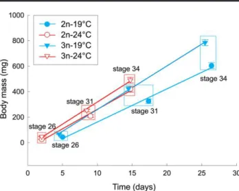
different. Comparisons of body masses at three developmental stages (fig. 3) by means of ANOVA (for each stage separately) revealed an increasing effect of the interaction between ploidy and temperature with advancing stage (F1, 6p 0.21, P p 0.662;
F1, 6p 3.11, P p 0.129; F1, 5p 11.46, P p 0.019, for stages 26,
31, and 34, respectively). The interaction was significant in stage 34 but not in previous stages. Body masses of diploid and triploid tadpoles in stage 34 did not differ at 247C (F1, 6 p 4.98, P p
0.067), in contrast with 197C, where triploids had significantly higher masses both in the same stage 34 (F1 ,5 p 30.28, P p
0.002) and at a similar age (in stage 31; F1, 6p 13.74, P p 0.010;
table 1). We concluded, therefore, that triploids enjoyed faster growth than diploids at 197C and the difference between two ploidies was less pronounced at 247C. This is supported by the comparison of the slopes of regression lines of body mass versus age in figure 3 (each fitted to three averages for each temper-ature/ploidy). Triploid tadpoles grew 31.5% and 13.0% faster than diploids at 197 and 247C, respectively, and growth rates of triploid tadpoles were similar at the two temperatures.
Body masses of triploid tadpoles at a given temperature, compared at the same developmental stage (26, 31, and 34), were always higher than in diploid individuals, except for the mentioned stage 34 at 247C (table 1). The mortality of tadpoles between hatching and stage 34 was very low, 1.4%5 0.5% on average (n p 16; eight crosses at each of the two tempera-tures) and was not affected by ploidy or temperature (F1, 6p
0.53, P p 0.495 and F1, 6 p 0.01, P p 0.941, respectively).
Metamorphosis
ANOVA revealed that ploidy and temperature significantly affected the body mass attained at metamorphosis (F1, 6 p
12.51, Pp 0.012 and F1, 6p 30.55, P p 0.001, respectively),
whereas there was no significant effect of the interaction
be-tween ploidy and the temperature (F1, 6p 1.74, P p 0.236).
Triploids were bigger at both temperatures, and both diploids and triploids were bigger at 197 than at 247C (fig. 4). Compar-ison of body masses at two temperatures separately revealed a significant difference between the two ploidies at 197 but not at 247C (table 1).
The age at metamorphosis was significantly dependent on the temperature (F1, 6p 361.30, P ! 0.001), but ploidy did not
affect the larval development time (F1, 6p 0.49, P p 0.511).
Both diploid and triploid tadpoles developed much faster at a higher temperature (fig. 4). The interaction between ploidy and temperature was not significant (F1, 6p 4.01, P p 0.092).
The mortality of tadpoles between stage 34 and meta-morphosis differed between the two temperatures but not be-tween ploidies (F1, 6p 27.75, P p 0.002 and F1, 6p 0.17, P p
0.692, respectively, with a nonsignificant interaction between the two factors, F1, 6p 0.53, P p 0.495). Mortality was higher
at 247C (32.8% 5 3.8%) than at 197C (6.3% 5 2.3%). Discussion
Cell Size
The results of our study showed that both ploidy and tem-perature significantly affected cell size. Triploid individuals, both tadpoles and froglets, had larger cells of the tested tissues compared to diploids (figs. 1, 2), which was, at least in part, a consequence of the larger genome of triploid cells. LLR trip-loids have 43% more DNA in erythrocyte nuclei than LR dip-loids (Ogielska et al. 2004). The large size of erythrocytes in polyploid animals is a well-documented fact, and measure-ment of erythrocyte size is the easiest method for distinguish-ing diploids from polyploids infish and amphibians (Austin and Bogart 1982; Polls Pelaz and Graf 1988; Matson 1990; Ballarin et al. 2004). However, only a few studies have reported that cells
Figure 2. Area of erythrocytes and cross-sectional area of hepatocytes in diploid and triploid froglets Pelophylax esculentus in relation to the temperature of the water in which they developed as larvae. The points are adjusted means (5SE) from two ANOVAs with ploidy and tem-perature as the main factors (see“Statistical Analyses”); the interaction terms were nonsignificant in these ANOVAs. N p sample size. Figure 1. Area of erythrocytes and cross-sectional area of epidermal
cells in diploid (2n) and triploid (3n) tadpoles of Pelophylax esculentus in relation to water temperature. The points are adjusted means (5SE) from two ANOVAs with ploidy and temperature as the main factors (see “Statistical Analyses”); the interaction terms were nonsignificant in these ANOVAs. Np sample size.
of other tissues are also larger in polyploid vertebrates than in their diploid counterparts (Fankhauser 1945; Swarup 1959; Suresh and Sheehan 1998). The larger hepatocytes and epi-dermal cells (unexplored tissues in Pelophylax esculentus un-til now) in triploids described in this study suggest that the whole body of polyploid P. esculentus is composed of larger cells, which is supported by the fact that cell size in amphibians is positively correlated between different tissues (Kozłowski et al. 2010).
Our results showed that water temperature strongly affected the size of erythrocytes and epidermal cells in tadpoles. Both
diploids and triploids had larger cells at lower temperature (fig. 1). This observation is consistent with research conducted on a wide range of diploid ectotherms that possessed larger cells at lower temperatures than their conspecifics reared at higher temperatures (van Voorhies 1996; Blanckenhorn and Llaurens 2005; Arendt 2007). It is not clear how temperature may induce changes in cell sizes. A recent study on Daphnia has shown that both nucleus and genome size increased in in-dividuals raised at 107C compared with those reared at 207C (Jalal et al. 2013). The authors of this study suggested that larger cell size at low temperature could be partly attributed to the enlarged nucleus and that DNA condensation was the most likely cause of the low-temperature response. It has been re-vealed that large-scale chromatin condensation occurs in on-togenesis for the control of the nucleocytoplasmic ratio at cell enlargement (Vinogradov 2005). More recently, Jalal et al. (2015) documented that both nucleus size and DNA condensation varied with temperature in Drosophila melanogaster, while DNA content appeared to be constant.
Tadpoles in our study were developing at two constant water temperatures, while in natural ponds tadpoles experience diurnal and seasonal variation of temperature. The average daily tem-perature range in ponds inhabited by P. esculentus in NE Poland was 5.17C in the beginning of tadpoles’ development and was 7.77C in the second half of development (A. Hermaniuk, un-published data). An interesting question is whether variable temperatures may have different impact on cell size of tadpoles than constant temperatures, although this subject requires fur-ther study. In a study on D. melanogaster, eifur-ther a higher mean temperature or daily variation of temperature (547C) caused flies to develop smaller cells relative to their body sizes, but the effect of thermal fluctuations was much weaker in magnitude than the effect of mean temperature (Czarnoleski et al. 2013). Interestingly, we did not observe any temperature effect on cell size in froglets a few months after metamorphosis when
Figure 3. Growth rate of diploid (2n) and triploid (3n) Pelophylax esculentus tadpoles in early phase of development at two water tem-peratures. Points are mean body masses of tadpoles at Gosner’s stages 26, 31, and 34 (adjusted means5 SE from ANOVA; see table 1); body masses are shown against time elapsed from stage 25, when tadpoles begin free swimming and independent feeding. The slopes of the lines at 247C are 30.7 and 34.7 mg d21in diploid and triploid tadpoles,
respec-tively, and at 197C are 26.0 and 34.2 mg d21, respectively.
Table 1: Body masses (mg) of diploid and triploid Pelophylax esculentus tadpoles in selected stages of development, reared at two water temperatures
Temperature (7C) and ploidy Stage 26 31 34 42 19: 3n 66.65 3.2 (20) 423.55 18.8 (34) 784.35 23.3 (21) 1385.95 59.0 (112) 2n 43.35 2.9 (38) 325.3 5 18.7 (34) 604.05 23.1 (23) 1124.35 59.3 (105) F 29.0 13.7 30.3 9.78 df 1, 6 1, 6 1, 5 1, 6 P .002 .010 .003 .020 24: 3n 47.45 1.9 (39) 252.8 5 10.6 (33) 490.15 22.5 (23) 1059.65 41.3 (78) 2n 26.45 2.0 (38) 208.5 5 10.8 (30) 419.15 22.5 (23) 923.35 41.4 (78) F 57.8 8.52 4.98 5.43 df 1, 6 1, 6 1, 6 1, 6 P !.001 .027 .067 .059
Note. Stage 42p metamorphosis. Values are adjusted means and standard errors from ANOVA, with ploidy and cross nested in ploidy as factors. Numbers of individuals are given in parentheses. Nonsignificant differences (P 1 0.05) between two ploidies at a given temperature are in bold.
animals shifted their environment from aquatic to terrestrial and were placed in common temperature (fig. 2). Cell size constraints (also TSR) seem to be most noticeable in aquatic habitats where the solubility of oxygen decreases with tem-perature. This may imply selection for smaller species at high water temperatures and also explain the gigantism of many polar taxa (Chapelle and Peck 1999). In marine invertebrates (e.g., in copepods and other major groups of crustaceans), the general pattern of enlarged adult body size at low temper-atures reflects enlarged cell and genome size, whereas among terrestrial invertebrates, various responses are common, including both larger and smaller body size in colder areas (Hessen et al. 2013).
Body Mass versus Cell Size in Tadpoles
Our measurements of cell size in tadpoles revealed a larger size in triploid than in diploid tadpoles and a larger cell size at lower than at higher temperature. Comparison of these mea-surements and the course of body masses in tadpoles indicates that cell size directly affects their body size. First, the body mass of triploid tadpoles was significantly larger than in dip-loids at all developmental stages at 197C and at the first stages at 247C (table 1). Second, both diploid and triploid tadpoles reared at lower temperature, besides having larger cells, reached a larger body mass at metamorphosis (fig. 4). The largest body size at metamorphosis found in triploid tadpoles reared at 197C was in agreement with the largest cell size of triploids devel-oping at that temperature. It should be noted that the large size of our P. esculentus triploid (LLR) tadpoles was not caused by the gene dosage effect, that is, by an excess of L genomes over R genomes, because the parental Pelophylax lessonae (LL) is smaller than P. esculentus (Berger 2008).
Earlier studies on P. esculentus have revealed that triploids (LLR) have a larger body length than diploids (LR) at the end of metamorphosis (Gosner’s stage 46; table A1). Different
results from ours and those of the above authors have been reported in a recent article by Pruvost et al. (2013), who reared tadpoles of P. esculentus complex at two temperatures, 187 and 247C. They did not find differences in body mass between diploid (LR) and triploid (LLR) P. esculentus at metamorpho-sis. Only LRR metamorphs tended to be heavier than diploids (LL and LR) at 247C, which was interpreted as a weight effect of the Pelophylax ridibundus genome (R), the largest species in the water frog complex. The difference between ours and Pru-vost et al.’s (2013) study is that we weighed tadpoles at the beginning of metamorphosis (stage 42) while Pruvost et al. did so at the end of metamorphosis (stage 46; after weight loss due to tail resorption). Nevertheless, body masses at stages 42 and 46 are strongly correlated irrespective of rearing temperature, and the loss of body mass at that time is an invariable per-centage of the maximum premetamorphic mass, at least in dip-loid frogs, including P. esculentus (Negovetic et al. 2001; Álvarez and Nicieza 2002; Orizaola and Laurila 2009a). The most plau-sible explanation for the discrepancy might be the very restrictive feeding of tadpoles in Pruvost et al.’s (2013) study. This led to a very long time to metamorphosis in diploid and triploid P. esculentus, for example, 35% longer in diploids reared at 247C as compared with diploids in our study at 247C. The prolonged development of tadpoles was associated with a very low body mass of metamorphs, about half of the mass recorded in our study at 247C (when corrected for body mass loss between stages 42 and 46 after Orizaola and Laurila 2009a).
Although there are examples of increased body size of poly-ploids versus dipoly-ploids in invertebrates (Weider 1987; Walsh and Zhang 1992), a larger size for triploid P. esculentus is exceptional among polyploid vertebrate ectotherms, as follows from the scarce studies on that group. Tetraploid frogs Neobatrachus and trip-loid Gasterosteus aculeatus (three-spine sticklebacks), despite their larger cells, are of approximately the same size as their diploid counterparts (Swarup 1959; Mahony and Robinson 1980). Like-wise, tri-, tetra-, and pentaploid eastern newts Notophthalmus viridescens are no larger at hatching and metamorphosis than diploid forms at the same developmental stages (Fankhauser 1945). It has also been found in the two latter studies that the increase in a size of cells is met by a decrease in cell number. The only example similar to our results of a positive correlation be-tween cell size (only erythrocytes were analyzed) and body size in polyploid vertebrates has been presented in tetraploid Cobitis biwae (spinous loach; Schultz 1980).
Low temperature had a dramatic impact on tadpoles by in-creasing the age when tadpoles reached metamorphosis and their body mass at that time. The larger body mass of diploid and triploid tadpoles at 197C at the end of their linear segment of growth (on stage 34; fig. 3) is at least partly explained by their larger cells (fig. 1). Similarly, an increased body mass of diploid metamorphs in the cold has been reported in numer-ous studies on larval development in amphibians, including P. esculentus (Licht and Bogart 1989; Negovetic et al. 2001; Watkins and Vraspir 2006; Pruvost et al. 2013). Studies of the contribution of cell size to body size at different temperatures in diploid vertebrates have reported very mixed results. Fish
Figure 4. Body mass at metamorphosis (Gosner stage 42) versus time to metamorphosis in diploid and triploid tadpoles of Pelophylax es-culentus. Adjusted means (5SE) from ANOVA. N p sample size.
are the only group that have been studied extensively in this respect, but the majority of studies have reported embryonic growth only, comparingfish at hatching (for review see Arendt 2007). Hatchlings incubated in colder water have usually been larger and have consisted of larger cells (Arendt 2007). In Di-centrarchus labrax (sea bass), low temperature increases the age but not body mass of metamorphs, and they are built of larger cells (Ayala et al. 2001). In the lizard Anolis carolinensis, larger cells have been produced in hatchlings from cooler treatments, but hatchling body size is unaffected by temperature (Goodman and Heah 2010).
To our knowledge, our study is the first to report an in-crease in the body mass of a polyploid vertebrate caused by low temperature and that the increase in cell size contributes to increased body mass. This effect was shown in tadpoles from the stage of independent feeding to metamorphosis. The only comparable example from vertebrates that we are aware of comes from Ambystoma. Large (triploid) eggs have given rise to hatchlings larger than from small (mainly diploid) eggs only at low temperature, and the body size of hatchlings from large and small eggs is larger at cold temperatures than at warm temperatures (significant interaction between egg size and tem-perature; Licht and Bogart 1989). Nothing has been reported, however, about the subsequent course of body size in these Am-bystoma larvae.
Growth Rate and Body Mass–Ecological Background
Characteristically, at the initial linear segment of growth, triploid tadpoles grew at the same rate at 197 and 247C (fig. 3). In con-trast, diploid tadpoles grew at a lower rate that was most pro-nounced at the lower temperature. This suggests that decreasing temperature has a lower impact on mechanisms controlling de-velopment in our LLR triploids than in diploids. Consequently, LLR triploids of P. esculentus are better adapted to cold envi-ronments, which may elucidate their high proportion in popu-lations at high latitudes (Plötner 2005). The most plausible ex-planation for the wide temperature tolerance in triploid tadpoles is their increased heterozygosity resulting from the additional genome; this heterozygosity may manifest, for example, in more different forms of enzymes available for polyploids (Otto and Whitton 2000). The other possibility is that a higher quantity of enzymes per cell improves the metabolic processes of the poly-ploids at low temperatures (Dufresne and Hebert 1998).
Also, the larger size of LLR triploids in comparison to dip-loids revealed in our study (table 1; figs. 3, 4) may have sig-nificant ecological consequences. Larger body mass at earlier stages of development reduces predation risk and enhances competitive ability in many species of tadpoles (Travis et al. 1985; Semlitsch 1990). Numerous studies have confirmed that size at metamorphosis is crucially important for amphibians and enhances survival and fecundity in later life stages (Morey and Reznick 2001; Altwegg and Reyer 2003). Therefore, larger triploid P. esculentus froglets may survive better than diploid froglets, assuming that differences in body mass between two ploidies at the beginning of metamorphosis (our study) and at
the end of metamorphosis (table A1) continue for longer in terrestrial life. This assumption is supported by the fact that diploid P. esculentus individuals metamorphosing at a large size are larger at maturity (Altwegg and Reyer 2003). Unfor-tunately, we could not test this assumption in our froglets, be-cause too many uncontrolled factors determined their growth rate.
Benefits arising from larger body size in amphibians be-come particularly important in the northern part of species ranges. Northern populations, in cold environments, undergo prolonged hibernation periods, and P. esculentus hibernates mainly on land (Holenweg and Reyer 2000). Metabolic rate in ectotherms is a function of ambient temperature; thus, hiber-nating froglets seem not to be at risk of depleting endogenous energy stores, as long as snow provides insulative cover and air temperature is stable. Variable winter temperatures with peri-odic spells of increased temperatures result in enhanced mor-tality of P. esculentus (Anholt et al. 2003). Increased metabolic rate at warmer temperatures may risk exhaustion of energy reserves before frogs can begin feeding again in spring. Al-though diploid and triploid P. esculentus froglets do not differ in metabolic rates when corrected for body mass (A. Herma-niuk and J. R. E. Taylor, unpublished data), larger individuals have lower mass-specific metabolic rates. Larger on average triploid froglets, with lower mass-specific rates, may survive longer on body reserves. Consistent with this reasoning, the larger P. esculentus has a higher overwinter survival rate than smaller P. lessonae in the same habitat (Anholt et al. 2003).
Freeze tolerance and freeze avoidance via supercooling are important mechanisms for dealing with subzero temperatures in a diverse array of ectothermic animals that are terrestrial hi-bernators (Ramlov 2000). Pelophylax esculentus exhibits modest supercooling with a crystallization temperature ranging from 20.87 to 21.47C and survives moderate freezing (Voituron et al. 2005). Freezing tolerance in P. esculentus is significantly cor-related with body mass, and larger individuals exhibit lower ice accumulation rates (expressed as percent of body water frozen vs. time; Voituron et al. 2005). This may pose a selective ad-vantage for the larger triploid P. esculentus compared to diploid individuals, enhancing winter survival in cold climates. The ob-servation that survival of P. esculentus from metamorphosis until the following spring is positively related to size at metamor-phosis (Altwegg and Reyer 2003) supports this reasoning.
The larger body size of triploid tadpoles may also have other selective advantages. It has been proved that P. esculentus frogs do not compensate their small size at metamorphosis by en-hancing their postmetamorphic growth (Altwegg and Reyer 2003). This might be especially true in northern habitats, where the season suitable for growth after metamorphosis is very short and body mass increase during terrestrial life might not be sufficient to ensure survival through the winter. This is pre-sumably connected with decreasing temperatures at the end of the growing season resulting in a lower activity of froglets and their arthropod prey. Under such conditions, attaining a high body mass at the end of the aquatic phase of life is of crucial importance.