Vestibular nuclear complex as a centre of sensorimotor integration
10
0
0
Pełen tekst
(2) Dorota Bukowska. The term sensorimotor integration means processing sensory information in the central nervous system and using it in motor activity. In the case of spinal reflexes a synaptic pattern constitutes a relatively simple way of translation of sensory information into motor commands. The brain mechanisms of sensorimotor integration are more complex and, in consequence, little known. Nevertheless, the process is well-explained regarding the vestibular system. The sense of balance is not prominent in our consciousness, unlike vision, hearing, smell or taste; however, it is essential for the posture, coordination of motor responses and eye movements. Proper posture and balance require continuous information about the position and motion of all body parts, including the head and the eyes. The vestibular apparatus in the inner ear and the vestibular nuclei in the brainstem, which constitute the vestibular system, fulfill this requirement. This article will indicate an ability of the vestibular apparatus to generate signals for change of the head position in space and of the vestibular nuclei to integrate these and other sensory signals. Finally, it will be shown that the vestibular nuclei are engaged in the motor function since they serve a variety of postural and ocular reflexes, which make the upright, bipedal posture as well as stabilize retinal images despite the head and body movements.. VESTIBULAR APPARATUS This vestibular apparatus is mirrorsymmetric. It lies within the membranous labyrinth in the inner ear. It comprises five receptor organs: saccule and utricle (called the otolithic organs), and three semicircular canals. The saccule and the utricle measure linear acceleration resulting both from the force of gravity (saccule) and body motion (utricle). The semicircular canals detect angular acceleration caused by rotations of the head or the body. Each of these structures is filled with the extracellular fluid (endolymph) and contains sensory receptor cells called the hair cells. In the saccule and the utricle the hair cells cluster in an elliptical patch, called the macula. The hair bundle of each hair cell is embedded in a gelatinous sheet – the otoconial membrane – which in its superficial part contains millions of dense particles (calcium carbonate crystals) called the otoconia. In the semicircular canals, the hairs of hair cells are embedded in a gelatinous diaphragm called the cupula, which extends across an enlargement of the canal, named the ampulla. The hair cells are present in a part of the ampulla called the ampullary crest [1]. Each hair cell of the vestibular apparatus has a single long kinocilium and 40-110 stereocilia, whose length decreases with distance from the kinocilium (Fig. 1). The morphological axis of polarity of the hair cell runs from the smallest. stereocilia. Depolarization Hyperpolarization. Figure 1. Hair receptor cells in the vestibular organ have the ability of mechanoelectrical transduction (Adapted from [4], modified). 10.
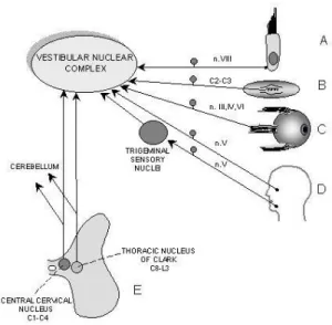
(3) Vestibular nuclear complex as a centre of sensorimotor integration. stereocilium to the kinocilium [2]. A condition for change in membrane potential of the hair cells is bending their hairs. The hair cells have the ability to transduce mechanical stimuli into receptor potentials. Deflection of the hair bundle toward the kinocilium causes depolarization of the hair cell and enhances the release of synaptic neurotransmitter – glutamate [3]. Deflection away from the kinocilium leads to hiperpolarization and reduction of neurotransmitter release. In consequence, these two situations increase or decrease the firing rate in the afferent fibres, respectively [4]. These fibres constitute the vestibular component (vestibular nerve) of the VIII cranial nerve (vestibulocochlear nerve) and make synapses with the hair cells. In the otolithic organs and semicircular canals a mechanism leading to hair bending is different, although both organs detect acceleration using the inertia of their internal contents. In the otolithic organs linear acceleration of the head causes shifting of an otoconial membrane that pulls the stereocilia, which undergo deformation. In the semicircular canals the head rotation causes a flexible flow of the endolymph against the rotation causing the deformation of the cupula. In result, the stereocilia bend in the opposite direction to the head rotation. AFFERENT CONNECTIONS TO THE VNC Sensory signals from the vestibular receptors about the linear and angular head acceleration are transmitted by the vestibular nerve directly to the vestibular nuclei and a specific part of the cerebellum, named the flocculonodular lobe, where glutamate or aspartate as excitatory neurotransmitters are released [3]. The vestibular nuclei constitute a complex of four nuclei that occupy a substantial part of the lower brainstem (medulla and pons) beneath the floor of the fourth ventricle. The vestibular nuclear complex (VNC) includes: the inferior (IV), medial (MV), lateral (LV) and superior (SV) vestibular nuclei. Each nucleus has a distinctive cytoarchitecture and a specific set of connections with the periphery and some regions of the brain and spinal cord [5]. The VNC as a sensory centre (Fig. 2A-E), apart from the primary vestibular afferents (A), receives sensory inputs from other receptors: proprioreceptors of the cervical (B) and extraocular muscles via the III, IV and VI cranial nerves (C) as well as mechanoreceptors (especially tactile. signals) of the face (D), transmitted by the trigeminal nerve (V cranial nerve) directly or indirectly with the synapse in the trigeminal sensory nuclei [6]. Cervical afferents come from levels C2-C3 and innervate MV and IV [7]. They send information from the muscle spindles and Golgi tendon organs of the dorsal and intervertebral cervical muscles which have a significant influence on the control of the head position and posture. In fact, transition of the dorsal roots of superior cervical segments result in posture disturbance which resembles symptoms after unilateral labirynthectomy, i.e. peripheral lesion of the vestibular apparatus. Moreover, IV, MV and LV are recipient of sensory information from the spinal cord (E), i.e. neurones of the central cervical nucleus and Clark nucleus, known as sources of the spinocerebellar fibres [8]. In addition, the VNC nuclei function in their own network of intranuclear (ipsilateral connections between one vestibular nucleus and another) and massive commissural connections (interconnections of the VNC nuclei). The latter allow the VNC to integrate the vestibular inputs of both sides [9, 10]. They arise from SV and MV, and exert inhibitory effects (by γ-aminobutyric acid and glycine) on canal-related neurones [3]. This is important in coordination of the vestibular organs of both sides. Strong reciprocal VNC connections can explain the phenomenon of vestibular compensation, i.e. a functional recovery from deficits associated with the unilateral labirynthectomy or vestibular nerve transection.. Figure 2. Vestibular neurones integrate sensory inputs from receptors on the periphery and from neurones of the spinal cord. 11.
(4) Dorota Bukowska. Apart from sensory information, the VNC neurones also integrate inputs from various brainstem centres, e.g. the reticular formation, inferior olive, interstitial nucleus of Cajal, locus coeruleus, nucleus prepositus hypoglossi [11, 12]. and from the hypothalamus of the diencephalon [13]. The largest projections yet are derived from the cerebellum, both cerebellar cortex and fastigial nucleus (Fig. 3).. Figure 3. Main afferent and efferent connections of the vestibular nuclei indicate their role in the sensorimotor integration. Figure 4. Vestibular nuclei receive inhibitory projections from the Purkinje cells of the cerebellar cortex and excitatory projections from the cerebellar fastigial nucleus (A). Photomicrographs of the retrogradingly labelled Purkinje cells in the posterior lobe cortex following injection of the WGA-HRP (horseradish peroxidase coupled with wheat germ agglutinin) into the medial vestibular nucleus (B). The Purkinje cell exerts a monosynaptic inhibitory (GABA-ergic) influence onto the vestibular neurone (C). D, F, G, E, dentate, fastigial, globose, emboliform cerebellar nuclei; Ve, vermis; FN, flocculonodular lobe. 12.
(5) Vestibular nuclear complex as a centre of sensorimotor integration. The sole output source of the processed information in the cerebellar cortex are axons of the Purkinje neurones (Fig. 4 A-C). They run to the cerebellar nuclei (where efferent pathways originate) and to the VNC. Afferents to the VNC arise mainly from most regions of the cerebellar vermis and from the flocculonodular lobe (Fig. 4A). Conspicuous projections come also from the cerebellar anterior and posterior lobes (Fig. 4B) [14, 15]. The Purkinje neurones are inhibitory. It has been shown in electrophysiological studies after stimlation of the Purkinje cell and intracellular recording in a vestibular neurone (Fig. 4C) (see [16] for more Refs). Direct inhibition of the VNC by these neurones may play an essential role in compensation of too strong impulses that are transmitted from the vestibular receptors to the VNC during active body movements. EFFERENT CONNECTIONS FROM THE VNC The VNC is not only a centre of sensory integration, but it is substantially engaged in motor functions. Vestibular neurones distribute information to the motoneurones of the spinal cord and motor nuclei of the extraocular muscles. Apart from the motor centres, the VNC projects to the cerebellar cortex and other centres at the supraspinal levels, e.g. the thalamus [17], red nucleus, inferior olive [18], cochlear nucleus [19], reticular nuclei [20] as well as at segmental levels (Fig. 3). Moreover, the VNC sends axons to some centres connected with autonomic functions: the hypothalamus [21], solitary tract nucleus, dorsal motor vagal nucleus, parabrachial nucleus and ventrolateral medullary reticular formation (see [22] for more Refs). These VNC pathways are suggested as substrate for vestibular contribution to phenomena as diverse as cardiovascular control, respiratory patterns, gastrointestinal function, anxiety disorders and motor sickness. In order to explain the motor function of the VNC, we will focus upon its central connections which play the most significant role in controlling of postural and eyes reflexes. According to classical studies [23] there are two pathways connecting the VNC with the spinal cord: the lateral vestibulospinal (LVST) and medial vestibulospinal (MVST) tracts. The LVST originates in LV and descends ipsilaterally to the lumbar segments in the ventral portion of the ventral funiculus. The MVST arises. from MV, and to a smaller extent from IV and LV, and descents bilaterally within the medial longitudinal funiculus only to the cervical segments. Both LVST and MVST terminate in the ventral horn (Rexed’s lamina VIII and rarely lamina IX) and intermediate gray matter (lamina VII), which remains in accordance with the main role of these pathways in the motor control. The LVST activates motoneurones mainly axial muscles and proximal muscles of the limbs, and thus influences the body posture. The MVST activates cervical motoneurones and controls the head position. It must be emphasized that exciting impulsation in the VNC neurones is permanently modulated by the inhibitory action of the cerebellar Purkinje cells. The significance of the cerebellovestibular inhibitory connections emerges from the fact that LVST and MVST neurones exert mono- or polysynaptic exciting of ipsilateral motoneurones of the extensor muscles and decrease of activity in the spinal mechanism of the flexor muscles. Therefore, straight reflexes are necessary to maintain equilibrium and proper body posture. More recent studies have proved that the vestibulospinal fibres are more widespread within the spinal cord than was previously thought. Axons from the LVN to the upper cervical cord descent not only ipsilaterally, but also contralaterally, and apart from the ventral horn, they terminate in lamina VII and even in the dorsal horn. Ipsilateral LVN projections are excitatory and make synapses with the dorsal and suboccipital extensor motoneurones. In contrast, contralateral projections inhibit motoneurones of these muscles [24]. Such connections play a role in the vestibular-neck reflex which stabilizes the head position. Moreover, LVST axones may extensively branch at many segmental levels throughout the entire length of the spinal cord. The multisegmental innervation of the spinal cord by LVST fibres suggests that a single LVST neurone may influence the activity of different types of interneurones along the spinal neuraxis and thus, in segment-specific manner, control movements of the head, trunk and limbs [25]. The findings from many laboratories suggest that LVST neurones modulate a variety of the spinal reflexes at different segmental levels and in this way influence the coordination of muscle action required for an ensemble of motor behaviors, including orientation and elaboration of body posture and movement. In addition, the vestibulospinal fibres originating in MV and IV can descent to the upper cervical levels not only within 13.
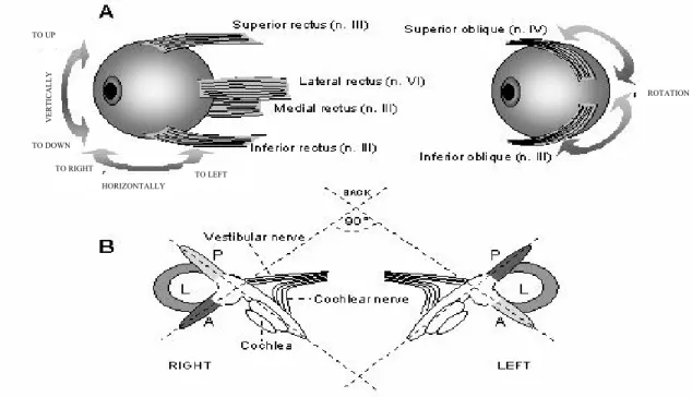
(6) Dorota Bukowska. the bilateral ventral funiculus, but also in the lateral, dorsolateral and dorsal funiculi. Fibres of the ventral funiculus make synapses in the ventral horn and the intermediate zone. Fibres of the lateral funiculus terminate in lamina VII and these of the dorsolateral and dorsal funiculi, terminate in the laminae IV and V as well as laminae I-VI (dorsal horn), respectively [26, 27]. Thus, the fibres contacting with different types of cervical neurones in the ventral horn, intermediate zone and dorsal horn are probably engaged in various functions, not only in the head movement control. For example, axons ending in the lamina V of the cervical dorsal horn might influence the processing of input from the cutaneous afferents, small-diameter muscle and joint afferents [28]. Also, the LVST axon collaterals coming from the cervical and lumbal levels can extend their terminal branches into lamina V. Thus, they can participate in the modulation of sensory transmission in the dorsal horn in addition to their well-known prominent influence on motor function via strong projections into the intermediate zone and the ventral horn [24]. The vestibulospinal pathways serve two reflexes: the vestibulocollic (vestibular-neck) and. vestibulospinal reflexes, which enable the skeletomotor system to compensate for the head movement. Each tilt or rotation of the head excites muscle motoneurones which maintain the head straight in relation to gravity. The vestibulocollic reflex acts on the neck muscles. When the body tilts foreward (e.g. in a situation when the body is pushed foreward without bending the neck), the neck extensor muscles contract raising the head up. When the body tilts backward, the motoneurones of the neck flexor muscles are excited. Tilting the body to the left or to the right causes extension of the ipsilateral limbs in order to counteract further tilting in this direction. On the other hand, the vestibulospinal reflex acts on limb muscles and prepares one for a potential fall while stumbling. Thus, tilting the head forward causes extension of the upper limbs and flexion of the lower limbs. In this way, a combination of movements is ensured which reduces the event of falling. Another function of the VNC as a motor centre is the control of eye movements reflexes that stabilize retinal images in spite of the head and body motions. The vestibuloocular and optokinetic reflexes which compensate for head movement. VERTICALLY. TO UP. ROTATION. TO DOWN TO RIGHT. TO LEFT HORIZONTALLY. Figure 5. Motor innervation of extraocular muscles responsible for eye movements in the skull. In parenthesis, n. III, IV and VI correspond to the oculomotor, trochlear and abducens motor nuclei (A). The semicircular canals display a bilateral symmetry and constitute three functional pairs. In the same plane lie the lateral horizontal (L) canals on both sides, and the anterior vertical (A) canal on one side and posterior vertical (P) canal on the opposite side (B). 14.
(7) Vestibular nuclear complex as a centre of sensorimotor integration. fulfill this task. The vestibuloocular reflex is decisive during rapid head movements and requires signals from the semicircular canals. The optokinetic reflex, in turn, processes information about sustained head rotations and depends on an optical input (see [29] for more Refs). Both reflexes generate conjugate eyes movements in the opposite direction to the head rotation in such a way that the retinal image does not undergo shifting. Eye movements in the skull are ensured by three pairs of extraocular muscles: two pairs of the rectus muscles (medial and lateral muscles and superior and inferior muscles) and one pair of the oblique muscles (superior and inferior muscles) (Fig. 5A). The extraocular muscles are innervated by the motoneurones of three cranial nerves: III (oculomotor), IV (trochlear) and VI (abducens) nerves. These motoneurones are controlled by the vestibular nuclei whose axones run in the medial longitudinal fasciculus. Eyes perform conjugate movements, which means that the muscle action of both eyes complement each other. For example, a contraction of the lateral rectus muscle of one eye is conjugated with a contraction of the medial rectus muscle of the other one. It results in the horizontal eyes movement. The semicircular canals are situated in three perpendicular plains and signalize the direction and velocity of the head rotation (Fig. 5B). Each of these plains lies each of the two pairs complementing extraocular muscles, e.g. the left and right lateral horizontal canals lie in the plain of the rectus muscles, medial and lateral. During head rotations, signals from each canals excite a pair of muscles, whose direction of activity is opposite to the direction of head rotation and simultaneously inhibits a pair of muscles whose direction of activity is in accordance with the direction of head rotation. These relationships can be presented easily on the basis of the horizontal vestibuloocular reflex (Fig. 6). For example, a head turning to the right excites the right horizontal canal. It gives rise to an increase of the discharge level in the right vestibular nerve and a decrease of he discharge level in the left vestibular nerve, proportionally to the head velocity. As a result, the left lateral and right medial rectus muscles are excited, and the left medial and right lateral rectus muscles are inhibited. The motor commands for this reflex are sent by the following neuronal network: Motoneurones of the lateral rectus muscle in the pons (abducens nucleus) receive monosynaptical input from the contralateral MV neurones. Motoneurones of the medial rectus. muscle in the midbrain (oculomotor nucleus) are driven by abducens interneurones and directly by the ipsilateral LV neurones. Both motoneurones and interneurones of the abducens nucleus receive the same vestibular signals, but the latter project to the oculomotor nucleus within the contralateral medial longitudinal fasciculus. These connections facilitate the horizontal eyes movement to the left, which compensates for the head rotation to the right (Fig. 6A). It should be stressed that during the rightward head rotation the rightward eyes movement is simultaneously inhibited by the afferents from the right horizontal canal. These afferents excite MV neurones which inhibit the motoneurones and interneurones of the right abducens nucleus. Such connections reduce the excitation of motoneurones for the left medial and right lateral rectus muscles (Fig. 6B). Moreover, the same head rotation causes a decrease of signals in the left horizontal canal and (basing on similar connections) a decrease of inhibition of the left lateral and right medial rectus muscles as well as a decrease in the excitation of the left medial and right lateral rectus muscles (not shown). Neuronal connections in the horizontal vestibuloocular reflex are decisive for coordination of the rectus muscles, medial and lateral, in a situation of gazing to the side. SOME EFFECTS OF THE VESTIBULAR LESIONS Lesions of the peripheral receptors (labyrinthectomy) or the vestibular nerve (vestibular neurectomy) lead to the appearance of characteristic symptoms of ataxia accompanied by severe deficits involving body posture, locomotor equilibrium, spatial orientation and oculomotor control (gaze stabilization). In most species after a unilateral lesion, the lack of symmetry occurs in distribution of the postural muscle tone. It is characterized by extension of the contralateral limbs and flexion of the ipsilateral limbs to the lesioned side. In addition, as a result of an ipsilateral increase and a contralateral decrease in the head extensor muscle tone, a considerable tilt of the head to the lesion is observed. During the first days after the lesion, standing is impossible. Typical ataxic gait shows an ipsilateral deviation with frequent falls to the lesion side. Spatial stabilization of the head is impaired and the ocular nystagmus toward the intact side is marked [5]. All 15.
(8) Dorota Bukowska. Figure 6. Neuronal network in the horizontal vestibuloocular reflex. Excitatory (A) and inhibitory (B) connections. SV, superior, LV, lateral, MV, medial, IV, inferior vestibular nuclei. these behavioral disorders are correlated with the imbalance in the vestibular influences, normally exerted bilaterally on the neck, trunk, limbs and extraocular muscles by the vestibulospinal and vestibuloocular pathways, respectively. Studies of astronauts during orbital flights in microgravity indicate the significant role of the vestibular connections originating from the otholitic organ. It indeed stops registering the head tilting, but maintains tonic postural activity in the antigravity muscles. In man the strongest antigravity muscles (i.e. supporting the body against gravity) are the extensors of the lower limbs and flexors of the upper limbs. It is interesting that in zero gravity a functional differentiation of the VNC nuclei can be comparable in several aspects to this resulting from experimental labyrinthectomy or pathology (see [5] for more Refs.). Experimental studies show that the vestibulospinal fibres are necessary for development of the decerebellar rigidity that displays the maximal extension of all limbs. It is induced by the rostral midbrain transaction, which interrupts descending rubrospinal fibres controlling flexor muscles, but abolished by lesion of LV or 16. interruption of the LVST [2]. Thus, the rigidity mechanism is connected with a tremendous increase of the extensor muscle tone, caused by an increased firing rate of gamma and alpha motoneurones excited by the vestibulospinal fibres. The extent of the decerebellar rigidity depends on the action of the cerebellar Purkinje cells which inhibit the LV neurones. Lesions from the cerebellar cortex regions which project to LV neurones abolish this inhibition and thus enhance the rigidity. An electrical stimulation of these cortical regions invokes an opposite effect. CONCLUSIONS In this article, the morphology and functioning of only some of vestibular connections have been presented. However, they reflect a functional variety of the vestibular system related to the maintenance of body equilibrium, orientation in a three-dimensional space and modification of the muscle tone. The above conclusions derive from the ability of the vestibular neurones to integrate the sensorimotor information. These neurones.
(9) Vestibular nuclear complex as a centre of sensorimotor integration. process information from several sensory systems in order to form suitable output motor response. As a result of the coordinated action of the VNC with above mentioned centres, a change of the head position leads to an immediate involuntary correction of the tonus muscles of the neck, limbs and trunk (realized through straight reflexes) as well as the compensatory movement of the eye balls to stabilize a retinal image. An action of the vestibular system is so efficient that in normal conditions it escapes our attention. However, when the system is disrupted or disordered characteristic symptoms appear such as disturbances of body posture and equilibrium in static position (astasia) and motion (abasia), pathology of eyes movement (nystagmus) and labyrinthine vertigo (Meniere’s syndrome). Because of the involvement in the autonomic function, it is also responsible for motion sickness. Thus, the vestibular nuclear complex is not only a very important sensory centre, but it also occupies a strategic position among the structures connected with the motor control. REFERENCES [1] Shepherd G.M., Neurobiology, part 14, The Sense of Balance, Oxford University Press, 1988, pp. 286-303. [2] Spencer R.F., The Vestibular System, (in:) P.M. Conn, ed., Neuroscience in Medicine, Philadelphia, 1995, pp. 501-509. [3] Raymond J., Dememes D., Nieoullon A., Neurotransmitters in vestibular pathways, Progress in Brain Research, 1988, 76: 29-43. [4] Goldberg M.E., Hudspeth A.J., The Vestibular System, (in:) E.R Kandel, J.H. Schwartz, T.M Jessell, eds., Principles of Neural Science, New York, 2000, pp. 801-815. [5] Lacour M., Borel L., Vestibular control of posture and gait, Archives Italiennes de Biology, 1993, 131: 81-104. [6] Buisseret-Delmas C., Compoint C., Delfini C., Buisseret P., Organisation of reciprocal connections between trigeminal and vestibular nuclei in the rat, Journal of Comparative Neurology, 1999, 409: 153-168. [7] Bankoul S., Neuhuber W.L., A cervical primary afferent input to vestibular nuclei as demonstrated by retrograde transport of wheat germ agglutininhorseradish peroxidase in the rat, Experimental of Brain Research, 1990, 79: 405-411.. [8] Matsushita M., Gao X., Yaginuma H., Spinovestibular projections in the rat, with particular reference to projections from the central cervical nucleus to the lateral vestibular nucleus, Journal of Comparative Neurology, 1995, 361: 334-344. [9] Ito J., Matsuoka I., Sasa M., Takaori S., Commissural and ipsilateral internuclear connections of vestibular nuclear complex of the cat., Brain Research, 1985, 341: 74-81. [10] Pompeiano O., Mergner T., Corvaja N., Commissural, perihypoglossal and reticular afferent projections to the vestibular nuclei in the cat. An experimental anatomical study with the method of the retrograde transport of horseradish peroxidase. Archives Italiennes de Biology, 1979, 116: 130-173. [11] Grottel K., Jakielska-Bukowska D., The reticulovestibular projection in the rabbit: an experimental study with the retrograde horseradish peroxidase method, Neuroscience Research, 1993, 18: 179-193. [12] Schuerger R.J., Balaban C.D., Organization of the coeruleo-vestibular pathway in rats, rabbits, and monkeys, Brain Research Review, 1999, 20: 189-217. [13] Takeda N., Morita M., Kubo T., Yamatodani A., Watanabe T., Tohayama M., Wada H., Matsunaga T., Histaminergic projection from the posteriori hypothalamus to the medial vestibular nucleus of rats and its relation to motion sickness, (in:) M.D. Graham and J. L. Kemink, eds., The Vestibular System: Neurophysiologic and Clinical Research, Raven Press, New York 1987, pp. 571-580. [14] Bukowska D., Cerebellovestibular projection from the posterior lobe cortex in the rabbit: an experimental study with the retrograde HRP method. I. Topographical relationships, Acta Neurobiologiae Experimentalis, 1995, 55: 23-34. [15] Bukowska D., Cerebellovestibular projection from the posterior lobe cortex in the rabbit: an experimental study with the retrograde HRP method. II. Zonal organization, Acta Neurobiologiae Experimentalis, 1995, 55: 35-47. [16] Ito M., The Cerebellum and Neural Control, Raven Press, New York, 1984. [17] Shiriyama T., Kayahara T., Yasui Y., Nomura J., Nakano K., The vestibular nuclei of the rat project to the lateral part of the thalamic parafascicular nucleus (centromedian nucleus in primates), Brain Research, 1995, 704: 130-134. [18] Matesz C., Bacskai T., Nagy E., Halasi G., Kulik A., Efferent connections of the vestibular nuclei in the rat: A neuromorphological study using PHA-L, Brain Research Bulletin, 2002, 57: 313-315.. 17.
(10) Dorota Bukowska. [19] Bukowska D., Morphological evidence for secondary vestibular afferent connections to the dorsal cochlear nucleus in the rabbit, Cells Tissues Organs, 2002, 170: 61-68. [20] Bukowska D., Grottel K., Secondary vestibular projection to the reticular formation of rabbit lower brainstem with reference to reciprocity, Archives Italiennes de Biology, 1997, 135: 205-218. [21] Matsuyama T., Kayahara T., Nomura J., Nakano K., Direct projections from the medial vestibular nucleus to the posterior hypothalamic area in the monkey (Macaca fuscata), Neuroscience Letters, 1996, 219: 199-202. [22] Balaban C.D., Vestibular nucleus projections to the Edinger-Westphal and anteromedian nuclei of rabbits, Brain Research, 2003, 963: 121-131. [23] Nyberg-Hansen R., Origin and termination of fibers from the vestibular nuclei descending in the medial longitudinal fasciculus. An experimental study with silver impregnation methods in the cat, Journal of Comparative Neurology, 1964, 122: 355-367. [24] Rose P.K., Ely S., Norkum V., Neuber-Hess M., Projections from the lateral vestibular nucleus to the upper cervical spinal cord of the cat: a correlative light and electron microscopic study of axon terminals stained with PHA-L., Journal of Comparative Neurology, 1999, 410: 571-585.. 18. [25] Kuze B., Matsuyama K., Matsui T., Miyata H., Mori S., Segment-specific branching patterns of single vestibulospinal tract anons arising from the lateral vestibular nucleus in the cat: a PHA-L tracing study, Journal of Comparative Neurology, 1999, 414: 80-96. [26] Bankoul S., Goto T., Yates B., Wilson V.J., Cervical primary afferent input to vestibulospinal neurons projecting to the cervical dorsal horn: an anterograde and retrograde tracing study in the cat, Journal of Comparative Neurology, 1995, 353: 529-538. [27] Donevan A.H., MacDonald J.A., Brennan P.A., Rose P.K., Morphology of single vestibulospinal collaterals in the upper cervical spinal cord of the cat. II. Collaterals originating from axons outside the ventral funiculi, Journal of Comparative Neurology, 1992, 322: 343-359. [28] Neuhuber W.L., Zenker W., Central distribution of cervical primary afferents in the rat, with an emphasis on proprioceptive projections to vestibular, perihypoglossal, and upper thracic spinal nuclei, Journal of Comparative Neurology, 1989, 280: 231-253. [29] Watanabe S., Kato I., Sato S., Norita M., Direct projection from the nucleus of the optic tract to the medial vestibular nucleus in the cat, Neuroscience Research, 1992, 17: 325-329..
(11)
Obraz
![Figure 1. Hair receptor cells in the vestibular organ have the ability of mechanoelectrical transduction (Adapted from [4], modified)](https://thumb-eu.123doks.com/thumbv2/9liborg/3117886.8873/2.892.109.802.792.1118/figure-receptor-vestibular-ability-mechanoelectrical-transduction-adapted-modified.webp)

+2

Powiązane dokumenty
Relacje pomiędzy nauczycielem a uczniami oparte powinny być na współpracy. Ta zaś wymaga, by we wspólnej pracy zachować szacunek dla wzajemnej autonomii i różno- rodności. W
By integrating moral, spiritual and social aspects, the process of education focuses on the holistic development of an individual in harmony with the world and himself..
Можно таким образом полагать, что выдвинутый Кедровым постулат переворачивания закреплен в пространстве
Kobiety i rodzice istotnie częściej niż mężczyźni i osoby bezdzietne są prze- ciwnikami stosowania kar fizycznych, zwolennikami prawnego ich zakazu, częściej chcą reagować
The assessment of the usefulness of social media in the dissemination of information about health and disease in relation to the e-health literacy of Polish
A non-exercising, control group (76.9±9.3 kg) showed very similar mean scores in handgrip strength and visuo-motor abilities during both examinations, therefore those data
onTK=that is the average number of the most common altmetric indicators for most highly cited lA articles written by Czech and molish
( 9 ) 13 airports in the Union are forecasted to be operating at full capacity eight hours a day every day of the year in 2030, compared to 2007 when only 5 airports were operating
