TRENDS
in
Sport Sciences
2014; 4(21): 185-194.ISSN 2299-9590
Numerous data have indicated that water permeability in living systems is greater than it could be explained by simple diffusion. Electron microscope observations have identified special structures presumed to be water channels. The molecular identity of the first water channel was determined in the early 1990s and named Aquaporin 1 (AQP1). It has been now well documented that aquaporins are members of a large family of small (about 28- kDa/ monomer) integral membrane proteins which exist as tetramers, with each subunit containing its own pore. Mammalian AQPs are believed to fold and assemble in the endoplasmic reticulum before being transported to the cell surface. To date 13 AQP’s have been identified in mammals (AQP0-AQP12); however, functional studies have identified a subgroup of AQPs, i.e. AQP 3, 7, 9 and 10, responsible for both water and glycerol transport, named aquaglyceroporins. Further studies have demonstrated that, apart from water and glycerol, AQPs 3, 7, 9 also transport ammonia and urea. Additionally, AQPs 11 and 12 named superaquaporins, were localized inside the cells, but until now their functions have not been fully elucidated. AQP defects in the human adipose tissue and liver are recognized as a possible cause of obesity. Numerous data indicate that AQPs contribute to carcinogenesis. Data on the effects of physical activity on AQP are scarce; however, it has been recently demonstrated that AQP expression in skeletal muscle and adipose tissue, but also in the brain, respond to physical stress. Thus, it seems possible that in near future AQP studies will provide more knowledge concerning preventive effects of physical exercise in medicine. Furthermore, there is a growing interest in chemicals affecting AQPs, and it could not be excluded that AQP-targeting drugs will be used in medical practice.
KEYWORDS: aquaporins, metabolism, aquaporin-related disease.
Received: 21 October 2014 Accepted: 12 November 2014
Corresponding author: grazyna.lutoslawska@awf.edu.pl The Józef Piłsudski University of Physical Education, Department of Biology and Biochemistry, Warszawa, Poland
What this paper adds?
The present paper discusses current views concerning the structure and functions of water channels (aquaporins – AQPs) in mammals. Data have indicated that distorted expression of AQPs brings about pronounced metabolic disturbances in the kidney, skin, eyes and lungs, but also in the adipose tissue and liver, the latter possibly contributing to the development of obesity. Physical activity was found to affect AQP expression; however, until now data concerning this issue are limited. In skeletal muscle AQPs are expressed mostly in fast twitch fibers, and in animals their knockout affects energy processes. Thus, analysis of AQPs function can shed light on our understanding of water function in metabolic processes, both in health and disease.
S
tudies of water transport in amphibian tissues have indicated that certain cells are more permeable to water than others. In other words, numerous data have demonstrated that in certain cells water permeability is greater than it could be explained by simple diffusion [1].Aquaporins in physiology and pathology
Further studies and electron microscope observations identified special structures in the amphibian bladder which were presumed to be water channels. Similar structures were identified in red blood cells, and they were shown to be inhibited by mercury salts and reversed by reducing chemicals. Thus, it has been suggested that water transport, at least partially, occurs with the contribution of proteins containing sulfhydryl groups [2]. The molecular identity of the water channel was determined by Agre in the early 1990s, and was named Aquaporin 1 (AQP1) [3]. In 2003 Agre was honored with the Nobel Prize, after his team’s experiments markedly extended our knowledge concerning water transport in living systems.
Aquaporin structure and regulation
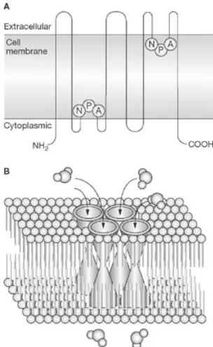
Aquaporins are members of a large family of small (about 28-kDa/monomer) integral membrane proteins which exist as tetramers with each subunit containing its own pore. The monomer structure of bovine AQP1 reveals six trans-membrane helices, each with highly conserved three amino-acid sequences (asparagine-proline-alanine – NPA) [4, 5] (Figure 1).
Mammalian AQPs are believed to fold and assemble in the endoplasmic reticulum before being transported to the cell surface, with NPA motifs playing a key role in plasma membrane targeting [6, 7]. However, the translocation of AQPs into the plasma membrane is precisely regulated by different stimuli such as hormones, neurotransmitters, amino acids, as well as by hypo- and hyper-tonicity [8] (Table 1).
Aquaporins as small molecule transporters in mammals
To date 13 AQPs have been identified (AQP0-AQP12) in mammals; however, functional studies have identified a subgroup of AQPs, i.e. AQP3, 7, 9 and 10, named aquaglyceroporins, responsible for both water and glycerol transport [9]. Moreover, further studies demonstrated that, except for water and glycerol, AQPs 3, 7, 9 also transport ammonia and urea [10] (Table 2). Additionally, AQPs 11 and 12, named superaquaporins, are localized inside the cells, but until now their functions have not been fully elucidated [11, 12]. Interestingly, AQP1 and 8 seem to be engaged in hydrogen peroxide transport [13]. This finding is of special importance, since in mammals, hydrogen peroxide is a signaling compound involved in the regulation of diverse functions such as immunity and vascular remodeling [14].
Figure 1. General structure of the Aquaporin family: A –
monomer; B – tetramer in cell membrane www.nature.com/ nrendo/journal/v4/n11//images/ncpendmet0980-f1.jpg
Table 1. Selected triggers of aquaporin translocation in
mammals [8]
AQUAPORIN Trigger Cell/Tissue
AQP1 Secretin Cholangiocytes
Hypotonicity Astrocytes
AQP2 Vasopressin Kidney tissue
Hypertonicity Collecting duct
AQP3 Isoprenaline Adipocytes
Hypotonicity Keranocytes
AQP4 Histamine Gastric cells
Glutamate Astrocytes AQP5 Hypertonicity Lung epithelial cells
VIP* Brunner’s gland
AQP7 Isoprenaline Adipocytes
AQP8 Hypertonicity Amnion epithelium
AQP9 Hyperonicity Astrocytes
Furthermore, there are experimental data suggesting that AQP1s transport carbon dioxide; however, this issue remains controversial [15, 16].
AQPs are thus multifunctional channels, and the function of a single AQP is difficult to define. However, recently, it has been found that, at least in amphibians, different AQPs exhibit a diverse range of selectivity for water, glycerol, ammonia and carbon dioxide. In consequence, by expressing specific combinations of AQPs different cells could control the movement of each of these substances [17]. It could not be excluded that a similar mechanism operates in mammals, which makes research on the AQP activity in different tissues and organs rather complicated.
Nonetheless, considering that the water flow across cell membranes is fundamental for all living systems many studies have been undertaken to elucidate the role of aquaporins in physiology and pathology. Special attention has been paid to the gastrointestinal tract, eye, salivary glands, skin, lungs and kidneys, whose physiological functions are significantly regulated by the water flow [18, 19].
Metabolic consequences of AQPs knockout in animals and inherited human defects
Several AQPs have been identified in humans and animals in different GT regions: AQP1, 3, 4, 6, 7, 8, 9 and 10 in the small intestine; AQP3 and AQP4 in the colonic epithelium [20, 21]. However, the functions of AQPs in GT have not been fully elucidated as yet. On the other hand, it has been found that AQP-3 deficient mice develop markedly more severe, experimental colitis
than their wild-type littermates [22]. Moreover, it has been shown that in rats the AQP8 mRNA expression is significantly elevated in different GT regions either after subtotal colectomy or massive intestinal resection [23, 24].
A significant role of AQP8 in GT has been suggested in humans, since the expression of AQP8 differs between healthy colonic epithelial cells and colorectal tumors [25]. Furthermore, an elevated AQP8 expression was noted in inflammatory bowel disease [26]. Six APQs (AQP0-AQP5) have been found in the eye in mice. They are located in different organ regions (corneal epithelium, conjunctiva, lens and ciliary epithelium, lens fiber cells and Müller cells) [27]. Individual AQPs in mice have been shown to be responsible for intraocular pressure regulation, corneal epithelium repair, corneal and lens transparency, retinal signal transduction, retinal swelling after injury and accelerated cataract formation. Moreover, genetic defects of AQP0 in humans lead to congenital inherited cataracts [28, 29]. AQP1, 3 and 4 are located in the lung and the airways, however, their knockout in mice does not affect lung function [30, 31]. In contrast, in rats, changes in AQP1 and 5 in the small airways and the alveoli were found in an ovoalbumin sensitization asthma model [32]. Moreover, in rare cases of human AQP1 deletion, decreased pulmonary vascular permeability was noted [33]. Recently, it has been found that a decreased AQP5 expression in the lung is observed in humans with chronic obstructive pulmonary disease (COPD), and that it correlates with the disease severity [34].
Early studies concerning the role of AQPs in the kidney demonstrated that in mice the lack of AQP1 fluid re-absorption in the proximal tubule is defective and that the urinary concentrating ability is severely impaired [35, 36]. It has been documented, however, that seven AQPs (AQP1-AQP4, AQP6, 7 and AQP11) are expressed in the kidney [37]. They are located in different kidney regions and their deletion brings about different metabolic disturbances with diabetes insipidus in response to AQP2 deletion, both in humans and animals [38, 39].
Aquaporins (AQP1-AQP7) are expressed in the salivary glands [40]. In humans and animals the AQPs activity in the salivary glands is markedly affected by age, radiation therapy, and it is depleted in Sjögren’s syndrome – a chronic autoimmune inflammatory disease
Table 2. Aquaporin permeability in mammals [5]
Aquaporin Water Glycerol Urea Ammonia
AQP0 +/–* – – Not detected
AQP1 + – – –
AQP2 + – – –
AQP3 + + +/– +
AQP4 + – – –
AQP5 + – – –
AQP6 +/– +/– +/– Not detected
AQP7 + + + +
AQP8 + – – +
AQP9 +/– + + +
AQP10 +/– + + Not detected
characterized by lymphocyte infiltration of the salivary and lacrimal glands. In an animal model of Sjöngren’s syndrome an altered AQP5 distribution was noted in the submandibular gland concomitantly with inflammation progress [41].
In mammals, the AQP3 water/glycerol transporter is responsible for skin hydration and skin wound healing, since both processes are depressed in AQP3 knockout mice [42]. On the contrary, glycerol replacement by topical, intra-peritoneal and oral routs corrects defective skin elasticity, hydration, and barrier function [43]. Moreover, it has been recently demonstrated that mutations in the AQP5 water channel in humans cause inherited palmoplantar keratoderma characterized by thickening of the stratum corneum on the palms and the soles [44]. Numerous data have indicated that AQP4 plays an important role in the brain, contributing to the regulation of water transmembrane movement at the blood-brain barrier and brain-cerebrospinal fluid interface [45]. In AQP4 knockout mice decreased CFS production and increased brain water content have been found together with ependymal layer disorganization [46]. Moreover, deletion of glial AQP4 brings about impairment of selected forms of spatial memory [47]. It was also found that AQP4 deletion in mice improved survival and neuroprotection following global cerebral ischemia due to reduced swelling [48]. Interestingly, it is suggested that in human neuromyelitis optica (NMO), an inflammatory demyelinating CNS disorder, and Baló’s disease, a rare variant of multiple sclerosis, are closely related to reduced AQP4 expression [49, 50].
Recent data have indicated several AQPs expressions in different regions of the male reproductive system suggesting their role in reproduction [51]. Furthermore, AQP8 seems to play an important role in pregnancy outcome in mice since its deletion induces a significant elevation in embryo number compared to wild controls [52]. Moreover, the distribution of and quantitative changes in amounts of AQP1, 5 and 9 have been found in the pig uterus during the estrous cycle and early pregnancy [53]. In addition, it was demonstrated that AQP4 is present in human follicles during ovulation and its and AQP3 expression markedly rise before follicle rupture [54]. In contrast, the late rise in AQP1 suggests its role in corpus luteum formation.
According to Rutkovsky et al. [55] AQP1, 3, 4 deletions in mice induce rather modest changes in the heart. On the other hand, AQP4 expression has been found to coincide with cardiac dysfunction [56]. In addition,
in AQP4 knockout mice abnormalities of calcium modulating proteins were noted increasing the risk of cardiac failure and arrhythmia [57]. Furthermore, it is worth noting that deletion of aquaglyceroporin 7 in mice significantly and negatively affects heart energy production. This finding is of special importance since it indicates a significant influence of glycerol on energetic processes in cardiomyocytes [58]. It is worth noting that AQP1 also plays a significant role in regulation of the cardiovascular system, since its deletion in mice brings about microcardia and low blood pressure [59]. However, AQPs have been also identified in the pancreas, liver, adipose tissue and skeletal muscle, i.e. in tissues whose disturbed functions contribute to distorted lipid and carbohydrate metabolism.
Aquaglyceroporins and pancreas
It has been demonstrated that β-cells in mice express aquaglyceroporin 7 [60]. Additionally, the immuno-histochemistry method revealed a complete overlap between insulin and AQP7 immunostaining in the pancreatic islet. AQP7-/ mice were healthy, however
their β-islet mass and insulin content was lower and triacylglycerols content was higher than in AQP7+/+
littermates. Moreover, in AQP7 knockout animals the β-islet was characterized by a higher glycerol kinase activity and glycerol content. There was no evidence of insulin resistance in AQP7-/- animals; however, their
circulating insulin was higher than in wild-type animals. Recently, Louchami et al. [61] have found a dual role of AQP7 in isolated mice β-islet. On the one hand, AQP7 regulates the entry and exit of glycerol across islet cell membranes, and secondly – directly or indirectly – it affects the stimulus-secretion pathway of insulin in pancreatic islets. However, the last assumption has to be yet fully elucidated. Data concerning the AQPs role in the human pancreas are limited; however, it has been found that AQP1 is over-expressed in the plasma membranes of pancreatic ducts in autoimmune pancreatitis [62].
Adipose tissue and a new role of aquaglyceroporins
In fasting conditions triacylglycerols stored in the adipose tissue are hydrolyzed to glycerol and free fatty acids (FFA) by hormone-sensitive lipase, and both are released into the bloodstream [63]. To maintain circulating glucose glycerol is taken up by the liver and kidney, both expressing glycerol kinase, and used for glucose production in the gluconeogenesis pathway [64].
However, for a long time our knowledge about the mechanism of glycerol transport across adipocyte cell membranes was limited. It is now well documented that in humans and animals glycerol transport from adipocytes into the bloodstream is regulated by aquaporin 7 [65, 66].
Further animal studies showed that young AQP7 knockout (AQP7 KO) mice had lower circulating glycerol and were characterized by impaired glycerol response to adrenergic stimulation and rapid reduction in circulating glucose due to limited glycerol availability for gluconeogenesis in the liver [67].
On the contrary, in older AQP7 KO mice the loss of glycerol efflux from adipocytes resulted in elevated glycerol kinase (E.C.2.7.1.30) activity and increased synthesis of glycerol 3-phoshate followed by excessive triacylglycerol synthesis and accumulation [68, 69]. In consequence, whole-body metabolic disturbances have been noted such as elevated circulating FFA, hyperglycemia, and insulin resistance.
Taking into account the worldwide obesity epidemic animal data concerning AQP7 knockout initiated numerous human studies examining the AQP7 role in human obesity. Marrades et al. [70] and Ceperuelo-Mallafrĕ et al. [71] found that AQP7 gene expression was markedly depressed in obese vs. lean subjects. A significant role of AQP7 in glycerol and TG accumulation in human adipocytes was confirmed in
vitro using isolated 3T3-L1 cells [72]. Furthermore,
in both subcutaneous and visceral fat there were positive associations between AQP7 expression and lipogenic and lipolytic enzymes genes [73].
Studies on the AQPs’ role in human adipose tissue have been in progress. For a long time AQP7 has been the only aquaporin associated with adipose tissue. Recently, it has been demonstrated that, different than in mice, in humans both superaquaporins, i.e. AQP10 and 11, are expressed inside subcutaneous and visceral adipocytes in the vicinity of lipid droplets [74, 75]. Furthermore, both AQPs are colocalized with perilipin, a lipid droplet protein contributing to the regulation of lipolysis, and this localization confirms their contribution to fat metabolism in adipose tissue [76, 77].
However, an overall AQPs’ contribution to the regulation of metabolic processes cannot be elucidated without the adipocyte-liver connection.
Aquaporins in the liver
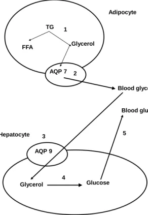
AQPs are expressed in mammalian heptocytes, intrahepatic bile ducts and gallbladder epithelium, which suggests that they contribute to the regulation of bile formation and transport but also to glucose and fat metabolism [78]. Special interest has been paid to aquaglyceroporin 9 activities in hepatocytes, since its gene expression responds to fasting and refeeding [79]. Additionally, it has been noted that AQP9 and AQP7 activities undergo coordinated regulation by insulin and, in consequence, AQP7-dependent glycerol efflux from adipocytes and AQP9-dependent glycerol uptake by hepatocytes play a crucial role in the regulation of liver gluconeogenesis and plasma glucose level [80] (Figure 2). Adipocyte TG FFA Glycerol AQP 7 Blood glycerol AQP 9 Glycerol Glucose 1 2 3 4 Hepatocyte 5 Blood glucose
Figure 2. Adipocyte glycerol release and gluconeogenesis
from glycerol in the liver under physiological conditions
1 – lipolysis; 2 – AQP7-mediated glycerol transport to the blood-stream; 3 – AQP9-mediated glycerol uptake by the liver; 4 – liver gluconeogenesis; 5 – glucose transport to the blood. AQP7 dele-tion in adipocytes brings about decreased glycerol transport into the blood, excessive glycerol accumulation in adipocytes and in-creased triacylglycerol synthesis, but also depressed sis in the liver. AQP9 deletion in the liver depresses gluconeogene-sis, increases circulating and adipocyte glycerol and triacylglycerol levels.
More information concerning the AQP9 role in hepatocytes was provided by authors of studies on AQP9 knockout mice [81, 82]. In AQP9 KO animals circulating glycerol and triacylglycerols were significantly elevated, while the glucose level was markedly lowered in comparison with their wild littermates. It suggests impaired liver gluconeogenesis due to limited glycerol uptake.
Moreover, it is worth noting that high triacylglycerol accumulation in the liver in response to disturbed aquaporin function is a possible cause of non-alcoholic fatty liver disease (NAFLD) in the rats on a high-fat diet [83]. Furthermore, it was also noted that AQP9 but also AQP8 and 11 are important for other vital liver functions, since it is postulated that in mice they contribute to liver regeneration after partial hepatectomy [84] (Table 3).
Function of aquaporins in skeletal muscle
AQP expression in skeletal muscle was identified in 1998 by Friegeri et al. [85], who demonstrated that in the rat AQP4 is expressed exclusively in fast-switch extensor digitorum longus (EDL) muscle but not in slow twitch soleus muscle. More detailed in vitro studies found that AQP4 was expressed in mice in the sarcolemma, but AQP1 is expressed in intramuscular capillary endothelial cells [86]. These findings enable a hypothesis that both AQP4 and AQP1 could be responsible for exercise-induced fast water transport from the blood into the muscle.
More data concerning the AQP4 function in the skeletal muscle were obtained from AQP4 knockout mice in which 19 proteins exhibited changed expression in comparison with wild-type animals [87]. Protein
identification revealed that 12 were up- and 7 were down-regulated, but all were engaged in energy metabolism and/or calcium handling (e.g. creatine kinase, parvalbumin). Additionally, physical treadmill training markedly elevated AQP4 protein content in rat muscles, which positively correlated with running speed during a single bout of exercise [88]. However, AQP4 mRNA was not elevated following training indicating posttranslational aquaporin regulation.
Until now data on effects of physical training on AQPs expression in humans have remained scarce. Lebeck et al. 2012 [89] revealed that – exclusively in females – AQP7 expression in subcutaneous fat increased 2.2-fold following training. Contrarily, a marked decrease in AQP7 expression has been found in men. These findings may be significant for our understanding of sex-related differences in substrate utilization during exercise. Moreover, the results of the same study revealed that genetic predisposition to type 2 diabetes was associated with lower AQP7 expression in subcutaneous abdominal fat.
More data concerning training effects on AQP are available in rats. Wang and Chen [90] have noted that a 6-week exhaustive swimming training in rats brings about decreased renal water reabsorption due to decreased AQP2 expression, and according to them, this may be the main cause of fatigue following extreme physical load. Furthermore, He et al. [91] have noted that in rats pre-training down-regulates AQP4 expression in the brain and, in consequence, ameliorates brain edema following ischemic stroke. Thus, it could be suggested that at least in rats regular physical activity plays a protective role in brain edema following stroke.
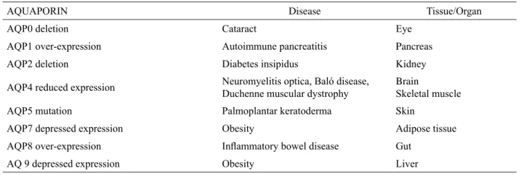
Table 3. Health consequences of disturbed AQP expression or deletion in humans
AQUAPORIN Disease Tissue/Organ
AQP0 deletion Cataract Eye
AQP1 over-expression Autoimmune pancreatitis Pancreas
AQP2 deletion Diabetes insipidus Kidney
AQP4 reduced expression Neuromyelitis optica, Baló disease, Duchenne muscular dystrophy Brain Skeletal muscle
AQP5 mutation Palmoplantar keratoderma Skin
AQP7 depressed expression Obesity Adipose tissue
AQP8 over-expression Inflammatory bowel disease Gut
Much more attention has been paid to the role of AQP4 in pathological skeletal muscle. It has been found that AQP4 expression is markedly reduced in the mdx mice model of Duchenne muscular dystrophy (DMD) [92]. In addition, there are also data suggesting decreased AQP4 expression in human DMD [93]. Less attention has been paid to the AQP1 function in skeletal muscle; however, it is postulated that this capillary-located endothelial cell aquaporin enhances muscle regeneration [94].
On the other hand, more attention should be paid to the role of exogenous glycerol during exercise, since water and glycerol mixtures are popular as hyperhydrating agents
[95]. However, data concerning glycerol drink effects on
metabolic variables are scarce suggesting protection of the cardiovascular system due to better hydration and no effects on anaerobic metabolism [96, 97].
The data presented above clearly suggest that studies on the structure and functions of AQPs shed a new light on metabolic processes which are of substantial importance for human health (Table 2). This is why research focusing on strategies that may affect the functions of AQPs has been understaken. Kong et al. [98] have found that carboxymethyl chitin (CM-chitin), a water-soluble chitin derivative, may exert an anti-obesity activity in isolated adipocytes by increasing glycerol efflux and decreasing triglyceride synthesis due to activation of AQP7 and AMP kinase. Additionally, it has been noted that gold-based compounds are effective in AQP protein inhibition, and according to researchers, are suitable for further research on anti-AQPs drugs [99, 100]. Interestingly, it has also been indicated that anti-AQP4 monoclonal antibodies are effective in preventing neuromyelitis optica (NMO) in mice [101]. Moreover, it is postulated that in the future AQPs may be drug targets in oncology due to their important role in cell proliferation and migration [102, 103]. Thus, it could not be excluded that aquaporin-mediated processes and their modulation by exogenous agents will open a new era in our understanding of body functions and in medical treatment of numerous serious diseases.
References
1. Agre P. The aquaporin water channels. Proc Am Thorac Soc. 2006; 3: 5-13.
2. Carbrey JM, Agre P. Aquaporins. Handb Exp Pharmacol. 2009; 190: 4-28.
3. Agre P, Sasake S, Chrispeels MJ. Aquaporins, a family of membrane water channels. Am J Physiol. 1993; 265: F461.
4. Borgnia M, Nilsen S, Engel A, et al. Cellular and molecular biology of the aquaporin water channels. Ann Rev Biochem. 1999; 68: 425-458.
5. King LS, Kozono D, Agre P. From structure to disease: the evolving tale of aquaporin biology. Nature Rev Mol Cel Biol. 2004; 5: 687-698.
6. Guang XG Su WH, Yi E, et al. NPA motifs play a key role in plasma membrane targeting of aquaporin-4. IUBMB Life. 2010; 62: 222-226.
7. Pitonzo D, Skach WR. Molecular mechanism of aquaporin biogenesis by the endoplasmic reticulum Sec61 translocation. Biochim Biophys Acta. 2006; 1758: 976-988.
8. Conner AC, Bill RM, Conner MT. An emerging consensus on aquaporin translocation as a regulatory mechanism. Mol Membr Biol. 2012; 18: 22-33.
9. Hara-Chikuma M, Verkamn AS. Physiological roles of glycerol-transporting aquaporins: the aquaglyceroporins. Cell Mol Life Sci. 2006; 63: 1386-1392.
10. Litman T, Søgaard R, Zeuthen T. Ammonia and urea permeability of mammalian aquaporins. Handb Exp Pharmacol. 2009; 190: 327-360.
11. Ishibashi K, Tnaka Y, Morishita Y. The role of mammalian superaquaporins inside the cell. Biochim Biophys Acta. 2014; 1840: 1507-1512.
12. Calvanese L, Pellegrini-Calace M, Oliva R. In silico study of human AQP11 and AQP12 channels. Protein Sci. 2013; 22: 455-466.
13. Bienert GP, Chaumont F. Aquaporin-facilitated transmembrane diffusion of hydrogen peroxide. Biochim Biophys Acta. 2014; 1840: 1596-1604.
14. Veal EA, Day AM, Morgan BA, et al. Hydrogen peroxide sensing and signaling. Molecular Cell. 2007; 26: 1-14. 15. Boron WF. Sharpey-Schafer lecture gas channel. Exp
Physiol. 2010; 95: 1107-1130.
16. Kaldenhoff R, Kai L, Uehlein N. Aquaporins and membrane diffusion of CO2 in living organism. Biochim Biophys Acta. 2013; 1840: 1592-1595.
17. Geyer RR, Musa-Aziz R, Qin X, et al. Relative CO2 / NH3 selectivities of mammalian aquaporins 0-9. Am J Physiol Cell Physiol. 2013; 304: C985-C994.
18. Agre P, King LS, Yasui M, et al. Aquaporin water channels – from atomic structure to clinical medicine. J Physol. 2002; 542: 3-16.
19. Verkman AS. More than just water channels: unexpected cellular roles of aquaporins. J Cell Sci. 2005; 118: 3225-3232.
20. Ma T, Verkman AS. Aquaporin water channels in gastrointestinal physiology. J Physiol. 1999; 517; 317-326.
21. Laforenza U, Gastaldi G, Polimeni M, et al. Aquaporin-6 is expressed along the rat gastrointestinal tract and upregulated by feeding in the small intestine. BMC Physiology. http://www.biomedcentral.com/1472-6793/9/18.
22. Thiagarajah JR, Zhao D, Verkman AS. Impaired enterocyte proliferation in aquaporin-3 deficiency in mouse model of colitis. Gut. 2007; 56: 1529-1535. 23. Nakano M, Koyama Y, Nogami H, et al. Enhanced
aquaporin 8 expression after subtotal colectomy in rat. Open Journal of Gastroenterology. 2013; http://www. scirp.org/journal/ojgasa.
24. Koyama Y, Kameyama H, Sakata J, et al. Aquaporin 8 mRNA expression after intestinal resection in rat. Open Journal of Gastroenterology. 2014; http://www.scirp.org/ journal/ojgasa.
25. Fischer H, Stenling R, Rubio C, et al. Differential expression of aquaporin 8 in human colonic epithelial cells and colorectar cancer. BMC Physiology. 2011; http://www.biomedcentral.com/1472-6793/1/1.
26. Zahn A, Moehle Ch, Langmann T, et al. Aquaporin-8 expression is reduced in ileum and induced in colon of patients with ulcerative colitis. Worl Gastroenterol. 2007; 21: 1687-1695.
27. Verkamn AS, Ruiz-Ederra J, Levin MH. Functions of aquaporins in the eye. Prog Retin Eye Res. 2008; 27: 420-433.
28. Chepelinsky AB. Structural function of MIP/aquaporin 0 in the eye lens: genetic defects ladto congenital inherited cataracts. Handb Exp Pharmacol. 2009; 190: 265-297.
29. Yu Y, Yu Y, Chen P, et al. A novel MIP gene mutation associated with autosomal dominant congenital cataracts in a Chinese family. BMC Medical Genetics. 2014; http://www.biomedcentral/com/1471-2350/15/6.
30. Kreda SM, Gynn MC, Fenstermacher DA, et al. Expression and localization of epithelial aquaporins in the human adults. Am J Resp Cell Mol Biol. 2001; 24: 224-234.
31. Verkman AS. Role of aquaporins in lung liquid physiology. Resp Physiol Neurobiol. 2007; 159: 324-330. 32. Ablimit A, Hasan B, Lu W, et al. Changes in water
channel aquaporin 1 and aquaporin 5 in the small airways and the alveoli in a rat asthma model. Micron. 2013; 45: 68-73.
33. King LS, Nielsen S, Agre P, et al. Decreased pulmonary vascular permeability in aquaporin-1-null mice. PNAS 2002; 99: 1059-1063.
34. Zhao R, Liang X, Zhao M, et al. Correlation of apical fluid-regulating channel proteins with lung function in human COPD lungs. PLOS One. 2014; 9: e109725.
35. Ma T, Yang B, Gillespie A, et al. Severely impaired urinary concentracting ability in transgenic mice lacking aquaporin-1 water channels. J Biol Chem. 1998; 273: 4296-4299.
36. Schnermann J, Chou CL. Traynor T, et al. Defective tubular fluid reabsorption in transgenic aquaporin-1 null mice. PNAS. 1998; 95: 9660-9664.
37. Noda Y, Sohara E, Ohta E, et al. Aquaporins in kidney pathophysiology. Nat Rev Nephrol. 2010; 6: 168-178. 38. Cheong HI, Cho SJ, heng SH, et al. Two novel mutations
in the aquaporin 2 gene in a girl with congenital diabetes insipidus. J Korean Med Sci. 2005; 20: 1076-1078. 39. Bockenhauer D, Bichet DG, et al. Inherited secondary
diabetes insipidus: concentrating on humans. Am J Physiol Renal Physiol. 2013; 304: F1037-F1042. 40. Delporte Ch. Aquaporins in salivary glands and
pancreas. Biochem Biophys Acta. 2014: 1840: 1524-1532.
41. Soyfoo MS, Konno A, Bolaky N, et al. Link between inflammation and aquaporin-5 distribution in submandibular gland in Sjögren syndrome. Oral Diseases. 2012; 18: 568-574.
42. Hara M, Ma T, Verkman AS. Selectively reduced glycerol in skin of aquaporin-3-deficient mice may account for impaired skin hydration, elasticity, and barrier recovery. J Biol Chem. 2002; 277: 46616-46621. 43. Hara M, Verkman AS. Glycerol replacement correct
defective skin hydration, elasticity, and barrier function in aquaporin-3-deficient mice. PNAS. 2003; 100: 7360-7365. 44. Blaydin DC, Lind LK, Plagnol V, et al. Mutations
in AQP5, encoding a water-channel protein cause autosomal-dominant diffuse nonepidermolytic palmoplantar keratoderma. Am J Human Genet. 2013; 93: 330-335.
45. Ballabh P, Braun A, Nedergaard M. The blood-brain barrier: an overview. Structure, regulation, and clinical implications. Neurobiol Dis. 2006; 16: 1-13.
46. Li X, Kong H, Wu W, et al. Aquaporin-4 maintains ependymal integrity in adult mice. Neurosci. 2009; 162: 67-77.
47. Skucas V, Mathew IB, Yang J, et al. Impairement of select forms of spatial memory and neuroprophin-dependent synaptic plasticity by deletion of glial aquaporin-4. J Neurosci. 2011; 27: 6392-6397.
48. Katada R, Akdemir G, Asavapanumas N, et al. Greatly improved survival and neuroprotection in aquaporin-4-knockout mice following global cerebral ischemia. FASEB. 2011; 28: 705-714.
49. Papadopoulos MC, Verkman AS. Aquaporin-4 and neuromyelitis optica. Lancet Neurol. 2012; 11: 535-544.
50. Matsuoka T, Suzuki SO, Iwaki T, et al. Aquaporin astrocytopathy in Baló disease. Acta Neuropathol. 2010; 120: 651-660.
51. Hermo L, Smith ChE. Thirsty business: Cell, region, and membrane specificity of aquaporins in the testis, efferent ducts, and epidydymis and factors regulating their expression. J Androl. 2011; 32: 565-575.
52. Sha XY, Xiong ZF, Liu HS, et al. Pregnant phenotype in aquaporin 8-deficient mice. Acta Pharmacol Sin. 2011; 32: 840-844.
53. Skowronski MT. Distribution and quantitative changes in amounts of aquaporin 1,5 and 9 in the pig uterus during estrous cycle and early pregnancy. Reprod Biol Endocrinol. 2010; 8: 109, http://www.rbej.com/ content/8/1/109.
54. Thoroddsen A, Dahm-Kähler P, Lind AK, et al. The water permeability channels aquaporins 1-4 are differentially expressed in granulosa and theca cells of the preovulatory follicle during precise stages of human ovulation. J Clin Endocrinol Metab. 2011; 96: 1021-1028. 55. Rutkovsky A, Valen G, Vaage J. Cardiac aquaporins.
Basic Res Cardiol. 2013; 108: 393-410.
56. Zhang HZ, Kim MH, Lim JH, et al. Time-dependent expression patterns of cardiac aquaporins following myocardial infarction. J Korean Med Sci. 2013; 28: 402-408.
57. Cheng YS, Tang Q, Dai DZ, et al. AQP4 knockout mice manifest abnormal expression of calcium handling proteins possibly due to exacerbating pro-inflammatory factors in the heart. Biochem Pharmcol. 2012; 83: 97-107.
58. Hibuse T, Maeda N, Nakatsuji H, et al. The heart requires glycerol as an energy substrate through aquaporin 7, a glycerol facilitator. Cardiovasc Res. 2009; 83: 34-41. 59. Montiel V, Gomez LE, Bouzin C, et al. Genetic deletion
of aquaporin-1 results in microcardia and low blood pressure in mouse with intact nitric oxide-dependent relaxation, but enhanced prostanoid relaxation. Pflügers Arch-Eur J Physiol. 2014; 466: 237-251.
60. Matsumura K, Chang BH-J, Fujimiya M, et al. Aquaporin 7 is a β-cell protein and regulator of intraislets glycerol content and glycerol kinase activity, β-cell mass, and insulin production and secretion. Mol Cel Biol. 2007; 27: 6026-6037.
61. Louchami K, Best L, Brown P, et al. A new role for aquaporin 7 in insulin sensitivity. Cell Physiol Biochem. 2012; 12: 65-74.
62. Ko SBH, Mizuno N, Yatabe Y, et al. Aquaporin 1 water channel is over-expressed in the plasma membranes of pancreatic ducts in patients with autoimmune pancreatitis. J Med Invest. 2009; (Suppl.): 318-321.
63. Lafontan M, Langin D. Lipolysis and lipid mobilization in human adipose tissue. Progess Lipid Res. 2009; 48: 275-297.
64. Landau BR, Hahren J, Chandramouli V, et al. Contribution of gluconeogenesis to glucose production in the fasted state. J Clin Invest. 1996; 98: 378-385. 65. Kuriyama H, Kawamoto S, Ishida N, et al. Molecular
cloning and expression of a novel aquaporin from adipose tissue with glycerol permeability. Biochim Biophys Res Commun. 1997; 241: 53-58.
66. Kishida K, Kuriyama H, Funahashi T, et al. Aquaporin adipose, a putative glycerol channel in adipocytes. J Biol Chem. 2000; 275: 20896-20902.
67. Maeda N, Funahashi T, Hibuse T, et al. Adaptation to fasting by glycerol transport through aquaporin 7 in adipose tissue. PNAS. 2004; 101: 17801-17806.
68. Hara-Chukuma M, Soharas E, Ri T, et al. Pogressive adipocyte hypertrophy in aquaporin-7-deficient mice. J Biol Chem. 2005; 280: 15493-15496.
69. Hibuse T, Maeda N, Funahashi T, et al. Aquaporin 7 deficiency is associated with development pf obesity through activation of adipose glycerol kinase. PNAS. 2005; 102: 10993-10998.
70. Marrades MP, Milagro FI, Martinez JA, et al. Differential expression of aquaporin 7 in adipose tissue of lean and obese high fat consumers. Biochem Biophys Res Commun. 2006; 339: 785-789.
71. Ceperuelo-Mallafrė V, Miranda M, Chacón R, et al. Adipose tissue expression of the glycerol channel aquaporin-7 gene is altered in severe obesity but not in type 2 diabetes. J Clin Endocrinol Metab. 2007; 92: 3640-3645.
72. Madeira A, Camps M, Zorzano A, et al. Biophysical assessment of human aquaporin-7 as a water and glycerol channel in 3T3-L1 adipocytes. PLOS One. 2013; 8: e83442.
73. Miranda M, Escotĕ V, Ceperuelo-Mallafrė V, et al. Paired subcutaneous and visceral adipose tissue aquaporin-7 expression in human obesity and type 23 diabetes: differences and similarities between depots. J Clin Endocrinol Metab. 2010; 95: 3470-3479.
74. Laforenza U, Scaffino MF, Gastaldi G. Aquaporin-10 represents an alternative pathway for glycerol efflux from human adipocytes. PLOS One. 2013; 8: e54474. 75. Madeira A, Fernàndez-Valedo S, Camps M, et al.
Human aquaporin-11 is a water and glycerol channel and localizes in the vicinity of lipid droplets in human adipocytes. Obesity. 2014; 22: 2010-2017.
76. Sztalryd C, Xu G, Dorward H, et al. Perilipin A is essentail for the translocation of hormone-sensitive lipase during lipolytic activation. J Cell Biol. 2003; 161: 1093-1103.
77. Wang H, Bell M, Sreenevasan U, et al. Unique regulation of adipose triglyceride lipase (ATGL) by perilipin-5, a lipid droplet-associated protein. J Biol Chem. 2011; 286: 15707-15715.
78. Portincasa P, Palasciano G, Svelto M, et al. Aquaporins in the hepatobiliary tract. Which, where, and what they do n health and disease. Eur J Clin Invest. 2007; 38: 1-10. 79. Kuriyama H, Shimomura I, Kishida K, et al. Coordinated
regulation of fat-specific and liver-specific glycerol channels, aquaporin adipose and aquaporin 9. Diabetes. 2002; 51: 2915-2921.
80. Rodriguez A, Catalàn V, Gómez-Ambrosi J, et al. Insulin-and leptin-mediated control of aquaglyceroporins in human adipocytes and hepatocytes is mediated via the PI3K/Akt/mTOR signaling cascade. J Clin Endocrinol Metab. 2011; 96: E586-E597.
81. Hashem M. Biochemical and expression studies on Aquaporin 9 (AQP9) in wild and AQP9 knockout mice. Veterinski Arhiv. 2010; 80: 93-112.
82. Calamita G, Gena P, Rosito A, et al. Biophysical assessment of aquaporin-9 as principal facilitative pathway in mouse liver import of gluconeogenic glycerol. Biol Cell. 2012; 104: 342-351.
83. Cai C, Wang C, Ji W, et al. Knockdown of hepatic aquaglyceroporin-9 alleviates high fat diet-induced non-alcoholic fatty liver disease. Int Immunopharmacol. 2013; 15: 550-556.
84. Hung KC, Hsieh PM, Hsu CY. Expression of aquaporins in rat liver regeneration. Scand J Gastroenterol. 2012; 47: 676-685.
85. Frigeri A, Nicchia GP, Verbavatz JM. Expression of aquaporin-4 in fast-twitch fibers of mammalian skeletal muscle. J Clin Invest. 1998; 102: 695-703.
86. Fregeri A, Nicchia GP, Balena R. Aquaporins in skeletal muscle: reassessment of the functional role of aquaporin-4. FASEB J. 2004; 18: 905-907.
87. Basco D, Nicchia GP, D’Alessandro A, et al. Absence of aquaporin-4 in skeletal muscle alters proteins involved in bioenergetic pathway and calcium handling. PLOS One. 2011; 6; e1925.
88. Basco D, Blaauw B, Pisani F. et al. AQP4-dependent water transport plays a functional role in exercise-induced skeletal muscle adaptations. PLOS One. 2013; 8: e58712.
89. Lebeck J, Østergård T, Rojek A. Gender-specific effect of physical training on AQP7 protein expression in human adipose tissue. Acta Diabetol. 2012; 49 (Suppl. 1): S251-S226.
90. Wang L, Chen Y. Effect of exhaustive swimming on the kidney urinary concentration function. Lecture Notes in Electrical Engineering. 2013; 259: 23-31.
91. He Z, Wang X, Wu Y, et al. Treadmill pre-training ameliorates brain edema in ischemic stroke via down-regulation of aquaporin-4: An MRI study in rats. PLOS One. 2014; 9: e84602.
92. Crosbie RH, Divico SA, Flanagan JD, et al. Characterization of aquaporin-4 in muscle and muscular dystrophy. FASEB J. 2002; 16: 943-949.
93. Wakayama Y. Aquaporin expression in normal and pathological skeletal muscle: A brief review with focus on AQP4. J Biomed Biotechnol. 2010; doi: 10.1155/2010/731569.
94. Wakayama Y. Skeletal muscle regeneration may be enhanced by over-expression of aquaporin 1 in intramuscular capillary endothelial cells. Med Hypotheses. 2006; 68: 856-859.
95. van Rosendal SP, Osborne MA, Fassett RG, et al. Guidelines for glycerol use in hyperhydration and rehydration associated with exercise. Sports Med. 2010; 40: 113-129.
96. van Rosendal SP, Strobel NA, Osborne MA, et al. Peformance benefits of rehydration with intravenous fluid and oral glycerol. Med Sci Sports Exerc. 2012; 44: 1780-1790.
97. Beis LY, Polyviou T, Malkova D. et al. The effects of creatine and glycerol hyperhydration on running economy in well trained endurance runners. J Int Soc Sports Nutr. 2011; 8, http://www.jissn.com/ content/8/1/24.
98. Kong Ch-S, Kim J-A, Bak S-S, et al. Anti-obesity effects of carboxymethyl chitin by AMPK and aquaporin-7 pathway in 3T3-L1 adipocytes. J Nutr Biochem. 2011; 22: 276-281.
99. Martins AP, Marrone. A, Cincetta A, et al. Targeting aquaporin function: potent inhibition of aquaglyceroporin-3 by gold-based compounds. PLOS One. 2012; 7; e37435.
100. Seeliger D, Zapater C, Krenc D, et al. Discovery of novel human aquaporin-1 blockers. ACS Chem Biol. 2013; 8: 249-256.
101. Trandtrantip L, Zhang H, Saadoun S, et al. Anti-aquaporin-4 monoclonal antibody blocker therapy for neuromyelitis optica. Ann Neurol. 2012; 71: 314-322. 102. Huber VJ, Tsujita M, Nakada T. Aquaporins in drug
discovery and pharmacotherapy. Mol Asp Med. 2012; 33: 691-703.
103. Papadopoulos MC, Sadoun S. Key roles of aquaporins in tumor biology. Biochim Biophys Acta. 2014 (doi: 10.1016.j.bbamem.2014.09.001).