Joanna Siuda
Katedra i Klinika Neurologii, Śląski Uniwersytet Medyczny w Katowicach
Oddział Neurologii, Uniwersyteckie Centrum Kliniczne im. prof. Kornela Gibińskiego, Śląski Uniwersytet Medyczny w Katowicach
Maja Patalong ‑Ogiewa
Oddział Neurologii, Uniwersyteckie Centrum Kliniczne im. prof. Kornela Gibińskiego, Śląski Uniwersytet Medyczny w Katowicach
Grzegorz Opala
Katedra i Klinika Neurologii, Śląski Uniwersytet Medyczny w Katowicach
Impairment of Episodic and Working Memory
as a Predictor of Dementia Development
in Mild Cognitive Impairment
Results From Four Years of Prospective Follow up
Abstract: Mild cognitive impairment (MCI) is a risk factor of dementia. Early impairment in epi‑ sodic memory is considered to be a predictor of progression to dementia. The goal of the study was to evaluate the utility of baseline cognitive testing of episodic and working memory in the risk assess‑ ment of the dementia development in MCI. We studied 55 MCI patients and 44 controls, assessed annually by a set of neuropsychological tests for four years. The Petersen criteria were used to diag‑ nose MCI, and DSM‑IV criteria were applied to diagnose dementia. Variant analysis revealed signifi‑ cant differences in the results of individual cognitive tests between MCI converters who developed Alzheimer’s disease (AD) at follow up, MCI non‑converters, and controls (p < 0,05). At baseline, MCI converters had significant impairment in all tests comparing to MCI non‑converters. APOE4 status had a significant influence on AD development in our MCI group (p = 0,0022). The study showed that significant impairment in baseline results of tests evaluating episodic (delayed recall) and working memory in MCI is a good predictor of dementia development in the future.Key words: memory, mild cognitive impairment, dementia
Background
Cognitive impairment is one of the most common neurological syndromes in the elderly. The identification of people with increased risk of developing demen‑ tia is nowadays the most important strategy for delaying or preventing dementia onset. Mild cognitive impairment, a clinical concept proposed in 1999 by Mayo
Clinic Group1, only slightly changed during following years2, supported by MCI
workgroup at the Alzheimer’s Association International Conference on Alzheimer’s Disease (ICAD) in July 2010, is known to be a good precursor of dementia3. Mild
cognitive impairment (MCI) has been described as a transitional state between nor‑ mal aging and dementia. MCI patients progress to dementia at a greatly accelerated rate compared to general population4. However not all MCI patients progress to
Alzheimer’s disease (AD) or decline in identical rates and a significant proportions of patients remain stable for several years or even improve. So far, known risk factors of the dementia development include: elderly age, low level of education, gait disor‑ ders, hippocampal atrophy and ApoE4 status5. Significant impairment of episodic
memory in the MCI is also considered as a predictor of progression to Alzheimer’s disease. The amnestic presentation is the most common presentation of MCI pro‑ gressing to AD6. There are also nonamnestic presentations including executive dys‑
function and visual‑spatial or language problems7. There has been growing interest
in how other cognitive processes are affected in the MCI and if their impairment could help to predict progression to the early stage of the AD.
1 R.C. Petersen et al.: Mild cognitive impairment: clinical characterization and outcome. “Archi‑
ves of Neurology” 1999, No. 56, p. 303–308.
2 B. Winblad et al.: Mild cognitive impairment: beyond controversies, towards a consensus.
“Journal of Internal Medicine” 2004, No. 256, p. 240–246; B. Dubois et al.: Research criteria for the
diagnosis of Alzheimer’s disease revising the NINDCS ‑ADRDA criteria. “Lancet Neurology” 2007,
No. 6 (8), p. 734–746.
3 Redefining Alzheimer’s Disease: NIA and Alzheimer’s Association Float New Draft Diagno‑ stic Criteria. (Alzheimer’s Association International Conference on Alzheimer’s Disease – ICAD,
2010). Honolulu 2010.
4 R.C. Petersen et al.: Mild cognitive impairment…; B. Winblad et al.: Mild cognitive impair‑ ment…; M.H. Tabert et al.: Functional deficit in patients with mild cognitive impairment: prediction of AD. “Neurology” 2002, No. 58, p. 758–764.
5 H. Amieva et al.: Annual rate and predictors of conversion to dementia in subjects presenting mild cognitive impairment criteria defined according to a population ‑based study. “Dementia and
Geriatric Cognitive Disorders” 2004, No. 18, p. 87–93; P.B. Rosenberg, D. Johnston, C.G. Lyket‑ sos: A clinical approach to mild cognitive impairment. “American Journal of Psychiatry” 2006, No. 163 (11), p. 1884–1890; R.C. Petersen et al.: Prevalence of mild cognitive impairment is higher in men. “Neurology” 2010, No. 10, p. 889–897.
6 R.C. Petersen et al.: Prevalence of mild cognitive impairment…
7 R.C. Petersen et al.: Apolipoprotein E status as a predictor of the development of Alzheimer’s disease in memory ‑impaired individuals. “JAMA” 1995, No. 273 (16), p. 1274–1278; H. Wolf et al.: The prognosis of mild cognitive impairment in the elderly. “Journal of Neural Transmission” 1998, No. 54,
p. 31–50; S. Tervo et al.: Incidence and risk factors for mild cognitive impairment: A population ‑based
three ‑year follow ‑up study of cognitive healthy elderly subjects. “Dementia and Geriatric Cognitive
Disorders” 2004, No. 17, p. 196–203; G. Tognoni et al.: From mild cognitive impairment to demen‑
tia: a prevalence study in a district of Tuskany, Italy. “Acta Neurologica Scandinavica” 2005, No. 112,
p. 65–71; G.G. Yener et al.: Diagnosis profile and comparison of risk factors in major types of demen‑
At the beginning of the disease, cognitive problems in AD do not have global nature, but they involve specific aspects of memory process – episodic memory. Episodic memory deficit is the earliest and the most severe cognitive deficit in AD but is also a feature of normal aging. Episodic memory refers to encoding (remem‑ ber), storage and retrieval (recall) the events embedded in context, that the events occurred, in spatio ‑temporal relations. It requires access to the context andpersonal participation in an event. Episodic memory (together with semantic memory) is a category of long ‑term, declarative, explicite memory systems. The key function of episodic memory is to allow an individual to remember personal and extraper‑ sonal past experiences and happenings8. Memory for past events is based on encod‑
ing and retrieval (recall) accompanied by specific contextual details (recollection or contextual memory) or on the feeling that an item is old or new in the absence of contextual details. The ability to recognize a previously experienced stimulus is supported by two processes: recollection of the stimulus in the context of other information associated with the experience, and a sense of familiarity with the fea‑ tures of the stimulus9.
Results from functional neuroimaging studies of episodic memory show the dis‑ sociable role of the medial temporal lobes regions supporting qualitatively different aspects of memory encoding and retrieval processing. Activity in the hippocam‑ pus and posterior parahippocampal gyrus is associated with recollection, whereas activity in the anterior parahippocampal gyrus is associated with familiarity. The hippocampus is critical for recollection but not familiarity. It is involved in encod‑ ing and retrieval the relation between memory items. It supports recollection by associating item and context information. The parahippocampal cortex (posterior parahippocampal gyrus) also contributes to recollection, it is selectively activated during the processing of contextual information of episodic memory. It supports recollection by encoding and retrieving contextual (especially spatial) information. By contrast, the entorhinal and perirhinal cortices (located in the anterior parahip‑ pocampal gyrus) contribute to the encoding and retrieval of a single item. It sup‑ ports familiarity by encoding and retrieving specific item information and is nec‑ essary for familiarity ‑based recognition10. A more recent work suggests that other
8 R. Perri et al.: Amnestic mild cognitive impairment: difference of memory profile in sub‑ jects who converted or did not convert to Alzheimer’s disease. “Neuropsychology” 2007, No. 21 (5), p. 549–558; S. Belleville et al.: Characterizing the memory changes in persons with mild cognitive
impairment. “Progress in Brain Research” 2008, No. 169, p. 365–375.
9 S. Baudic et al.: Executive function deficits in early Alzheimer’s disease and their relations with episodic memory. “Archives of Clinical Neuropsychology” 2006, No. 21, p. 15–21; S.M. Daselaar,
M.S. Fleck, R. Cabeza: Triple dissociation in the medial temporal lobes: recollection, familiarity,
and novelty. “Journal of Neurophysiology” 2006, No. 96 (4), p. 1902–1911.
10 R.A. Diana, A.P. Yonelinas, C. Ranganath: Imaging recollection and familiarity in the medial temporal lobe: a three ‑component model. “Trends in Cognitive Sciences” 2007, No. 11 (9),
brain structures make up a large ‑scale network that appears to support successful encoding and retrieval of episodic information11.
Retrieval processing are modulated by how items are encoded. Encoding processing refers to the memory as well as to the executive function. Episodic mem‑ ory encoding process requires strategy and plan of operation with contribution of executive functions as well as insight to own cognitive processes (frontal func‑ tions). Prefrontal area plays a precedent role in memory and learning process. Exec‑ utive functions are required to initiate, plan, control and manage the organization of complex cognitive functions. The efficiency of executive functions is reflected by capacity of working memory. Working memory is used to refer to a limited capacity system that is capable of storing and manipulating temporary informa‑ tion involved in the performance of complex cognitive tasks. It means an ability to keep information for a short ‑time and to manipulate them. Working memory encompasses aspects of attention and executive control. Working memory assumes a central role of executive controller (central executive system), of limited atten‑ tional capacity, aided by visual (visuo ‑spatial sketchpad) and verbal (phonologi‑ cal loop) temporary storage systems12. Two functional components make up the
phonological loop: a phonological short‑term store, from which verbal informa‑ tion traces spontaneously fade after a few seconds and the articulatory rehearsal mechanism. Visuo ‑spatial sketchpad is comprised of distinct visual and spatial components: visual and spatial temporary stores where visual and spatial infor‑ mation are represented and separate rehearsal mechanisms for visual and spatial information which rehears the contents of the both stores and can be used to plan movement. The main functions of the central executive system is to be co ‑ordina‑ tion of the subsidiary systems, attention set ‑shifting, focusing, switching and divid‑ ing attention, inhibition and updating and manipulation of information. Work‑ ing memory is strongly involved in context through episodic buffer, where from executive system take necessary information. An episodic buffer places working memory at the interface of episodic memory and executive function. In episodic buffer, information from separate conscious memory systems (visuo ‑spatial and verbal temporary storage system and long ‑term memory) are integrated into single
11 D.A. Wolk, B.C. Dickerson: Fractionating verbal episodic memory in Alzheimer’s disease.
“NeuroImage” 2011, No. 54 (2), p. 1530–1539.
12 J.D. Huntley, R.J. Howard: Working memory in early Alzheimer’s disease: a neuropsycholo‑ gical review. “International Journal of Geriatric Psychiatry” 2010, No. 25 (2), p. 121–132; S.E. Price
et al.: Learning and memory in amnestic mild cognitive impairment: contribution of working memory. “Journal of the International Neuropsychological Society” 2010, No. 16 (2), p. 342–351; N. Unsworth:
Variation in working memory capacity, fluid intelligence, and episodic recall: a latent variable exa‑ mination of differences in the dynamics of free recall. “Memory & Cognition” 2009, No. 37 (6),
p. 837–849; N.S. Foldi: Getting the hang of it: preferential gist over verbatim story recall and the roles
of attentional capacity and the episodic buffer in Alzheimer disease. “Journal of the International
memory chunks. Episodic buffer functions ‑strategic retrieval and manipulation of information. Neurological correlates of working memory: left supramarginal gyrus and left hemisphere speech areas, such as Broca’s area and premotor cortex, (verbal working memory), frontal, posterior parietal and occipital cortex (visuo ‑ ‑spatial working memory): premotor and right superior parietal cortex for spatial storage and rehearsal, inferior parietal areas for object storage, dorsolateral pre‑ frontal cortex (central executive system function) with left parietal cortex during the manipulation of information and dorsolateral prefrontal cortex and anterior cingulate gyrus during dual task paradigms needed attention.
The neuropsychological test battery is essential to diagnose MCI and cognitive testing may also help to predict the possibility of dementia development. Since new NINCDS ‑ADRDA research criteria for the diagnosis of Alzheimer disease were proposed by Dubois in 200713, the utility of baseline results of cognitive testing
has attracted great interest from researches seeking to identify individual at high risk of developing dementia before the occurrence of structural deficit. The MCI workgroup presenting new clinical and cognitive criteria for MCI due to AD at the Alzheimer’s Association International Conference on Alzheimer’s Disease 2010 in July 2010 emphasize that “episodic memory impairment is clearly the most com‑ mon impairment seen in the prodromal phase to AD”, but other domains may be impaired14. They have not at this point incorporated the terms amnestic or nonam‑
nestic MCI. According to new assumptions, meeting the clinical and cognitive cri‑ teria for the disorder without structural and molecular biomarkers should be suf‑ ficient for the diagnosis MCI of a neurodegenerative etiology. For diagnosis MCI of the Alzheimer type, the patient should meet clinical and cognitive criteria for the disorder, plus should have positive findings from 1 of the structural or functional biomarkers, such as MRI evidence of hippocampal atrophy, or decreased FDG PET in temporoparietal cortex15.
Aim, material and methods
The main goal of the present study was to estimate the utility of the baseline cog‑ nitive testing, without using disease specific biomarkers and neuroimaging tech‑ niques, in predicting dementia development in future. We have also tried to find the most accurate neuropsychological set of tests being a good predictor of dementia
13 B. Dubois et al.: Research criteria… 14 Redefining Alzheimer’s Disease…
15 Ibidem; J. Hort et al.: EFNS guidelines for the diagnosis and management of Alzheimer’s di‑ sease. “European Journal of Neurology” 2010, No. 17, p. 1236–1248.
development in the future and finally we wanted to establish the rate of progression to dementia among our population of MCI patients.
Patients were recruited prospectively from out ‑patient clinic of Neurology Department in Katowice. Control group was mainly recruited from patients relatives. The final sample included: 55 amnestic ‑MCI patients (30 male and 25 female) and 44 (25 male and 19 female) cognitively unimpaired controls matched for age, gender and education. After formal approval of the Local Ethics Commit‑ tee informed written consent was obtained from all participants. All participants underwent medical, neurological and neuropsychological examination.
Mini ‑Mental State Examination (MMSE) and Clock Drawing Test (CDT) were used to evaluate general cognitive functioning. Extensive neuropsychological test‑ ing assessing memory was performed in each subject: episodic memory was assessed with Rey Auditory Verbal Learning Test (RAVLT Total Learning, long delayed recall [20 minutes] and recognition), for verbal ‑auditory modality and Rey ‑Osterreith Complex Figure (ROCF Copy and short delayed [3 minutes] recall) for nonverbal visuo ‑spatial modality evaluation; Serial Seven Test (SST) was used to assess verbal working memory and Trail Making Test B (TMT B) was used for visuo ‑spatial and verbal working memory estimation reflecting executive functions. Trail Making Test A (TMT A) was assessed for cognitive speed and attention estimation16.
Beck Depression Inventory (BDI) and Montgomery ‑Asberg Depression Rating Scale (MADRS) were used to assess depressive symptoms. Patients with depression were excluded from the study17.
A Mayo Clinic Group criteria by Petersen were used to diagnose MCI18. Patients
with other medical, psychiatric or neurological diagnosis that could interfere cog‑ nitive status were excluded from the study.
The cognitive status in the control group was assessed with MMSE and CDT19.
Subject was classified as cognitively normal when has had no subjective complaints on memory impairment and there were no objective cognitive impairment confirmed by MMSE (27–30 pts) and CDT (done without errors). Patients were examined at baseline and then were followed up annually for four years. Dementia was diagnosed
16 E. Strauss, E.M.S. Sherman, O. Spreen: A Compendium of Neuropsychological Tests: Administration, Norms and Commentary. [Oxford ], Oxford University Press 2006; M.D. Lezak,
D.B. Howieson, D.W. Loring: Neuropsychological Assessment. New York – Oxford, Oxford Uni‑ versity Press 2004.
17 A.T. Beck et al.: An inventory for measuring depression. “Archives of General Psychiatry”
1961, No. 4, p. 53–63; S.A. Montgomery, M. Asberg: A new depression scale designed to be sensitive
to change. “British Journal of Psychiatry” 1979, No. 134, p. 382–389. 18 R.C. Petersen et al.: Mild cognitive impairment…
19 M.F. Folstein, S.E. Folstein, P.R. McHugh: Mini ‑mental state. A practical method for gra‑ ding the cognitive state of patients for the clinician. “Journal of Psychiatric Research” 1975, No. 12,
p. 189–198; K.L. Schulman, R. Shedletsky, I.L. Silver: The challenge of time: Clock drawing
and cognitive function in the elderly. “International Journal of Geriatric Psychiatry” 1986, No. 1,
according to the DSM ‑IV criteria20 and Alzheimer’s disease was diagnosed accord‑
ing to the criteria of the National Institute of Neurological and Communicative Dis‑ orders and Stroke and the Alzheimer’s Disease and Related Disorders Association (NINDS ‑ADRDA)21 and for vascular dementia the criteria of the National Institute
of Neurological Disorders and Stroke with Association Internationale pour la Recher‑ che et l’Enseignement en Neurosciences (NINDS ‑ARIEN) were used22. No other type
of dementia was developed by our MCI patients. Genetic testing of apolipoproteine E gene (APOE) polymorphism was performed. DNA was first isolated with QIAamp DNA Blood Mini Kit by QIAGEN following standard procedures. DNA was then amplified using polymerase chain reaction (PCR) with Taq Polimerase by Innogenet‑ ics. For detection of APOE allele in 112 and 158 codons INNO ‑LiPA ApoE test based on phenomenon of reverse hybridization described by Saiki et al. was used23. The final
product was a purple precypitat corresponding to APOE allele. CT or MRI scanning of the brain was performed to exclude subjects with symptomatic cognitive impairment caused by brain pathologies such as stroke, tumor, subdural hematoma etc.
Statistical analyses were performed by experienced statistician with Statis‑ tica PL (release 6.9). Demographic data were analyzed with variant analysis or t ‑test for qualitative variables. Spearman correlation test or Mann ‑Whitney U test were used to analyze the results of our study. p < 0,05 was accepted as statistically significant.
Results
After four years of follow up 10 (6 male and 4 female) of the original MCI patients demonstrated significant cognitive decline sufficient to diagnose demen‑ tia and constituted the Progressive MCI group (PMCI). 9 patients were diagnosed as having dementia of Alzheimer type and 1 patient with vascular dementia. The remaining 45 (24 male and 21 female) MCI patients showed no change in cogni‑ tive status and were included in the Stable MCI group (SMCI) (table 1). An over‑ all rate of conversion to dementia was 18,1% for four years, making 4,52% annual rate of progression.
20 Diagnostic and Statistical Manual of Mental Disorders: DSM‑IV. Washington, American
Psychiatric Association 1994.
21 G.C. Roman et al.: Vascular dementia: diagnostic criteria for research studies. Report of the NINDS ‑ARIEN International Workshop. “Neurology” 1993, No. 43, p. 250–260.
22 R.K. Saiki, C.H. Levenson, H.A. Ehrlich: Genetic analysis of amplified DANN with immo‑ bilized sequence specific oligonucleotide probes. “Proceedings of the National Academy of Sciences”
1989, No. 86, p. 6230–6234.
Table 1. Demographic data of studied groups
Demographic parameter Total MCI Progressive MCI* Stable MCI* Controls p ‑value
Number 55 10 45 44
Female (n/%) 25 (45,45%) 4 (40%) 21 (46,67%) 19 (43,18%) NS** Male (n/%) 30 (54,55%) 6 (60%) 24 (53,33%) 25 (56,82%) NS** Age (years + SD) 70,75 + 5,73 73,20 + 3,82 70,20 + 5,96 68,68 + 7,76 NS*** Education (years + SD) 13,45 + 3,26 14,10 + 3,28 13,31 + 3,28 14,45 + 2,83 NS***
*MCI group divided according to cognitive impairment progression after four years of follow up. ** Pearson’s Chi ‑square test.
*** Mann ‑Whitney U test, NS – statistically non significant.
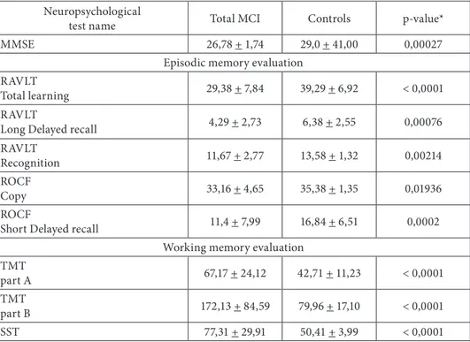
Variant analysis revealed significant differences between MCI group as a whole and cognitively normal group results in all kind of the neuropsychological tests assessing memory used in the study (table 2). MCI patients were impaired in their overall level of cognitive functioning and showed memory deficits under immediate and delayed recall conditions, recognizing description and working memory.
Table 2. Comparison of MCI and control group according to the neuropsychological tests results from the baseline
Neuropsychological
test name Total MCI Controls p ‑value*
MMSE 26,78 + 1,74 29,0 + 41,00 0,00027
Episodic memory evaluation RAVLT
Total learning 29,38 + 7,84 39,29 + 6,92 < 0,0001
RAVLT
Long Delayed recall 4,29 + 2,73 6,38 + 2,55 0,00076
RAVLT
Recognition 11,67 + 2,77 13,58 + 1,32 0,00214
ROCF
Copy 33,16 + 4,65 35,38 + 1,35 0,01936
ROCF
Short Delayed recall 11,4 + 7,99 16,84 + 6,51 0,0002
Working memory evaluation TMT
part A 67,17 + 24,12 42,71 + 11,23 < 0,0001
TMT
part B 172,13 + 84,59 79,96 + 17,10 < 0,0001
SST 77,31 + 29,91 50,41 + 3,99 < 0,0001
MMSE – Mini Mental State Examination (pts.), RAVLT – Rey Auditory Verbal Learning Test (number of words), TMT – Trail Making Test (sec.), ROCF – Rey ‑Osterrieth Complex Figure (pts.), SST – Serial Seven Test (sec.).
At baseline, MCI converters (PMCI) had significant impairment in MMSE (p = 0,027), RAVLT long delayed recall (p = 0,029), ROCF short delayed recall (p = 0,040) and TMT A&B (p = 0,031) scores comparing to MCI non ‑converters (SMCI) (table 3).
Table 3. Comparison of the baseline neuropsychological tests results and the risk of developing dementia of Alzheimer’s type
Neuropsychological
test name Total MCI Progressive MCI Stable MCI p ‑value*
MMSE 26,78 + 1,74 25,6 + 1,71 27,04 + 1,65 0, 0275
Episodic memory evaluation RAVLT
Total learning 29,38 + 7,84 26,1 + 4,86 30,11 + 8,22 NS
RAVLT
Long Delayed recall 4,29 + 2,73 2,8 + 1,40 4,62 + 2,85 0,0299 RAVLT
Recognition 11,67 + 2,77 11,2 + 3,19 11,78 + 2,7 NS
ROCF
Copy 33,16 + 4,65 31,7 + 6,36 33,49 + 84,02 NS
ROCF
Short Delayed recall 11,4 + 7,99 7,1 + 5,53 12,36 + 8,18 0,0402 Working memory evaluation
TMT
part A 67,17 + 24,12 83,67 + 17,26 63,87 + 24,08 0,0230
TMT
part B 172,13 + 84,59 208,89 + 53,57 164,78 + 88,12 0,0318
SST 77,31 + 29,91 89,73 + 9,34 74,56 + 27,18 NS
MMSE – Mini Mental State Examination (pts.), RAVLT – Rey Auditory Verbal Learning Test (number of words), TMT – Trail Making Test (sec.), ROCF – Rey ‑Osterrieth Complex Figure (pts.), SST – Serial Seven Test (sec.).
* Mann ‑Whitney U test (comparing Progressive MCI to Stable MCI), NS – statistically non significant.
The frequency of APOE4 allele carriers was higher in the MCI group than in controls (n = 25, 45,45%, Pearson’s Chi ‑square test; p = 0,0187). After dividing total MCI group into PMCI and SMCI statistically significant difference was also found in APOE4 status (n = 7, 70%, Pearson’s Chi ‑square test, p = 0,0022) (table 4).
Table 4. APOE status in the examined groups
APOE status Total MCI Progressive MCI Stable MCI Controls
N 55 10 45 44
APOEε4 carriers 25 (45,45%)* 7 (70%)** 18 (40%) 10 (22,73%) APOEε4 non carriers 30 (54,55%) 3 (30%) 27 (60%) 34 (77,27%) * p=0,0187, Pearson’s Chi ‑square test, comparing MCI to controls.
Discussion
During four years of follow up 18,1% (n = 10) of MCI patients developed demen‑ tia, while 81,9% (n = 45) remained stable. Our annual rate of conversion was 4,52%, what is a concordance with data from the literature24. Our MCI group was not rep‑
resentative for general population because only patients referred to our department on the basis of memory complaints were included into the study. Most of them were well educated (academic education). In this case even minor deficits may interfere high intellectual functioning than in people with only primary education. Prob‑ ably highly educated people are also more aware of the need of early diagnosis of cognitive impairment. That is why in our group we could not verify the influence of education level on dementia development, but data from the literature suggest that there is such relation, and that low education could be a risk factor for demen‑ tia development25.
There are many studies indicating that the frequency of depression in the MCI is over 30%26 and that depression has an influence on development of cognitive
impairment27. That is why, to collect as homogenic group as possible, we excluded
people with any depressive symptoms, even if the depression criteria was not ful‑ filled. On this basis we avoided including people whose cognitive problems were a consequence of depression.
APOEε4 allele is a well known genetic risk factor for dementia of Alzhei‑ mer’s type. It was estimated that in the MCI there are also more APOEε4 carri‑ ers than in general population28. In our MCI group there were 45,45% (n = 25)
APOEε4 allele carriers and in the control group 22,73% (n = 10) and it was statis‑ tically significant (Pearson’s Chi ‑square test, p = 0,01875). This data are consistent with the literature, also in Polish population29. In our MCI group, subjects who
developed dementia after follow up were more likely to be the APOEε4 carriers than those who were stable at that time (Pearson’s Chi ‑square test, p = 0,0022).
24 R.C. Petersen et al.: Prevalence of mild cognitive impairment…
25 H. Amieva et al.: Annual rate and predictors of conversion to dementia…; G. Tognoni et al.: From mild cognitive impairment to dementia: a prevalence study in a district of Tuskany, Italy. “Acta
Neurologica Scandinavica” 2005, No. 112, p. 65–71.
26 J.L. Gatz et al.: Do depressive symptoms predict Alzheimer’s disease and dementia? “Journal
of Gerontology” 2005, No. 60 (6), p. 744–747.
27 Ibidem; T. Gabryelewicz et al.: The rate of conversion of mild cognitive impairment to de‑ mentia: predictive role of depression. “International Journal of Geriatric Psychiatry” 2007, No. 22,
p. 563–567.
28 M.R. Farlow et al.: Impact of APOE in mild cognitive impairment. “Neurology” 2004, No. 63,
p. 1898–1901.
29 Ibidem; R.C. Petersen et al.: Apolipoprotein E status as a predictor…; R.J. Caselli et al.: Cognitive domain decline in healthy apolipoprotein E ε4 homozygotes before the diagnosis of mild cognitive impairment. “Archives of Neurology” 2007, No. 64 (9), p. 1306–1311.
It confirms that APOEε4 allele may predispose to early dementia development in the MCI group.
Episodic memory is selectively impaired in early, preclinical phases of amnestic manifestation of Alzheimer’s disease. It is suggested that in early phase of Alzhei‑ mer’s type dementia other cognitive dysfunctions are also present30. Neuropsy‑
chological evaluation is a key element of mild cognitive impairment diagnosis and important component of diagnostic criteria of Alzheimer’s disease. Most neuropsy‑ chological tests are not selectively assessing many aspects of memory and other cognitive functions and many factors may interfere the neuropsychological tests results31. Methods assessing episodic memory are based on encoding and learning
processes. At the beginning of learning process episodic memory is started first followed by semantic memory processes. Evaluation of memory process is concen‑ trated on immediate and delayed retrieval, free recall and recognition. Learning and encoding influence memory reproduction abilities. Memorizing process needs mnemonic strategy with engagement of executive functions. Plateau in consecutive immediate recall trial means lack of strategy indicating frontal but not temporal lobe dysfunction.
In this study encoding strategies during learning are not presented in final results, but recent researches suggest that subjects with amnestic MCI demonstrate diminished use of strategic encoding strategies during learning, related to the stra‑ tegic function of the episodic buffer, confirming the presence of executive functions impairment32. Delayed recall worse than delayed recognition discrimination may
be the result of not only selectively impaired hippocampal contextual memory, but also the defect of strategy. That is why evaluation of executive function is also impor‑ tant in neuropsychological diagnosis of memory disorders. Performing specific task assessing different aspects of memory is also depended from visuo ‑spatial and lan‑ guage abilities, that is why we have found different modality in memory systems in our study. Final effect of evaluating memory processes depends on many aspects not always clearly indicating episodic memory deficit, which is most reliably assessed by delayed recall trials.
Until now there are not wildly accepted cut ‑off points for neuropsychological tests in MCI diagnosis criteria, but the impairment is typically 1.0 to 1.5 stand‑ ard deviations below the mean of the individual, adjusted for age and education. In Poland the major problem is very low number of neuropsychological tests stand‑
30 R.C. Petersen et al.: Prevalence of mild cognitive impairment…; R. Perri et al.: Amnestic mild cognitive impairment…; S. Belleville et al.: Characterizing the memory changes…; S. Baudic
et al.: Executive function deficits…; S.M. Daselaar, M.S. Fleck, R. Cabeza: Triple dissociation in
the medial temporal lobes…
31 E. Strauss, E.M.S. Sherman, O. Spreen: A Compendium of Neuropsychological Tests…;
M.D. Lezak, D.B. Howieson, D.W. Loring: Neuropsychological Assessment…
32 R. Perri et al.: Amnestic mild cognitive impairment…; S. Belleville et al.: Characterizing the memory changes…
ardized and validated for Polish population requiring individual attitude to particu‑ lar patient. Making diagnosis in individual patient seen for the first time becomes a challenge and always needs taking into consideration someone’s intra ‑individual cognitive functioning.
In our study we have found that the baseline evaluation of cognitive functions in further converters to Alzheimer’s disease in comparison to stable MCI group, neu‑ ropsychological tests results were significantly lower in delayed recall tests than in encoding and recognition processes evaluation. Besides significantly lower results in cognition speed and attention in converters, encoding and recall process was com‑ parable in both group (stable and progressive MCI). Comparing total MCI group to controls we found significantly worse encoding, retrieval and executive functions representing working memory. Our results confirm the previously published data, that mild cognitively impaired patients with memory plus other cognitive domain deficits, rather than those with pure amnestic MCI, constituted the higher ‑risk group for development of dementia.
Conclusions
Significant impairment in baseline results of tests evaluating episodic memory (especially in delayed recall subtests) and working memory in MCI may be a good predictor of Alzheimer’s type of dementia development in the future. Cognitive tests battery still must be expanded to allow as accurate diagnosis as possible. MCI must be identified by using a more detailed procedure for the assessment of cog‑ nitive decline than the evaluation of memory alone. There is no appropriate set of neuropsychological tests for MCI diagnosis, so far.
Literature
Amieva H. et al.: Annual rate and predictors of conversion to dementia in subjects presenting mild
cognitive impairment criteria defined according to a population ‑based study. “Dementia and Geria‑
tric Cognitive Disorders” 2004, No. 18, p. 87–93.
Baudic S. et al.: Executive function deficits in early Alzheimer’s disease and their relations with epi‑
sodic memory. “Archives of Clinical Neuropsychology” 2006, No. 21, p. 15–21.
Beck A.T. et al.: An inventory for measuring depression. “Archives of General Psychiatry” 1961, No. 4, p. 53–63.
Belleville S. et al.: Characterizing the memory changes in persons with mild cognitive impairment. “Progress in Brain Research” 2008, No. 169, p. 365–375.
Caselli R.J. et al.: Cognitive domain decline in healthy apolipoprotein E ε4 homozygotes before the
diagnosis of mild cognitive impairment. “Archives of Neurology” 2007, No. 64 (9), p. 1306–1311.
Daselaar S.M., Fleck M.S., Cabeza R.: Triple dissociation in the medial temporal lobes: recollec‑
tion, familiarity, and novelty. “Journal of Neurophysiology” 2006, No. 96 (4), p. 1902–1911. Diagnostic and Statistical Manual of Mental Disorders: DSM‑IV. Washington, American Psychia‑
tric Association 1994.
Diana R.A., Yonelinas A.P., Ranganath C.: Imaging recollection and familiarity in the medial
temporal lobe: a three ‑component model. “Trends in Cognitive Sciences” 2007, No. 11 (9), p. 379–
386.
Dubois B. et al.: Research criteria for the diagnosis of Alzheimer’s disease revising the NINDCS‑
‑ADRDA criteria. “Lancet Neurology” 2007, No. 6 (8), p. 734–746.
Farlow M.R. et al.: Impact of APOE in mild cognitive impairment. “Neurology” 2004, No. 63, p. 1898–1901.
Foldi N.S.: Getting the hang of it: preferential gist over verbatim story recall and the roles of atten‑
tional capacity and the episodic buffer in Alzheimer disease. “Journal of the International Neu‑
ropsychological Society” 2011, No. 17 (1), p. 69–79.
Folstein M.F., Folstein S.E., McHugh P.R.: Mini ‑mental state. A practical method for grading
the cognitive state of patients for the clinician. “Journal of Psychiatric Research” 1975, No. 12,
p. 189–198.
Gabryelewicz T. et al.: The rate of conversion of mild cognitive impairment to dementia: predictive
role of depression. “International Journal of Geriatric Psychiatry” 2007, No. 22, p. 563–567.
Gatz J.L. et al.: Do depressive symptoms predict Alzheimer’s disease and dementia? “Journal of Gerontology” 2005, No. 60 (6), p. 744–747.
Hort J. et al.: EFNS guidelines for the diagnosis and management of Alzheimer’s disease. “European Journal of Neurology” 2010, No. 17, p. 1236–1248.
Huntley J.D., Howard R.J.: Working memory in early Alzheimer’s disease: a neuropsychological
review. “International Journal of Geriatric Psychiatry” 2010, No. 25 (2), p. 121–132.
Lezak M.D., Howieson D.B., Loring D.W.: Neuropsychological Assessment. New York – Oxford, Oxford University Press 2004.
McKhann G. et al.: Clinical diagnosis of Alzheimer’s disease: report of NINCDS ‑ADRDA Work group
under the auspices of Department of Health and Human Services Task Force on Alzheimer’s disease.
“Neurology” 1984, No. 34, p. 939–944.
Montgomery S.A., Asberg M.: A new depression scale designed to be sensitive to change. “British Journal of Psychiatry” 1979, No. 134, p. 382–389.
Perri R. et al.: Amnestic mild cognitive impairment: difference of memory profile in subjects who conver‑
ted or did not convert to Alzheimer’s disease. “Neuropsychology” 2007, No. 21 (5), p. 549–558. Petersen R.C. et al.: Apolipoprotein E status as a predictor of the development of Alzheimer’s disease
in memory ‑impaired individuals. “JAMA” 1995, No. 273 (16), p. 1274–1278.
Petersen R.C. et al.: Mild cognitive impairment: clinical characterization and outcome. “Archives of Neurology” 1999, No. 56, p. 303–308.
Petersen R.C. et al.: Prevalence of mild cognitive impairment is higher in men. “Neurology” 2010, No. 10, p. 889–897.
Price S.E. et al.: Learning and memory in amnestic mild cognitive impairment: contribution of
working memory. “Journal of the International Neuropsychological Society” 2010, No. 16 (2),
p. 342–351.
Redefining Alzheimer’s Disease: NIA and Alzheimer’s Association Float New Draft Diagnostic Crite‑ ria. (Alzheimer’s Association International Conference on Alzheimer’s Disease – ICAD, 2010).
Roman G.C. et al.: Vascular dementia: diagnostic criteria for research studies. Report of the NINDS‑
‑ARIEN International Workshop. “Neurology” 1993, No. 43, p. 250–260.
Rosenberg P.B., Johnston D., Lyketsos C.G.: A clinical approach to mild cognitive impairment. “American Journal of Psychiatry” 2006, No. 163 (11), p. 1884–1890.
Saiki R.K., Levenson C.H., Ehrlich H.A.: Genetic analysis of amplified DANN with immobilized
sequence specific oligonucleotide probes. “Proceedings of the National Academy of Sciences” 1989,
No. 86, p. 6230–6234.
Schulman K.L., Shedletsky R., Silver I.L.: The challenge of time: Clock drawing and cognitive
function in the elderly. “International Journal of Geriatric Psychiatry” 1986, No. 1, p. 135–140.
Strauss E., Sherman E.M.S., Spreen O.: A Compendium of Neuropsychological Tests, Administra‑
tion, Norms and Commentary. [Oxford ], Oxford University Press 2006.
Tabert M.H. et al.: Functional deficit in patients with mild cognitive impairment: prediction of AD. “Neurology” 2002, No. 58, p. 758–764.
Tervo S. et al.: Incidence and risk factors for mild cognitive impairment: A population ‑based three‑
‑year follow ‑up study of cognitive healthy elderly subjects. “Dementia and Geriatric Cognitive
Disorders” 2004, No. 17, p. 196–203.
Tognoni G. et al.: From mild cognitive impairment to dementia: a prevalence study in a district of
Tuskany, Italy. “Acta Neurologica Scandinavica” 2005, No. 112, p. 65–71.
Unsworth N.: Variation in working memory capacity, fluid intelligence, and episodic recall: a latent
variable examination of differences in the dynamics of free recall. “Memory & Cognition” 2009,
No. 37 (6), p. 837–849.
Winblad B. et al.: Mild cognitive impairment: beyond controversies, towards a consensus. “Journal of Internal Medicine” 2004, No. 256, p. 240–246.
Wolf H. et al.: The prognosis of mild cognitive impairment in the elderly. “Journal of Neural Trans‑ mission” 1998, No. 54, p. 31–50.
Wolk D.A., Dickerson B.C.: Fractionating verbal episodic memory in Alzheimer’s disease. “Neu‑ roimage” 2011, No. 54 (2), p. 1530–1539.
Yener G.G. et al.: Diagnosis profile and comparison of risk factors in major types of dementia: an

!["A Short History of the Gout and the Rheumatic Diseases", W. S. C. Copeman, Berkeley-Los Angeles 1964; "The Conquest of Tuberculosis", Selman A. Waksman, Berkeley-Los Angeles 1964 : [recenzja]](data:image/gif;base64,R0lGODlhAQABAIAAAP///wAAACH5BAEAAAAALAAAAAABAAEAAAICRAEAOw==)