REVIEW ARTRICLE
TRENDS
in
Sport Sciences
2013; 3(20): 135-139.ISSN 2299-9590
The use of surface electromyography for diagnosis of muscle
dysfunction with pain symptoms
JULIUSZ HUBER1, ALEKSANDRA KULCZYK1, PRZEMYSŁAW LISIŃSKI2,3, JOANNA LIPIEC1,3Received: 25 July 2013 Accepted: 13 August 2013
Corresponding author: zpnr@wp.pl
1 University of Medical Sciences, Poznań, Department of
Patho-physiology of Locomotor Organs, Poland
2 University of Medical Sciences, Poznań, Department and Clinic
of Physiotherapy, Rheumatology and Rehabilitation, Poland
3 University of Medical Sciences, Poznań, Department and Clinic
of Rehabilitation, Poland
Surface electromyography (sEMG) is generally considered by neurologists as an unacceptable diagnostic tool for examination of changes in the activity of muscle motor units in patients with non-specifi c back pain.
The aim of this review is to demonstrate the usefulness of neurophysiological fi ndings for the application of sEMG in differentiation of root-confl ict and non-root-confl ict sources of muscle pathologies with pain as the main symptom.
In the fi rst experiment carried out on 30 patients with clinically recognized myofascial pain, an attempt was undertaken to fi nd out whether surface electromyographic (sEMG) readings during relaxation and maximal contraction revealed differences in the activity of muscles with or without trigger points (TrPs) detected by palpation. In the second experiment carried out on 40 offi ce workers similar methodologies of clinical and neurophysiological examination were used, however, with the aim to verify a hypothesis about the dysfunction of cervical and shoulder girdle muscle motor units as the cause of cervicogenic headache (CEH).
The results of both experiments led to the following conclusions: 1. Surface EMG performed at rest and during maximal contraction is a precise diagnostic tool that can be used for detection of changes in the activity of motor units in patients with myofascial pain syndrome and cervocogenic headache; 2. Surface EMG readings at rest, with an amplitude exceeding 25μV, may be helpful for evaluation of increased muscle tension, which leads to a decrease of the activity of muscle motor units during maximal contraction.
KEY WORDS: surface electromyography, muscle resting state, maximal contraction, muscle pain.
Introduction
S
urface electromyography (sEMG), commonly used for clinical purposes, seems to be fairly unpopular among neurologists and traditional neurophysiologists, who prefer intramuscular (needle) electromyography (nEMG) examination as the golden standard for confi rmation of neurogenic or myogenic changes in muscle motor units of patients with various movement disorders. It seems that this approach originated fromWhat was previously known on the presented topic?
According to “Simons D.G., Travell J.G., Simons
L.S., “Travell & Simons’ Myofascial Pain and Dysfunction”. The Trigger Point Manual. Upper Half of Body. Baltimore: Lippincott Williams & Wilkins; 1999” as the ... . “Bible” …of evaluation
the TRPs points with palpation and the current opinions on EMG examinations, the recognizing of muscles involved with myofascial pain syndrome is …“diffi cult”… . Neurologists believe, that only nEMG studies are reliable enough to reveal any changes in muscles’ motor units activity, while sEMG recordings are doubtful in recognizing the myofascial pain syndromes within certain muscles showing the pain symptoms. Clear “Myofascial pain syndrome” should not be accompanied with changes in C5-T1 motor transmission revealed in ENG neurophysiological studies.
HUBER, KULCZYK, LISIŃSKI, LIPIEC
a paper published by Pullman et al. in 2000 [1], which discussed the use of sEMG in the evaluation of patients with neuromuscular diseases, low back pain and motor control disorders. More than 2,500 original articles, reviews and books have been published on the scope of sEMG application, its benefi ts and risks and the extent to which sEMG techniques vary. Researchers have also assessed the strengths and weaknesses of sEMG in specifi c clinical applications and concluded that … “sEMG is considered unacceptable as a clinical tool in the diagnosis of neuromuscular disease and evaluation of patients with low back pain”… and … “sEMG is considered an acceptable tool for kinesiologic analysis of movement disorders and evaluation of gait and posture
disturbances”. Pullman et al. summarized to some degree the outstanding work of Basmanijan and DeLuca [2], but they were stimulated by more extensive studies by Merletti et al. as well as their followers [3, 4]. Results of studies on healthy volunteers led to the development of methods of non-invasive acquisition and processing of sEMG readings, which started to be performed since the pioneer works of Fritz Buchtal (1907-2003) [5]. Surprisingly, the available PubMed data for years 2000-2013 indicate a great interest among rehabilitation specialist s, physical t herapist s, or t hoped ist s, neurologists and neurosurgeons in non-invasive sEMG a ssessment of t reat ment ef fect iveness of patients with different movement disorders. A simple keyword search using “surface EMG” and “treatment” resulted in 1,590 records. Moreover, some of these studies are devoted to such complicated neurologic disorders as stroke [6], or to evaluation of treatment of patients with symptoms of specifi c or non-specifi c back pain [7]. On the basis of results of earlier research [8, 9], the aim of the present study is to provide convincing evidence that both surface EMG as well as needle EMG examinations offer a valuable insight into the pathological
activities of certain groups of muscles in patients with the chronic spine-related muscle pain and cervicogenic headache.
Patients with chronic muscle pain
The fi rst experiment showed that spine-related muscle pain can affect muscle strength and motor unit activity in 30 patients with clinically recognized myofascial pain, and that sEMG readings during relaxation and maximal contraction reveal differences in the activity of muscles with or without trigger points (TrPs) detected by palpation. It also included an analysis of the possible co-appearance of characteristic spontaneous activity in nEMG readings alongside TrPs (Fig. 1).
Patients with non-specific cervical and back pain were preliminarily assessed with the use of clinical, neuroimaging and electroneurographic examinations. Muscle pain was measured with a visual analog scale (VAS = 6), and strength with Lovett’s scale (3.5); trigger points were detected bilaterally by palpation (in 55% of all patients). A Keypoint System (Medtronic A/S, Skovlunde, Denmark) was used for neurophysiological tests. For comparison, 30 healthy volunteers were also examined in the same way as the patients. Both sEMG and nEMG at rest and during 5-second maximal contraction were used to examine motor unit activity, mostly on the basis of readings from painful trapezius muscles. Examinations were also performed bilaterally in the gluteus medius, tensor fasciae latae and lumbar erector spinae muscles.
The mean sEMG amplitude obtained from the trapezius and cervical or lumbar axial muscles at rest increased > 25 μV in patients, as compared with 15 μV in healthy subjects (p = 0.05). This was indicative of increased muscle tension, which in turn decreased the muscle ability of maximal contractions (Fig. 2A). Mean values of sEMG amplitudes during maximal contractions were decreased
Figure 1. A. Localization of pain radiation in a patient with spine-related muscle pain; B. Placement of surface electrodes
on the trapezius muscle at rest and during maximal contraction; C. EMG examination with a needle inserted in a painful trapezius muscle; D. Detection of active trigger points using palpation
bilaterally in patients (723 μV) in comparison with the reference values (1606 μV) (p = 0.05) (Fig. 2B).
Electroneurographic (ENG) examinations of impulses in peripheral parts of motor fi bers (compound muscle action potentials – CMAP) within suprascapular and femoral nerves were performed in all participants in order to exclude consequences of radiculopathies (the studied patients suffered only from non-specifi c cervical and low back pain). No changes in motor fi ber transmission were observed in any of the patients (N = 30) or healthy volunteers (N = 30) when parameters of amplitudes and latencies were compared in the CMAP.
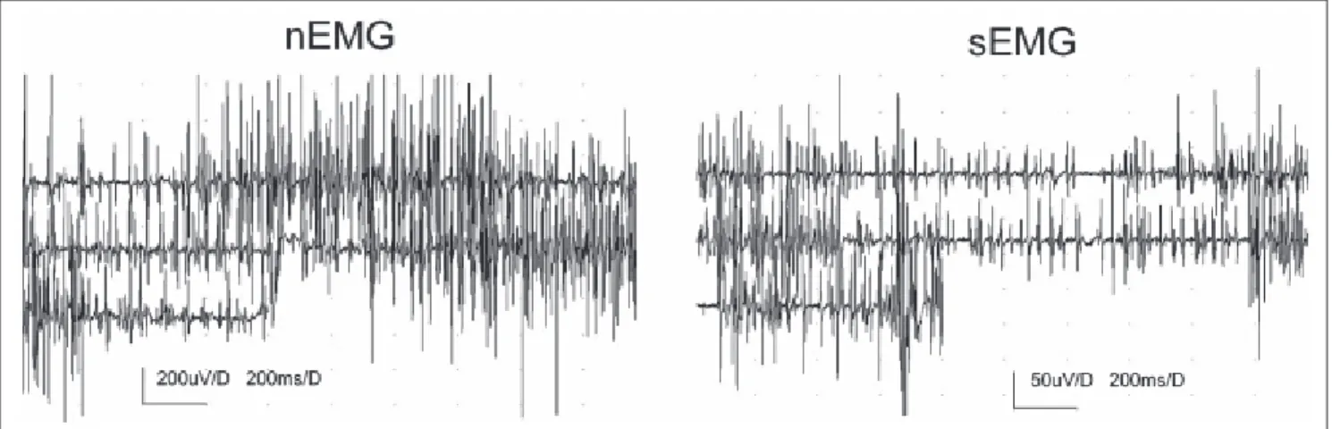
During the nEMG analysis it was assumed that the spontaneous activity typical for muscles with active TrPs would not show the synchronous pattern characteristic for high-frequency firing, and that it would have a different morphology from end-plate potentials, denervation potentials (both fi brillations and positive sharp waves), fasciculations and pseudomyotonic discharges. The readings of spontaneous activity from muscles with TrPs were exclusively asynchronous with a mean frequency of 28 Hz. They consisted mainly of polyphasic potentials with a duration of 16.4 ms and average amplitude of 362 μV.
This fi ring was transient and some records (7/13 cases) showed to be interrupted by a period of “bioelectrical silence”. Sometimes fi rings could be also observed in simultaneous sEMG readings (Fig. 3).
The conclusions of the fi rst experiment were that: 1. Complex neurophysiological and clinical diagnostic examinations may help explain the possible etiology of non-specifi c cervical and back pain arising from active trigger points; 2. The EMG revealed that the symptom of muscle pain depends on the progression of myofascial syndrome.
Studies on patients with cervicogenic headache
In the second experiment similar methodologies of clinical and neurophysiological examinations were used, however, the aim was to verify the hypothesis about dysfunction of cervical and shoulder girdle muscle motor units as the reason for cervicogenic headache (CEH). The performed diagnosis of mainly unilateral CEH with the coexisting myofascial pain syndrome met the criteria of The International Classifi cation of Headache Disorders from 2004.
Clinical methods included evaluation of unilateral headache intensity, using the Visual Analog Scale (VAS = 6.8) in temporal, frontal or occipital areas,
Figure 2. A. sEMG on the trapezius muscle at rest; B. sEMG on the trapezius muscle during maximal contraction lasting 5
seconds. Measurements were performed on a healthy volunteer (left) and on a patient with myofascial pain syndrome (right). Mean amplitudes in the populations of healthy volunteers and patients are shown for comparison
HUBER, KULCZYK, LISIŃSKI, LIPIEC
radiating mainly to the trigeminal innervation. The range of cervical movement (ROM) was measured with a goniometer. The most limited movement was fl exion at 29o in comparison to the normal range in healthy
volunteers at 42o. Pain sensation in the neck and shoulder
girdle muscles during their passive elongation (PE) was scored as present (1 – yes) or absent (0 – no), and for the trapezius muscle it amounted to 14/20. Trigger points were found bilaterally with the predominance of CEH side incidence – 17/20 for the trapezius muscle. The strength of muscles was tested with Lovett’s scale and it was scored at about 3.4. Surface EMG readings from the neck and shoulder girdle muscles were bilaterally analyzed at rest and during maximal contraction. The statistical analysis revealed a negative correlation between values of amplitudes in resting EMG and maximal contraction EMG in patients. Positive correlations were found between the increase in resting EMG amplitudes for more than 30 μV and high VAS scores as well as between high-amplitude resting EMG incidence and the increased number of TrPs. Incidence of radiculopathies in patients with CEH was excluded on the basis of clinical and neuroimaging results as well as electroneurographical examinations of the transmission of motor fi bers in suprascapular and axillar nerves. CMAPs were normal in the parameters of amplitudes and latencies. Incidence of trigger points found mostly in the trapezium muscle coexisted with the cervicogenic headache intensity.
The experiment results led to the following conclusions: 1. Muscle pain in the neck and shoulder girdle muscles meeting the criterion of myofascial pain syndrome coexisted with cervicogenic headache in examined
patients; 2. Dysfunction of the trapezius muscles motor units was most responsible for CEH etiology, and again, like in the fi rst experiment, the same resting vs. maximal contraction pattern of motor unit dysfunction was detected.
General conclusions
1. Surface EMG performed at rest and during maximal contraction is a precise diagnostic tool that can be used for detection of motor units activity changes in patients with spinal-related myofascial pain syndrome and cervocogenic headache.
2. Surface EMG at rest with an amplitude above 25 μV may be helpful for assessment of increased muscle tension, which in turn leads to decreased motor units activity during maximal contraction (Fig. 4).
What this study adds?
The present article demonstrates the results of an sEMG study documenting relationships between the functional state of muscles at rest and during maximal contraction in healthy subjects and patients with myofascial pain syndrome. Muscle related disorders with pain as the main symptom, most frequently arising from overloading, involve increased muscle tension with a simultaneous decrease of motor unit activity during maximal contraction. Palpation does not always precisely reveal increased muscle tension when postisometric relaxation procedures should be implemented.
Figure 3. Results of nEMG (left) and sEMG (right) performed on the trapezius muscle with active TrPs in a patient with
Figure 4. Diagram illustrating relationships between conditions of proper muscle tension and maximal muscle contraction
(left), and relationships when these two parameters are abnormal (right)
References
1. Pullman SL, Goodin DS, Marquinez AI, et al. Clinical utility of surface EMG. Report of the therapeutics and technology assessment subcommittee of the American Academy of Neurology. Neurology. 2000; 55: 171-177. 2. Basmajian JV, De Luca CJ. Muscles Alive, (5th edition).
Williams and Wilkins, Baltimore. MD 1985.
3. Merletti R, Parker P. Electromyography. Physiology, engineering and noninvasive applications. John Wiley & Sons Inc. Hoboken. New Jersey. 2004.
4. Kulczyk A, Lipiec J, Huber J, Zagłoba-Kaszuba A, Wytrążek M, Stryła W, Warzecha D. Standards of the surface electromyography examinations for assessment of muscle motor unit activity for physiotherapy purposes. In: Huber J, Wytrążek M, Joanna J, Kulczyk A, eds., Current topics on clinical neurophysiology, physiotherapy and manual therapy. Coll Educ Ther Publ House. Poznań 2011: 106-119.
5. Cram J R , Kasman GS. I nt roduct ion to su r face electromyography. Gaithersburg, Aspen Publishers; 1998.
6. Lisiński P, Huber J, Samborski W, Witkowska A. Neurophysiological assessment of the electrostimulation procedures used in stroke patients during rehabilitation. Int J Artif Organs. 2008; 31, 1: 76-86.
7. Huber J, Lisiński P, Samborski W, Wytrążek M. The effect of early isometric exercises on clinical and neurophysiological parameters in patients with sciatica: An interventional randomized one-blinded study. Isokinetic Ex Sci. 2011; 19, 3: 207-214.
8. Wytrążek M, Huber J, Lisiński P. Changes in muscle activity determine progression of clinical symptoms in patients with chronic spine-related muscle pain. A complex clinical and neurophysiological approach. Functional Neurology. 2011; 26, 3: 141-149.
9. Huber J, Lisiński P, Polowczyk A. Reinvestigation of the dysfunction in neck and shoulder girdle muscles as the reason of cervicogenic headache among offi ce workers. Di Rehab. 2012; 35 (9-10): 793-802.