The Henryk Niewodniczański Institute of Nuclear Physics
Polish Academy of Sciences
Characterization of cell surface structure and its relation to
cytoskeleton elasticity in cancer cells
Justyna Bobrowska
Thesis submitted for the Degree of Doctor of Philosophy in Physics
Prepared under the supervision of
dr hab. Małgorzata Lekka (thesis supervisor)
dr hab. Jakub Rysz (auxiliary supervisor)
2
You must have long-range goals to keep you
from being frustrated by short-range failures.
3
Acknowledgements
Undertaking this PhD has been a truly life-changing experience and it would not have been possible to do without all the support and guidance that I received from many people. I am lucky to have met them.
First of all, I would like to express my deepest gratitude to my thesis supervisor Assoc. Prof. Małgorzata Lekka. I am grateful for her help, encouragement, constructive criticism, and guidance. With her immense knowledge and excitement in regard to research, she shows me what does it mean to be a real researcher.
I would also like to thank my co-supervisor, Assoc. Prof. Jakub Rysz, for his help, insightful comments and expertise that greatly assisted the research.
My deepest thanks to all my colleagues from the Division of Applications of Physics and Interdisciplinary Research (IFJ PAN) headed by Prof. Wojciech Kwiatek and the Department of Advanced Materials Engineering (UJ) under the supervision of Prof. Andrzej Budkowski. Working with you, as a team, was a real honour for me. Kasia and Szymon, you were the best labmates I could have. I would like to thank Mrs. Joanna Wiltowska-Zuber and Mrs. Joanna Pabijan for their laboratory support and Dr Kamil Awsiuk for his help with PCA.
I am also grateful to Assoc. Prof. Franciszek Krok and Dr Benedykt R. Jany for their help with recording SEM images and to Dr Jonathan Moffat, and Prof. Mike Reading for sharing PTMS spectra.
Sincere thanks to the group of Prof. Peter Hinterdorfer from the University of Linz, where I spent a few months as an intern.
For my family and friends for their love and patience. Especially, during last months, when my duties were consuming all my time, but you never said a word. I know I owe you.
And, finally, to my husband Piotr, who always believes in me, even when I do not. Without you I would not be here.
4
Abstract
The alterations observed in tumour cells include the number of processes introducing abnormalities in cellular morphology, structure and growth profiles. Despite continuous efforts, the molecular mechanism of the metastasis is still not understood completely. That is the reason why there is an urgent need for the search of new scientific approaches in the cancer progression investigations. The development of various biochemical and biological methods increases the chance to detect cancer, however, in past decades, single cell biomechanics has gained large significance since certain diseases are known to manifest in altered biomechanical properties. Stiffness of single cells is one of the major properties that changes during cancerous progression. Studies have demonstrated that biomechanics can bring both data describing mechanisms underlying cancer progression and tools for its detection and diagnosis at the single cell level. However, one of the emerging directions is to correlate cellular biomechanics with biochemical and biophysical properties of single cells.
The main objective of the presented thesis is to study how single cell deformability is linked with cellular surface properties, and how these changes correlate with cancer progression. Thus, the elasticity of melanoma cells was measured by means of atomic force microscopy (AFM). Measurements were carried out for three groups of cells encompassing cells originating from primary tumour sites i.e. from radial and vertical growth phases (RGP and VGP, respectively), and those derived from skin and lung metastasis. The results were compared with properties of melanocytes (cells from which melanoma originates). The surface properties were determined using time of flight secondary ions mass spectrometry (ToF SIMS). The use of ToF SIMS has required to develop a dedicated sample preparation protocol enabling measurements of biological material in the high vacuum environment.
The final results show the correlation between single cell deformability and surface biochemical properties of melanoma cells. They confirm the hypothesis that cancer progression causes alterations in the morphological and mechanical properties of cancerous cells and these differences are connected with changes in the cellular surface composition.
5
Streszczenie
Zmiany obserwowane w komórkach nowotworowych obejmują szereg procesów wprowadzających zaburzenia w ich morfologii, strukturze i wzroście. Pomimo ciągłych badań, molekularny mechanizm procesu przerzutowania wciąż nie został do końca wyjaśniony. Dlatego wciąż istnieje potrzeba poszukiwania nowych rozwiązań w badaniach dotyczących nowotworów. Rozwój zaawansowanych metod biochemicznych i biologicznych zwiększa szanse na wczesne wykrywanie zmian nowotworowych. Jednakże, w ostatnich dekadach, biomechanika pojedynczej komórki zyskuje na znaczeniu, ponieważ okazuje się, że wiele chorób wpływa na zmianę właściwości biomechanicznych już na poziomie komórkowym. Sztywność pojedynczych komórek zmienia się podczas progresji nowotworowej. Liczne badania wykazują, że biomechanika może dostarczać zarówno danych opisujących mechanizm progresji nowotworu jak i narzędzi do jego detekcji na poziomie pojedynczej komórki. Niemniej jednak, jednym z istotnych kierunków badań jest korelacja biomechaniki z biochemicznymi i biofizycznymi właściwościami komórek.
Głównym celem niniejszej rozprawy jest zbadanie w jaki sposób elastyczność komórki jest związana budową biochemiczną jej powierzchni i czy zmiany te są skorelowane ze stopniem progresji nowotworowej. W tym celu, elastyczność komórek czerniaka została wyznaczona za pomocą mikroskopii sił atomowych (AFM). Pomiary zostały wykonane dla komórek wywodzących się z linii komórkowych pochodzących z pierwotnego ogniska czerniaka - z radialnej i wertykalnej fazy wzrostu, oraz z przerzutu czerniaka do skóry i płuc. Jako linię referencyjną wykorzystano prawidłowe melanocyty. Budowę biochemiczną powierzchni komórek zbadano za pomocą spektrometrii mas jonów wtórnych za analizatorem czasu przelotu (ToF SIMS). Wykorzystanie ToF SIMS wymagało opracowania protokołu preparatyki umożliwiającego pomiary materiału biologicznego w warunkach wysokiej próżni. Uzyskane rezultaty, przedstawione w niniejszej rozprawie, wskazują na związek pomiędzy deformowalnością a budową biochemiczną powierzchni komórek czerniaka. Tym samym potwierdzają hipotezę, że progresja nowotworowa powoduje różnice w morfologii i nanomechanice komórek nowotworowych, które są związane ze zmianami w budowie biochemicznej ich powierzchni.
6
Table of contents
List of abbreviations………8
1. Introduction ………9
2. Fundamentals and state of the art………...12
2.1 Melanoma progression……….12
2.2 Basic terms used in cellular biomechanics ……….………14
2.3 Significance of cellular deformability ………16
2.4 Surface properties …...………18
2.5 Aims of the thesis ………22
3. Materials………..………..23
3.1 Cell cultures……….23
3.2 Sample preparation for AFM measurements………24
3.3 Sample preparation for PTMS experiments…………...………..25
3.4. Fluorescent staining………..………..25
3.5 Sample preparation for ToF SIMS experiments ……….………25
3.6 Summary………..………33
4. Experimental methods………...34
4.1 Atomic Force Microscopy – basic principles………..34
4.2 Fluorescence microscopy……….38
4.3 Scanning electron microscopy (SEM)………..38
4.4 Time-of-flight secondary ion mass spectrometry (ToF SIMS)……….39
4.5 Photothermal Microscopy (PTMS)………..41
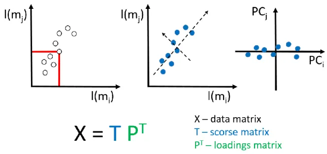
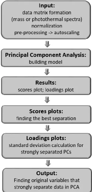
4.6 Principal Component Analysis – basic principles………42
4.7 Summary………..48
5. Functionality of ToF SIMS measurements………49
5.1 Objectives………....49
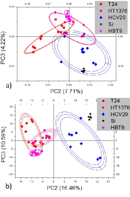
5.2 Deformability of human bladder cells……….………49
5.3 Mass spectra of human bladder cancer cells………50
5.4 Surface chemistry based differentiation of human bladder cancer cells…………..51
5.5 Molecular masses derived from PCA analysis……….53
7
6. Morphological and mechanical properties of melanoma cells from VGP (WM115) and skin
metastasis (WM266-4)………..55
6.1 Objectives………....55
6.2 Morphology of melanoma cells in low and high cellular densities……….55
6.3 Topography of melanoma cell surface……….58
6.4 Nano-mechanical properties of melanoma cells………..63
6.5 Summary………..66
7. Surface chemistry in melanoma cells from VGP (WM115) and skin metastasis (WM266-4) ……….. 68
7.1 Objectives………....68
7.2. PCA of mass spectra recorded for WM115 and WM266-4 melanoma cells………...……….68
7.3 Differentiation of melanoma cell lines based on photothermal spectra (PTMS)…..74
7.4 Summary..………79
8. Physico-chemical properties of melanoma cells from various stages of cancer progression..81
8.1 Objectives………....81
8.2 Elasticity of melanoma cells………..………..81
8.3 PCA of mass spectra for melanoma cells………..………..84
8.4 Summary..………89
9. Summarizing biomechanical and biochemical characteristics in melanoma cells…………91
10. References………...96
List of figures………...110
8
List of abbreviations
1205Lu - melanoma cell line derived from the lungs metastasis of WM793 cells A375P - melanoma cell line derived from the lungs metastasis
AC - surface area of an average single cell AN - surface area of an average single nucleus AFM - atomic force microscope
ECM – extracellular matrix EMEM - cell culture medium
HCV29 – non-malignant epithelial cells of ureter
HEMa-LP - human, epidermal, adult melanocytes - primary cell line HTB-9 - urinary bladder carcinoma
HT1376 - urinary bladder carcinoma N/C - nucleus-to-cell ratio
PBS - phosphate buffer saline PC - principal component
PCA - principal component analysis PTMS - photothermal microspectroscopy RGP - radial growth phase
RPMI - cell culture medium SD - standard deviation
SEM - scanning electron microscope T24 - transitional cell carcinoma
ToF SIMS - time of flight secondary ions mass spectrometry VGP - vertical growth phase
WM115 – melanoma cell line from vertical growth phase (VGP)
WM266-4 - melanoma cell line derived from the skin metastasis of WM115 cells WM239 - melanoma cell line derived from the skin metastasis
WM35 - melanoma cell line from radial growth phase (RGP) WM793 - melanoma cell line from vertical growth phase (VGP)
9
1. Introduction
Nowadays, the development of advanced spectroscopic techniques, working at the nanoscale, achieved through enhancing the sensitivity and through coupling with other complementary techniques, enables measurements of biological samples at the single cell level. Many of the processes, occurring at the cellular level, play an important role in the variety of biological functions at macroscale. The example is the carcinogenesis that starts with the alterations occurring in single cells. These changes encompass, among others, distinct cellular morphology, cytoskeleton organization, biochemical composition, biophysical and biomechanical properties. Such an observation leads to the conclusion that cancer progression is a complex process that cannot be explained with a single biomarker. Therefore, the employment of various techniques, including molecular biology and physical approaches, delivers distinct characteristics of cancer-related changes in single cells.
Since several decades, the atomic force microscopy (AFM) can serve not only as a tool for the measurements of the cellular morphology with a high resolution but also, working in the force spectroscopy mode, it delivers the quantitative description of mechanical properties of living cells in conditions close to their physiological environment. Thus, within the frame of the presented thesis, the goal was to characterize mechanical properties of various melanoma cells lines originating from distinct stages of melanoma progression, namely, from radial/vertical growth phases and from metastasis to skin and lung. The obtained results were compared to properties of melanocytes being the cells of origin for malignant melanoma.
On the other hand, the altered biomechanics is only one of many distinct features characteristic for cancerous cells. The other changes include modifications in adhesive properties of single cells occurring at the cell surface. Thus, one can expect that, instead of searching for specific molecules responsible for malignant phenotype, the alterations in the overall composition of cell’s surface are distinct enough to be identified using techniques probing sample surface and, further, to be used as a diagnostic marker. Among various techniques used to study the composition of cell surface, a time of flight secondary ions mass spectrometry (ToF SIMS) appears to be the optimal one to measure the biophysical/biochemical changes on the surface of cancerous cells. The recorded information is derived from a layer of a few nm, that is comparable to the thickness of the cell membrane. In this thesis, the ToF SIMS technique was applied to measure the surface composition of individual melanoma cells in an attempt to detect these changes occurring at the sub-cellular and cellular levels. The goal was
10
to study whether there is a correlation between biomechanical and biophysical/biochemical properties of cell surface.
The layout of the presented thesis is the consequence of the proposed research plan.
Chapter 2 introduces the basic information on melanoma progression, basic terms used in
biomechanics of cells, followed by significance of cellular deformability and chemical properties. The Chapter 2 ends with the presentation of the aims of the thesis. Subsequently, in
Chapter 3, the biological samples are characterized together with the applied methodology of
the sample preparation. In particular, the protocol devoted to ToF SIMS sample preparation was developed as a one of specific work objectives. Its validation involved the AFM based topography imaging after each step of preparation protocol and cellular structure and surface visualization by fluorescence and environmental scanning electron (ESEM) microscopes. The basics for all employed experimental methods are shortly described in the Chapter 4. Mass spectra of biological samples are very complex, that is why various statistical methods are usually used to resolve characteristic fingerprints in these samples. Here, the principal component analysis (PCA) was chosen, as it allows for the search of the biggest variances in the analysed data. Moreover, PCA was performed for the whole collected range of mass spectra without a pre-definition of any particular mass peaks a priori. Using such an approach, it is possible to determine those masses that cause the strongest differentiation between studied cell populations, afterwards. Next chapter, i.e. Chapter 5, contains the results of ToF SIMS measurements demonstrating the functionality of this technique. It was applied to determine chemical properties of human bladder cells. These cells are characterized by large deformability difference occurring between non-malignant and cancerous bladder cells. The large deformability of these cells was accompanied by visible separation among the studied cell lines. Characterizing cells that are clearly distinguishable by cellular morphology, biomechanical, biophysical, and biochemical properties with the use of various techniques bears traces of proof-of-concept approach, thus, in further studies melanoma cells were chosen (Chapters 6–8). First, two types of cells were analysed, namely, WM115 cells originating from vertical growth phase and from the metastasis of WM115 to skin (WM266-4 melanoma cells). Their nanomechanical characterization in relation to both, surface structure and actin cytoskeleton organization, is presented in Chapter 6. The measurements of surface chemical composition with identified molecular masses causing the strongest differentiation between these cell lines is presented in the Chapter 7. These studies were verified by the use of photothermal microspectroscopy (PTMS). The latter technique delivers the photothermal spectra that resemble the infrared spectra observed in Fourier Transform Infrared Spectroscopy (FTIR). The final chapter,
11
Chapter 8, presents the results of biomechanical and biophysical characterization of various
melanoma cells. The findings show that the alterations of mechanical properties observed in various melanoma groups (radial/vertical growth phase, metastasis to skin, metastasis to lung, melanocytes) are accompanied by the alterations in their surface composition.
Results, included in the presented thesis, were obtained thanks to the close collaboration of two research groups, namely, between the Department of Biophysical Microstructures at the Institute of Nuclear Physics (Polish Academy Sciences) and the Department of Advanced Materials Engineering at the Institute of Physics (Jagiellonian University).
This work was partially supported by National Science Centre (NCN) Project Number
DEC-2013/11/N/ST4/01860. The author is grateful to Polish National Science Center (NCN)
for the financial support of the ETIUDA scholarship no DEC-2015/16/T/ST4/00358 and to the Institute of Nuclear Physics PAS for a healthy work environment and KNOW for PhD scholarship.
12
2. Fundamentals and state of the art
2.1 Melanoma progression
Cutaneous melanoma is the most aggressive and lethal malignancy of the skin. Despite advances in melanoma treatment [1], mortality from melanoma is still increasing [2]. Thus, there is an urgent need to develop prognostic biomarkers that can differentiate between malignant and non-malignant skin lesions and that can identify melanoma patients with high-risk primary lesions to facilitate greater surveillance [3]. That is why, the discovery of biomarkers and their application, in conjunction with traditional cancer diagnosis, staging, and prognosis, could improve early diagnosis and patient care [4]. Melanoma arises from melanocytes [5]. Melanocytes are neural crest-derived cells endowed with defined morphological and biochemical markers. They are mainly located within the basal layer of the epidermis, just superficial to the basement membrane. Normal melanocytes produce and subsequently transfer pigment-producing melanosomes to neighbouring keratinocytes, which are thought to protect the keratinocytes against UV radiation. When the functioning of melanocytes is impaired, the UV irradiation can lead to the formation of melanoma.
Melanoma is characterized by high rate of invasion resulting at various metastatic sites in, for example, skin and lung. Such behaviour of cells requires the interaction of the tumour cells with the extracellular matrix (ECM) trough cell adhesion, cell migration, and cell-mediated tissue proteolysis [6]. During melanoma development and progression, cancerous cells encounter several basal membranes (BMs) being the sheets of ECM mainly composed of laminins, type IV collagen, nidogens and perlecan [7–9]. The BMs are lost or penetrated by tumour cells during invasion and metastasis: firstly melanoma cells penetrate the dermo-epidermal junction, and invade the BM of dermal lymphatic vessels, entering the lymphatic circulation and lodging into regional lymph nodes. After obtaining access to the blood circulation system, the melanoma cells interact with the blood vessel wall and invade target organs, causing metastasis [6].
Tumour growth and dissemination involve specific interactions with tumour cell surface adhesion receptors and multiple adhesive components of the ECM. The receptors that mediate the cellular adhesive interactions with the ECM are derived from a large family of heterodimeric molecules referred to as the integrins [10]. Today it is clear, that these molecules play key roles in the regulation of tumour cell migration and survival in dependency of their ligands.
13
Alterations in the number and composition of integrins is a result of changes in surface receptors linked with cancer progression [11]. In addition, many proteins participating in normal biochemical processes are glycosylated i.e. they have attached oligosaccharide moieties. The variations of this glycosylation pattern often lead to changes in their functions. It has been reported for many tumours that the plasma membrane oligosaccharides linked to proteins or lipids are altered during cancer transformation. Changes in the expression and the structure of oligosaccharides seem to be a characteristic feature of the malignant transformation. All together, these characteristics can be attributed to alterations in adhesion and communication between cancerous cells and the normal cells in their nearest environment. Malignant transformation is also characterized by disruption of cytoskeletal organization, decreased adhesion, and altered adhesion–dependent responses. It has been already reported that the deformability of a cell in response to external forces is mainly determined by cytoskeleton and governed by adhesive molecules, usually linked to the cell cytoskeleton [12,13].
Melanoma has been already classified into well-described clinical stages as illustrated in the Figure 2.1.
Figure 2.1. Schematic model for melanoma progression. [5]
Starting from benign lesions to highly metastatic ones. At the first step, due to oncogenic mutations, melanocytes can transform, through nevus stages, to a flat tumour, growing horizontally (radial growth phase, RGP). It is followed by the acquisition of the ability to invade deeper layers of the skin (vertical growth phase, VGP), what finally leads to the metastasis [14].
14
The transition, from radial to vertical growth phase, is a crucial step in the melanoma progression. It is correlated with the diversity of alterations that allow for distinguishing between RGP and VGP melanoma cells. As it was already described in the literature, the VGP melanoma cells change their phenotype, for example, BRAF and NRAS mutations towards more invasive one that lead to further metastatic progress [15,16]. Cells derived from vertical growth phase express surface receptors involved in the metastasis. These molecules are a part of the large family of heterodimeric receptors called integrins, that play crucial roles in the regulation of cell survival and migration [10,17].
Around 60% of melanoma cases had tumour thickness below 1 mm, it is important because they are frequently diagnosed as a non-invasive melanoma and there is a clear need for novel screening strategies [18]. Nevertheless, the incidence of thin metastasizing melanomas is still increasing [19]. It creates an urgent necessity for the determination of other significant criteria and novel melanoma characteristics, molecular as well as mechanical ones, especially on the correlation between their deformability and metastatic potential. It is already well-known, that melanoma cells deformability is related to their metastatic potential, for example, the more invasive melanoma cells, the larger deformability was observed [20].
2.2 Basic terms used in cellular biomechanics
There are many various theoretical and phenomenological models describing the properties and behaviour of distinct materials under the influence of external forces [21–23]. However, the mechanistic description of biological systems, in particular single cells, is not easy. In most cases, cells are approximated as a homogenous material without internal structure [24]. Currently applied models describe viscosity and elasticity of cells in a sufficient way to deliver semi-quantitative parameters enabling direct comparison between various populations of cells [25,26]. The mechanistic nature of any materials can be determined through the nature of deformation generated by the external force F. It can be purely elastic, viscoelastic or purely plastic. Most of the deformed biological objects reveal viscoelastic behaviour as observed phenomenologically [27–29], however, theoretical models are not easy to be applied. Thus, very frequently only elastic contribution is studied.
Purely elastic material has an ability to resist an external force and to return to its original size and shape when the stress is removed. In contrary, purely plastic materials undergo irreversible changes of shape in response to external forces. The remaining types of the deformations include a combination of the elasto-plastic behaviour.
15
Depending on the value of the external force acting upon the material, different deformation modes may occur. An elastic region, where deformation is reversible, occur up to a certain extent of the strain, the yield strength, where plastic deformation begins.
As it was mentioned above, biological materials, like living cells are not purely elastic. However, in order to the simplifying the theoretical models, usually, they are approximated in such a way that only the elastic contribution is analysed. Then, the Young’s modulus parameter is defined to describe their mechanical properties. Nevertheless, the limitations of this approach should be remembered.
In the linear elastic region the deformation of the material can be described using a Hooke’s law:
𝜎 = 𝐸𝜀
where: σ – the applied stress, E - a material constant called the Young's modulus, ε – fractional strain.
In the Figure 2.2 an ideal stress-strain curve in the elastic region is presented.
Figure 2.2. A stress-strain curve in the elastic region.
The Young’s modulus is the basic parameter describing the elastic properties of any investigated material. It is defined as the ratio of the stress along an axis (described as force per unit area) to the strain along that axis (calculated as deformation of the initial length) in the range where Hooke’s law is working.
During stretching, a material tends to contract in the directions transverse to the direction of stretching. In this case, the fraction of relative contraction to relative expansion is called the Poisson ratio. The majority of the materials have Poisson’s coefficient value between 0 and 0.5.
16
For living cells, Poisson’s ratio equal to 0.5 is usually assumed, as for a perfectly incompressible material deformed elastically at small strains [30,31].
2.3 Significance of cellular deformability
Alterations in the mechanical properties have been shown to accompany molecular changes in the cytoskeleton, as observed in various diseased cells, also in cancerous cells [32,33]. Adhesive interactions, involved in metastatic processes and occurring either between cells (by cadherins) or between cells and their environment (involving integrins) that reflect their individual properties, are mostly governed by the binding/unbinding of appropriate molecules present on a cell surface that frequently require the participation of actin cytoskeleton in their functioning [34]. In all types of eukaryotic cells, the structure responsible for maintaining cytoskeleton network is not only the 3D support, but it also participates in the diversity of cellular processes [35]. What is more, the mechanical properties of cells strongly depend on the cytoskeleton [36]. The cytoskeletal network participates also in the cellular signalling by various surface receptors [37]. As it is already proven, cancerous cells differ from normal ones in many features, such as growth, morphology and cellular adhesion. Also the mechanical resistance maintained by the cytoskeletal network and its organization is altered in cancerous cells in comparison the normal ones [38]. It is well known that the organization of cytoskeleton alters during tumour progression [39,40]. That is the reason why the research on the correlation between cellular structures and nano-mechanical properties of cells would improve the identification of new, potential biomarkers.
The technological progress, made over the last few decades, in the fields of bioengineering, nanotechnology and medicine allows for the determination of the properties of individual cells at the single cell level. Moreover, it is now possible to study the pathological changes occurring within a single cell. Among a variety of scientific apparatuses, atomic force microscope (AFM) seems to be an ideal tool in the research of biological materials ranging from single proteins to living cells [41,42]. It has been shown that the cellular stiffness can be a marker of oncogenic process in various cancer types [43,44]. Different properties of cells derived from the early and late stages of tumour progression may become an indicator of the oncogenic transformation and can be used not only in the diagnosis but also in the cancer treatment and therapy [45]. Taking this into account, studies of cellular biomechanics in the conjunction with the analysis of cellular structure are essential for the progress of advanced diagnostic approaches and the anti-cancer drugs development.
17
Initially, AFM was mainly used for non-organic samples investigations. However, soon after its invention, it was realized that it can be successfully applied in the measurements of a biological material [46,47]. A significant advantage of AFM, in comparison to other high-resolution microscopic techniques, like electron microscopy, is the possibility to measure biological samples in the environment close to their physiological conditions. It is realized by using a liquid cell-container filled with medium solution with submerged sample in the bottom. Thanks to that fact, measurements on living cells in medium can be performed. Moreover, AFM is not only an excellent tool for measuring the topography of the analysed samples, but also, when working in the force spectroscopy mode, it can give the information about mechanical properties of studied material. Soon after its invention, it was found that AFM can be used for the measurements of nanomechanical properties of biological materials, such as elasticity [46,47].
In 1999 Lekka et al. determined the elastic properties of normal and cancerous human bladder epithelial cells. They showed that the average Young's modulus values for the cancerous cells are much lower than for the benign cell line, what was connected with poorly developed cytoskeletal elements in metastatic cells [46]. Further research in this field confirmed these results and, in present times, it is known that cancer progression correlates with larger deformability of the cancerous cells. The alterations in the cytoskeletal structure in cancerous cells, revealing in the higher deformability, were confirmed in several studies. Ketene et al. showed that ovarian cells are stiffer when they are benign. Moreover, the increase in cell deformability directly correlates with the progression of a transformed phenotype from a non-tumorigenic cell to a malignant one. The decrease in the level of actin in the cytoskeleton and its organization is directly associated with the changes in cellular biomechanical properties [48]. They also indicated that further work is needed to enhance the understanding of the complex architecture of living cells and the interaction of molecular and biomechanical events. The cytoskeletal elements are anchored in the cellular membrane in the focal adhesions - the macromolecular assemblies through which the cytoskeletal elements are connected with the extracellular matrix (ECM). The changes in the cytoskeleton structure in cancerous cells will cause the alterations in the amount and molecular composition of focal adhesion sites in cellular membrane. Lekka et al. in 2006 showed that among many alterations, cancerous cells reveal the expression of different oligosaccharides in cellular membrane than normal ones [49]. By using functionalized AFM tip it was shown i. e. that the amount of sialic acid in cancerous cells is higher than in normal cells, but normal cells have much higher amount of the high-mannose-type glycans on their surface.
18
2.4 Surface properties
Cancer progression is a multistep process leading to alterations in cellular cytoarchitecture and biophysical properties that are frequently associated with biochemical changes [32,50]. Various studies on cancer progression have shown a wide range of alterations in chemical composition, leading to a conclusion that a general, single change of a specific molecular compound does not exist [3]. Therefore, the characterization of (bio)chemical cancer-related changes in a quantitative manner with respect to normal or benign and cancer cells defines new target area for novel diagnostic approaches.
There are various biochemical methods used to study properties and expression of particular molecule types. The research groups that investigate the overall chemical properties of various biological samples, hope that their deviations from a normal-functioning state will give rise to alterations detectable by methods sensing the chemical composition [52]. These various techniques can be classified as those analysing the molecular masses of molecules or their fragments emitted from the superficial layers of sample surface (mass spectrometry techniques) [53] and as those recording the absorption spectra originating from vibrational oscillations of chemical bonds present in the studied material (infrared based ones such as Raman [54] or Fourier transformed infrared [55] spectroscopies).
Time-of-Flight Secondary Ion Mass Spectrometry (ToF-SIMS) is an excellent tool for analysis of chemical composition of various materials belonging to the former of the abovementioned classes of techniques. ToF-SIMS spectrometer is characterized by its high transmission and mass resolution [56]. It enables to detect different masses simultaneously. Moreover, it is also possible to perform surface maps with good lateral resolution (even 0.1 μm) [57]. The huge advantage of this technique is that, besides information on elements, ToF-SIMS can also be used to probe surface species of higher molecular mass with an extremely high sensitivity [58]. An another advantage of this technique is that measurements are possible without the addition of any additives. Although the experiment performed by J. J. Thomson in the beginning of the 20th century [59], when he observed a release of particles induced by ion bombardment, may be considered as the origin of the mass spectrometry itself, the work of A. Benninghoven in late 1960s [60] could be regarded as the beginning of static SIMS spectrometry in a form used nowadays. The method was primarily applied to inorganic materials but a development of apparatuses allows now for an analysis of biological samples. The technique was originally developed for the analysis of solid state materials in 1960s and soon it was applied for biological samples. In 1970 Galle et al. studied the emission of sodium
19
from red blood cells [61]. SIMS is the first mass spectrometric technique that enables chemical imaging [62]. A. Benninghoven was the pioneer who used this technique to localize ions on a surface [63]. Shortly after its development, the mass resolution and the transmission efficiency of SIMS were improved by Chait and Standing so that this technique, coupled with a time-of-flight analyser, was used to measure masses of biological compounds [64].
The ToF-SIMS is capable of elemental and molecular analysis of complex chemistry on the sub-cellular scale [65]. However, the high vacuum requirements of SIMS prevent the analysis of live cells because of their water content. Therefore, sample preparation protocols are critical in preserving the biochemical architecture of biological samples and extracting meaningful information [66,67]. Biological samples like cells have to be dried or frozen. It is extremely important to remove any contamination from culture medium and to preserve cellular structure intact. The first attempt to achieve this was to apply the same methodology as for scanning electron microscopy for which it has been shown that drying protocols can preserve the biochemical structures [68]. These typically involve drying under argon or freeze drying. Before drying, either chemical (with e.g. parafomaldehyde) or physical (by cryofixation) fixation is necessary to prevent cell degradation, which would be evident in SIMS images as redistribution of diffusible ions like Na+ or K+ [67]. The popular solution is a cryogenic freezing [62]. This approach requires a careful temperature control. An another solution is a chemical fixation using, for example, paraformaldehyde and alcohol substitution. Although it was noticed that drying may cause a cell collapse [62]. Frozen hydrated protocols have also received a lot of interest, possibly due to the fact that chemical fixatives and the drying procedure can potentially distort chemical specificity [69].
The development of cluster and polyatomic ion beams, resulting in the increase of secondary ion yield and broadening of the mass range of molecules, and also highly sensitive ToF analysers, make possible the measurements of molecular composition of extremely complex biological samples, such as cells. However, the lack of reference spectra of relevant bio-compounds is hampering the use of ToF SIMS technique in bioscience [70]. Due to extensive research carried out over the last few decades, the knowledge about organic compounds emitted from cells or tissue surfaces upon primary ions bombardments is still expanding. So far, mass spectra of amino acids [71–73] and lipids [74–77] were established to study protein and lipid contents on cell or tissue surfaces. However, despite this, ToF SIMS data recorded for complex biological samples are still difficult to be interpreted due to the phenomenon of fragmentation of large biomolecules. Moreover, the cellular mass spectra are very similar to each other and, basically, differ only by the maxima intensities, but not, as one
20
could wrongly expect, due to the presence of a unique mass that would be characteristic solely for one cell type. Thus, there is currently no possibility to identify the sample by looking at the ToF SIMS spectrum only [78]. That is the reason why multivariate analysis, such as principal component analysis (PCA), is widely applied to find spectral differences between biological samples [69].
Nowadays, through the extensive research, ToF-SIMS is becoming an important tool; not only for the development of novel chemotherapeutic agents, but also for the diagnosis and prognosis of cancer. Much of the literature pertaining to this area describes the use of this technique to map certain ions within cancerous cells or tissue sections [79,80]. However, increasingly, studies are focusing on the use of multivariate, interdisciplinary techniques to characterize the various cellular differences associated with carcinogenesis. A significant part of the literature on this subject has been provided from dynamic SIMS studies. A particularly active group in this area, Chandra and colleagues, have provided the basis for many of the more recent investigations. After adapting the cryogenic technique to SIMS [81], they have been able to productively apply dynamic SIMS to cancer research. Investigations of various inorganic ions, in cancerous (MCF-7) and normal (MCF-10A) breast cell lines, revealed differences in the ratios of nuclear and cytoplasmic calcium concentrations [52]. Following this, as a part of the same investigation, the same group then progressed on to investigate boron neutron capture therapy (BNCT) of cancer [82]. Quong et al. investigated genotoxic heterocyclic amines produced during certain cooking methods [83]. The most important of these amines is 2-amino-1-methyl-6-phenylimidazo[4,5-b]pyridine (PhIP). The group was able to analyse the distribution of this carcinogen in human breast cancer cells using ToF-SIMS and a dye, specific to the outer leaflet of the cell membrane. This particular study was strongly reliant on the ability of the ToF-SIMS to obtain molecular information, specifically, from large membrane molecules. Furthermore, and more importantly, Fartmann et al. demonstrated the ability of static SIMS to examine molecular distribution without the addition of dyes or isotopic labels [84]. Interestingly, ToF-SIMS was applied to investigate the effects of hederacolchiside A1 (Hcol-A1) – a novel chemotherapeutic agent [85]. The group utilized a range of techniques to assess the ability of Hcol-A1 to permeabilize human melanoma cell membranes. Principally, ToF-SIMS was used to monitor the interaction of the compound with cholesterol and phospholipids. Through the use of imaging ToF-SIMS, the group illustrated the destruction of the cell after prolonged Hcol-A1 exposure. Finally, ToF-SIMS is widely used to investigate the molecular composition of normal and cancerous cell lines. Valuable information regarding the natural history of the disease and metastasis has been obtained from such experiments. One
21
particular research group used this technique to discriminate between malignant and non-malignant prostate cancer cells [86,87]. Baker et al. utilized three prostate cancer cell lines: PC-3, LNCaP and PNT2-C2 - the latter being the non-malignant example [88]. A 40KeV C60+
primary ion beam was used for the analysis to generate 135 spectra. The complexity of the spectral signatures associated with biological materials, as mentioned previously, is widely known. Interpretation of their spectra was, therefore, aided by the chemometric analysis. Subsequently, the greatest mass spectral differences were rapidly identified. Due to the non-destructive nature of static SIMS, the group was able to discriminate the cell lines according to the phospholipid contents of their cell membranes. This was then illustrated using Principal Component-Discriminant Function Analysis (PC-DFA).
Another use of SIMS results is addressed to the quantitative analysis of chemical alterations in mass spectra between cell lines in order to find differences between them. This approach has already been applied to differentiate three lines of human breast cancer cell types by Kulp et al. [78]. They tested lysed cells and a mass selection range of m/z 58–500. They used ToF-SIMS to classify single cells based on their spectra. For this purpose they applied statistical data reduction on the SIMS data sets. They proved that it is possible to distinguish breast cancer cell lines based on the knowledge about their chemical composition. These results showed also that it is not a single change or modification in a chemical composition that explains the progression of cancer, but rather it is a spectrum of changes that defines the carcinogenesis. In the present times, it is also possible to acquire SIMS images of distribution of certain compounds on cell membranes. Depth profiling can be made, for example, with C60
ion beam and followed by analysis with a Bi3+. Nygren et al. used this approach to perform 3D
imaging of single thyroid tumour cells [89]. They detected and imaged distribution of phosphocholine and inorganic ions in a cellular membrane. In the same study it was also shown that high intensity of potassium can be seen in a cellular membrane. These results may indicate that potassium is compartmentalized in cells of a thyroid tumour. It is clear from the literature, that ToF-SIMS is becoming the valuable tool for the researchers focused on biochemistry. It is important to recognize the strengths and weaknesses of ToF-SIMS. Through this recognition it is hoped that the technique can be used as a significant help in differentiation between cell lines with different metastatic potential.
22
2.5 Aims of the thesis
Melanoma arises from a disruption of melanocytes that, firstly, grow horizontally forming a superficial tumour (i.e. radial growth phase). Later on, cells invade deep layers of the skin transforming into vertical growth phase [14]. Next, such cells evolve into more invasive ones, leading to metastasis. The cellular conversion from healthy melanocytes to highly invasive melanoma cells is linked with genetic changes that are strongly accompanied by alterations in the expression of cell surface molecules [90], in the actin cytoskeleton organization [91], and indirectly in cellular deformability [92]. Thus, the main objective of the presented thesis is to study the relationship between cellular deformability obtained by means of AFM with alterations in surface chemical compositions measured by ToF SIMS in melanoma cells. The underlying hypothesis assumes that cancer-related changes generated during melanoma progression, namely, cellular deformability and surface chemical properties, are mutually correlated. As the first step of this work, the elaboration of the sample preparation protocol for ToF SIMS measurements, which are carried out in high vacuum conditions, was performed, followed by the acquisition of mass spectra of single cells from human bladder cancer. Bladder cells were chosen due to the gathered knowledge on their mechanical and biochemical properties. These served as basis for the optimisation of ToF SIMS experiments and data analysis using principal component approach (PCA). As next steps, the two melanoma cell lines were characterized using the proposed approach, namely, WM115 cells originated from vertical growth phase and WM166-4 ones derived from skin metastasis. Both cell lines were established from the same patient. Thus, the sample preparation protocol for ToF SIMS experiments was optimized, cellular surface was characterized using both atomic force and fluorescent microscopes, deformability of these cells was quantified through the AFM elasticity measurements, and finally, the mass spectra were recorded and analysed using PCA. The obtained results were validated by applying a data analysis scheme which was analogous to the interpretation of photothermal spectra measured by photothermal microspectroscopy (PTMS). The obtained validation of the proposed methodology, enabled to measure mechanical and chemical properties of seven melanoma cell lines and to compare them to melanocytes. The latter results were used to correlate the deformability of cells with the chemical composition of their surface.
23
3. Materials
3.1 Cell cultures
The studies performed within the frame of the presented thesis were carried out using cells of human bladder cancer and melanoma cell lines. All cell lines have been cultured for many years at the Department of Biophysical Microstructures (NZ55) at the Institute of Nuclear Physics in Cracow (Poland).
3.1.1 Human bladder cancer cell lines.
As a model system four cell lines representing human bladder cancer were chosen:
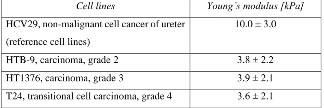
(1) non-malignant epithelial cells of ureter (HCV29, this cell line was established at the Fibiger Institute, Copenhagen, Denmark);
(2) urinary bladder carcinoma (HTB-9, ATCC, LGC Standards); (3) transitional cell carcinoma (HT1376, ATCC, LGC Standards); and (4) transitional cell carcinoma (T24, ATCC, LGC Standards).
The HCV29 cells represent a non-metastatic cancer while three other cell types are characterized by metastatic phenotype with different histological grades1. The higher the grade, the more invasive cancerous cells are. The histological grades for the studied human bladder cancer cell lines are: HTB-9 – grade II, HT1376 – grade III/IV and T24 – grade IV.
All cultures were carried out in 25 cm2 culture flask (Sarstedt) in the incubator (NuAire) providing 37⁰C and 95% air/5% CO2 conditions. The HCV29 and T24 cells were grown in
RPMI-1640 medium (Sigma) supplemented with 10% fetal bovine serum (FBS, Sigma). The HTB-9 cells were cultured also in RPMI-1640 medium containing 10% of FBS, but additionally supplemented with 1% HEPES (4-(2-hydroxyethyl)-1-piperazineethanesulfonic acid, Sigma), and 1% sodium pyruvate (Sigma). The culture of HT1376 cells required Eagle’s medium (EMEM, LGC Standards) supplemented only with 10% FBS (LGC Standards). No antibiotics were involved in cell cultures.
1 The histological grade describes a degree of abnormality in cancerous cells (differences in appearance and function in comparison to healthy cells).
24
3.1.2 Human melanoma cell lines.
To study the properties of cancerous cells, characterized by small differences in their morphology and physic-chemical properties, melanoma cell lines were chosen. In addition, all cell lines originate from very close stages of cancer progression. The measured cell lines were obtained from ESTAB Melanoma Cell Bank:
(1) WM35 cells – these cells were originally isolated from a patient’s skin diagnosed with radial growth phase (RGP) primary melanoma,
(2) WM115 cells – cells derived from a 55 year old female skin melanoma at a vertical growth phase (VGP) in the primary melanoma site,
(3) WM793 cells –cells were established from the vertical growth phase (VGP) of a primary skin melanoma lesion,
(4) WM266-4 cells – cells were established from a cutaneous skin metastasis detected in the same patient as WM115 cells,
(5) WM239 cells – cells were collected from a cutaneous skin metastasis,
(6) 1205Lu cells – cells originated from a lung metastasis diagnosed in the same patient as WM793 cells,
(7) A375P – cells were derived from a solid malignant tumour located in the lung.
Since melanoma develops from melanocytes, as a reference cell line, human epidermal melanocytes from adult skin (primary cell line HEMa-LP, ATCC, LGC Standards) were used. All melanoma cell lines, used here, were cultured in the RPMI-1640 medium (Sigma) supplemented with 10% fetal bovine serum (FBS, Sigma). The HEMa-LP melanocytes required a special, dedicated medium i.e. MEDIUM 254 (GIBCO). Also, in melanoma cell cultures, there were not any antibiotics involved.
3.2 Sample preparation for AFM measurements
For elasticity measurements using atomic force microscopy (AFM), cells were seeded on glass coverslips placed in the Petri dishes (Sarstedt) filled with the corresponding culture medium. The culture time was set to 48 hours. Afterwards, prior to AFM measurements, glass coverslips with cells were immersed into the AFM liquid cell setup, filled with corresponding fresh culture medium and placed on the AFM piezo-scanner.
25
3.3 Sample preparation for PTMS experiments
For photothermal microscopectroscopy (PTMS), cells were cultured in 25 cm2 culture flasks (Sarstedt) until they reached 70-80% of confluency (corresponding to around 3 millions of cells per 1 ml). Cells were trypsinized with 0.05% EDTA-trypsin solution (Sigma) for 5 minutes. Next, cells were centrifuged at 1800 rpm for 4 minutes in an Eppendorf tube (with volume of 1.5 ml). The supernatant was removed, the culture medium was added and cells were gently mixed. Next, cells were again centrifuged. After the culture medium was removed the phosphate buffered saline (PBS, Sigma) was added to a cell pellet, located at the bottom of the Eppendorf tube. Cells were centrifuged and, again, supernatant was removed in gentle way to keep a cell pellet unbroken. Afterwards, a 2.5% glutaraldehyde solution in PBS was added to each tube for 2 hours, followed by delicate twice rinsing in the sterile PBS buffer. Such prepared cells pellet was used for the PTMS measurements.
3.4 Fluorescent staining
Cells cultured on glass coverslips, immersed in the Petri dish with culture medium, were washed with phosphate buffered saline (PBS, Sigma), and the 3.7% solution of paraformaldehyde (Fluka) was added to fix them at room temperature for 20 minutes. After removing the fixative, cells were incubated with the 0.2% solution of Triton X-100 at 4ºC for 5 minutes, followed by rinsing them with PBS buffer. To visualize the actin filaments, coverslips were incubated with Alexa-Fluor 488 conjugated with phalloidin (0.033 µM in PBS, Molecular Probes) for 35 minutes, and washed again with PBS. The cell nuclei have been stained with Hoechst solution (1 mg/ml in PBS, Sigma) for 15 minutes, followed by washing in PBS buffer.
3.5 Sample preparation for ToF SIMS experiments
Although the sample preparation for AFM-based elasticity measurements is well established at the Department of Biophysical Microstructures, the sample preparation for ToF SIMS experiments required more elaboration. This is due to the fact that these experiments
26
have to be carried out in high vacuum conditions. Thus, a protocol specific for ToF SIMS sample preparation was developed2.
3.3.1 Substrates for cell culture for ToF SIMS.
To avoid harmful charge deposition on substrates, glass coverslips could not be used as a support for cell cultures. Instead, highly doped silicon was used as a substrate for cell growth due its conductive properties. Here, commercially available silicon wafers (Si-Mat, Germany) were cut into squares with the size of 1 cm × 1 cm. Such prepared substrates were cleaned with pressurized nitrogen and sterilized with a UV lamp for 1 hour on both sides. Afterwards, cells were seeded on these surfaces. Then, silicon slices were moved to Petri dishes (Sarstedt) that were filled with the corresponding culture medium and, further, cultured for 48 h in the CO2
incubator providing 95%/5% air/CO2 atmosphere. After 48 h of culture, silicon substrates with
cells were washed with phosphate buffered solution (PBS, Sigma) and underwent further steps of sample preparation protocol.
3.3.2 Drying protocol for silicon substrates containing single cells.
After 48 hours of culture on silicon substrates, cells were fixed using 3.7% solution of paraformaldehyde (Fluka) in PBS for 20 minutes, at room temperature (RT). After rinsing them in PBS buffer, steps leading to salt removal were applied (Figure 3.1).
To remove salt, two dilutions were prepared from initial PBS solution containing 150 mM concentrations of NaCl and 27 mM of KCl (pH 7.4). Dilutions were prepared by adding deionized water (Cobrabid purification system, 18 MΩ/cm2) at ratios 1:2 and 1:4. Samples with
cells were washed for 1 minute in each of them. Next, they were immersed in deionized water. As a subsequent part of the protocol, dehydration stage was carried out. Here, to be sure that all water molecules will be removed from the cellular samples, six dilutions of ethyl alcohol (POCH Gliwice) were prepared at concentrations of 40%, 50%, 60%, 70%, 80%, 90%.
2 The ToF SIMS specific sample preparation protocol has been published in the papers by (1) Bobrowska et al.
Analytical Biochemistry 2016, doi:10.1016/j.ab.2016.06.011 and (2) Bobrowska et al. Data in Brief, 2016, doi:10.1016/j.dib.2016.07.052.
27
Figure 3.1. Schematic illustration of the protocol used for sample preparation for ToF SIMS experiments (image adapted from Bobrowska et al. Analytical Biochemistry, 2016,
doi:10.1016/j.ab.2016.06.011).
Afterwards, silicon wafers with cells were immersed subsequently for 30 seconds in each solution, starting from the 40% one. At the end, the anhydrous ethyl alcohol (99.8%, POCH Gliwice) was used for 30 seconds, too. At the final step of the protocol, samples were air dried for 20 minutes in RT. Such prepared samples were transferred into the lock-in chamber of the SIMS apparatus in a vacuum sealed vessel to maintain a sterile environment. All protocol steps were carried out under the laminar flow chamber (NuAire) providing sterile conditions. What is more, for a single batch of experiments, all samples were prepared at once to avoid any differences stemming from the preparation process.
3.3.3 Monitoring changes on cell surface during the preparation protocol.
To study the effects of fixation, salt removal, dehydration and drying steps on cells, the AFM images of cell surface were recorded after each step of the sample preparation protocol. Measurements were carried out for two melanoma cell lines: WM115 from primary tumour site and WM266-4 from skin metastasis (Figure 3.2).
28
Figure 3.2. The surface topography and deflection images recorded for WM115 (a-f) and WM266-4 (g-l) melanoma cells: (a&g) living cell, (b&h) fixed cell with paraformaldehyde,
(c&i) cell in the deionized water, (d&j) dehydration step with 70 % ethyl alcohol solution, (e&k) dehydration step with anhydrous alcohol, and (f&l) dried sample in air (images adapted from Bobrowska et al. Analytical Biochemistry, 2016, doi:10.1016/j.ab.2016.06.011).
29
Images of living cell surface topography were collected in the culture medium (Figure 3.2 a&g). Next, changes of cellular surfaces were visualized after fixation with paraformaldehyde in PBS buffer (Figure 3.2 b&h). The effect of subsequent salt removal was measured for samples immersed in deionized water (Figure 3.2 c&i, for WM115 and WM266-4 cells, respectively). Dehydration effect on cell surface is visualized at two steps i.e. after immersing samples in 70% ethanol solution (Figure 3.2 d&j) and after treatment of the cells with anhydrous ethyl alcohol (Figure 3.2 e&k). Finally, the surface of dried cells (after 15 minutes) was imaged in ambient conditions (Figure 3.2 f&l).
Shapes of melanoma cells remain unchanged independently of the step of the preparation protocol at which the images of the cells were recorded. However, the cellular surface was altered depending on the undergoing preparation step. The largest changes were observed between living cell and fixed cell surfaces. The paraformaldehyde fixation manifested in the less clear image of cell surface. Actin filaments visible previously were less clear but the position of cell nucleus remained stable (the highest fragment of the cell). Such types of images seemed to be characteristic untill the moment when dehydration stage was applied. At these steps, cell membranes collapsed and the localization of cell nucleus began to be clearly visible. Drying of a sample did not changed the resulting cellular surface significantly. To roughly quantify these changes, the height of the cells was measured. Initially, the height of living cells was in the range from 3.5 µm to 4.0 µm. It increases after the paraformaldehyde fixation and deionized water washing, followed by the decrease during dehydration steps. Drying of the sample resulted in the decrease of a cell height below 1 µm (Figure 3.3 a&b, for WM115 and WM266-4 cells, respectively).
a) b)
Figure 3.3. The comparison of cross-sections for WM115 (a) and WM266-4 (b) cells (images adapted from Bobrowska et al. Analytical Biochemistry, 2016, doi:10.1016/j.ab.2016.06.011).
30
The highest points in Figure 3.3 a & b indicate the location above cell nucleus, however, its exact structure is hidden by the cell membrane. After fixation with paraformaldehyde, the height of cells increases from 3.5 ÷ 4.0 µm to about 4.0 ÷ 4.5 µm. Interestingly, after salt removal and washing with deionized water, the height of WM115 cells increases further to ~5 µm while for WM266-4 cells it remains comparable to cells fixed with paraformaldehyde. The proposed way of water molecules removal with ethyl alcohol dilutions followed by rinsing the cells with anhydrous alcohol resulted in their height comparable with that observed for living cells. As it was expected, drying of cells leads to significant drop of cells’ height, being now of the order of 0.5 ÷ 1.0 µm.
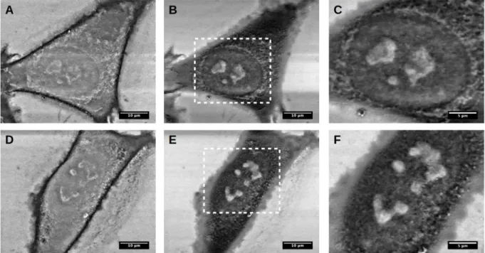
The samples with cells, cultured on silicon substrates and prepared using abovementioned protocol, were used for ToF SIMS experiments. In this technique, the ion bombardment may induce changes to cellular surface that could result in hidden image of surface chemical properties in cells. To verify whether the applied ion beam damages the cellular surface, its visualization was performed by means of scanning electron microscopy (SEM) working in low vacuum conditions under 100 Pa water pressure. In such a way, it was possible to obtain high spatial resolution images of single melanoma cells without any coatings (Figure 3.4 a-f).
Figure 3.4. SEM images of WM115 (a, b and c) and WM266-4 (d, e and f) melanoma cells cultured on bare silicon surface, recorded at 2kV (a, d) and 5 kV (b, c, e and f). Images adapted from Bobrowska et al. Analytical Biochemistry, 2016, doi:10.1016/j.ab.2016.06.011.
31
SEM images were recorded at two values of electron accelerating voltage, namely, 2 kV and 5 kV. This enabled to image either cell surface (i.e. cell membrane) or deeper parts of a cell (cell nucleus). The former images show corrugations on cell surfaces that veil underlying cell nucleus (Figure 3.4 b&e). Higher accelerating voltage allows to visualize cell nucleus with nucleoli accompanied by numerous mitochondria (Figure 3.4 c&f).
The images recorded by AFM and SEM validated the proposed protocol for single cells preparation methodology. Cellular morphological and structural characteristics can be preserved in the protocol involving fixation, dehydration and drying steps. In parallel, these results demonstrate the complementarity of various microscopic techniques (as AFM and SEM) applied to visualized morphological and structural characteristics of cells. The results show that the proposed methodology of sample preparation for single cells is suitable to study biological samples without using freezing based methods and it can be used in spectroscopic and microscopic techniques operating in vacuum conditions.
3.3.4 Effect of the various media composition
The medium composition is usually cell type specific since it has to provide various constituents required for proper growth of cells. Thus, the surface chemical properties measured using ToF SIMS on dry samples can be influenced by the constituents of culture media attached to cell surface. To verify the effect of culture media composition, in the presented studies, the silicon surfaces were exposed to three different compositions of culture media corresponding to those in human bladder cell cultures, i.e. to EMEM (used in HT1376 cells culture), to RPMI 1640 (used for HCV29 and T24 cells growth), and to RPMI 1640 supplemented with HEPES and sodium pyruvate (applied in the culture of HTB-9 cells). Silicon surfaces were kept in the corresponding culture media for 24 h at 37 °C in an CO2 incubator. Afterwards, they underwent
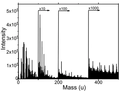
the steps of ToF SIMS specific sample preparation protocol followed by ToF SIMS measurements. The collected mass spectra were analysed in the same way as mass spectra recorded for bladder cells (as described in the Methods). The exemplary mass spectrum of the silicon surface exposed to RPMI 1640 culture medium is shown in Figure 3.5.
32 0 200 400 0 1x10⁴ 2x10⁴ 3x10⁴ 4x10⁴ 5x104 x100 x1000 Int en sity Mass (u) x10
Figure 3.5. The exemplary positive mass spectrum of the silicon substrate exposed to the RPMI 1640 cell’s culture medium.
The effect of culture medium is negligible in the case when all cell lines are grown in the same culture medium, but it could be considerable for cells cultured in various media. The types of melanoma cells chosen for these studies were grown in the culture medium composed of RPMI 1640 medium supplemented with 10% fetal bovine serum. Thus, the effect of culture media compositions in this case is negligible. Only melanocytes (normal HEMa-LP cells) required a special, dedicated medium. For bladder cells, three distinct culture media were applied since their composition is optimized for the best conditions for cellular growth.
The ToF SIMS positive mass spectrum of bare silicon surface, treated in a similar way as silicon with cells, possesses the organic compounds on its surface, which are resulting from the deposition of medium components. Independently of the culture medium type, analogous complex spectra are observed. Thus, to visualize the effect of the culture media compositions, the PCA was applied according to description in Methods. Autoscaling was used as a pre-processing method (Figure 3.6).
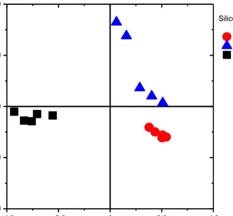
33 -40 -20 0 20 40 -40 -20 0 20 40
Silicon in culture media EMEM
RPMI 1640 + 10% FBS RPMI 1640
PC2 (22,26%)
PC1 (63,46%)
Figure 3.6. Scores plot of the 2nd principal component (PC2) versus the 1st principal component (PC1). Three different culture media were studied: RPMI 1640, RPMI 1640
supplemented with 10% foetal bovine serum, and EMEM.
The results of PCA show that mass spectra of silicon surfaces are grouped depending on the culture medium composition. These findings demonstrate that ToF SIMS technique is very sensitive and, simultaneously, they indicate the importance of measuring the reference sample during ToF SIMS experiments, especially, if this technique is applied to study single cells.
3.6 Summary
The use of various experimental techniques to analyse cell structure involves the application of distinct sample preparation protocols. Some of them, such as cell cultures, are conventional, but very often some protocols have to be optimized for a specific type of measurement. The SIMS measurements frequently employed a relatively complicated preparation method that involves freezing in liquid nitrogen in various ways. Namely, the sample preparation for SIMS involves either freeze fraction of single cells or frozen tissue slices. In the presented thesis, two questions were addressed. First, whether it is possible to develop a fixative-based sample preparation protocol and, second, whether it allows one to distinguish between cells originating from various stages of cancer progression. In this research, the functionality of the TOF-SIMS technique to study biological samples without using freezing based methodology for sample preparation is demonstrated.
34
4. Experimental methods
4.1 Atomic Force Microscopy – basic principles
Atomic force microscopy (AFM) is one of the scanning probe based techniques that enables imaging of surfaces with a sub-nanometric resolution. In the AFM, a flat spring (i.e. cantilever with a probing tip mounted at its free end) is used to scan surface of the sample (Figure 4.1).
a) b)
Figure 4.1. (a) Atomic Force microscope - working principle. (b) An image of the AFM apparatus from Park Systems – a model Xe120 integrated with inverted optical microscope
equipped with fluorescence functionality.
The tip curvature radius of the probing tip varies between few to tens of nanometres. When probing tip is placed in a close proximity to the investigated surface, forces acting between its end and material’s surface deflect the cantilever. The deflection is measured by an optical system consisting of a laser and a position sensitive detector (usually by a photodiode). A precise movement of the sample is realised by a piezo-scanner. All measurements presented in this thesis were carried out using a commercially available atomic force microscope model XE120 manufactured by Park Systems (Korea). This device is integrated with an inverted optical microscope Olympus IX71 used to localize place of interest for the AFM measurements, as well as to collect fluorescence images.
35
4.1.1 Surface topography measurements.
The surface topography measurements were carried out for both living and dried cells using AFM working in contact mode. Two types of V-shaped silicon nitride cantilevers were applied: (1) PNP-TR-customized (Nanoworld) characterized by a nominal spring constant of 0.03 N/m, and (2) MLCT (Bruker) characterized by nominal spring constants of 0.01 N/m (microlevel type C) and 0.03 N/m (microlever type D). The set point range varied between 0.2 nN and 0.8 nN (adjusted during the surface topography acquisition), while the scan rate range was set between 0.3-1 Hz, depending on the scan size. During the AFM measurements of living cells, glass coverslips with cells immersed in a Petri dish, were mounted on the piezoelectric scanner. Cells were imaged in the RPMI-1640 culture media supplemented with 1% HEPES to stabilize pH during the measurements. AFM images were analysed by means of XEI – software provided by a manufacturer, dedicated to XE 120 set-up and WSxM 5.0 software [93].
4.1.2 Force-distance curves.
The elasticity measurements are realized using AFM working in force spectroscopy mode. Here, cantilever is moving towards to and backwards from the sample surface. During this movement cycle, the deflection of the cantilever is registered as a function of relative scanner position (Figure 4.2). Such relation is later on converted into force versus tip-sample distance curve called shortly force-distance curve. Each force-distance curve consists of an approach curve and a retract curve. These curves consist of characteristic regions:
base line that is the deflection of the cantilever recorded when AFM probing tip is far away from the investigated surface. At this distance the interaction forces are negligible,
contact point that is the a point when the AFM probing tip touches the sample surface,
a region when cantilever is deflected due to interaction forces acting between the probing tip and the sample surface. For stiff substrates, only cantilever deflection is measured. For soft samples, the recorded deflection contains contribution from sample surface deformation. During approach, the deflection