Introduction
Paratrichodina incisa (syn: Trichodinella sp., Tripartiella sp.) has been found on the gills of Nemachius barbatulus by Lom in 1959 [1], though he defined Paratrichodina (Trichodinidae: Para tri cho dina) in 1963 [2]. More than ten species in Para -trichodina have been found so far from various freshwater and marine fishes throughout the world, such as P. globonuclea Lom, 1963 [2], P. obliqua Lom, 1963 [2], P. phoxini Lom, 1963 [3, 4], P. alburni (Vojtek, 1957) Lom, 1963 (Syn: Tri cho -di na alburnui Vojtek, 1957) [4]; P. degiustii Lom et Haldar, 1976 [4], P. corlissi Lom et Haldar, 1977 [5], P. erectispina Lom et Haldar, 1977 [5], P. ura -le nsis Kashkovsky et Lom, 1979 [6], P. voikarensis Kashkovsky et Lom, 1979 [6], P. africana Ka zub -ski et El-Tantawy, 1986 [7, 8], P. lizae Asmat, 2002 [9], P. bassonae Amlan et Probir, 2006 [10], and so on. In China, Para tri cho di na globonuclea and P. obli qua have been found from marine fishes [11, 12].
The Paratrichodina yangtzeus sp. n. was found
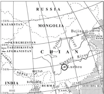
by the author on the gills of Silurus asotus Linnaeus, 1758 collected in the year 2006 from the Yangtze River at Luzhou, Sichuan, China (28°51'20.41"N 105°23'25.43"E), which is the longest river in China (Fig. 1). The collected Silurus
Description of Paratrichodina yangtzeus sp. n. (Ciliophora:
Trichodinidae) from the freshwater fishes in the Yangtze
River, China
Yinheng Hu
Luzhou Vocational Technical College, Luzhou 646005, Sichuan, P. R. China; E-mail: huyhen@163.com
ABSTRACT. Paratrichodina Lom, 1963 comprises more than ten species. Paratrichodina has been found so far in
various freshwater, marine fishes and amphibians worldwide. This report provides the first record of Paratrichodina from freshwater fishes in China. Smears with trichodinids were air-dried and then the slides with trichodinid ciliates were impregnated with Klein dry silver impregnation technique in order to reveal details of the adhesive disc. All measurements are presented in micrometres and follow the system proposed by Lom. Detailed descriptions of the denticles are presented in accordance with the method proposed by Van As and Basson. Denticulate ring of the studied specimens is constituted by loosely arranged denticles and the adoral ciliary spiral is between 180 to 200°. So the studied specimens were assigned to Paratrichodina. The presently described trichodinid resembles P. erectispina Lom et Haldar, 1977 and P. voikarensis Kashkovsky et Lom, 1979, but it is clearly distinguished from P. erectispina by the shape of denticle and some other measurements. Based on its unique characteristics, a new Paratrichodina species is erected, and named Paratrichodina yangtzeus sp. n.
Key words: Ciliophora, Trichodinidae, Paratrichodina yangtzeus sp. n., Silurus asotus, Yangtze River, China
Fig 1. Map of sampling localities where fishes were collected from the Yangtze River
asotus Linnaeus, 1758 are infected only by this kind of trichodinid, with a 100% infection rate. Each fish contains a large number of trichodinids.
Materials and methods
The host fishes, Silurus asotus Linnaeus, 1758 were collected from the Yangtze River at Luzhou, Sichuan, China in 2006. Gill scrapings were made from the hosts. Smears with trichodinids were air-dried and then the slides with trichodinid ciliates were impregnated with Klein dry silver impre -gnation technique [13] in order to reveal details of the adhesive disc. All photomicrographs and illustration drawings were made with the help of camera (Motic DMBA300B) at 100× magnifi -cation with oil immersion lens and computer software Motic Images Advanced 3.2 and CorelDRAW X3. The statistics were obtained with
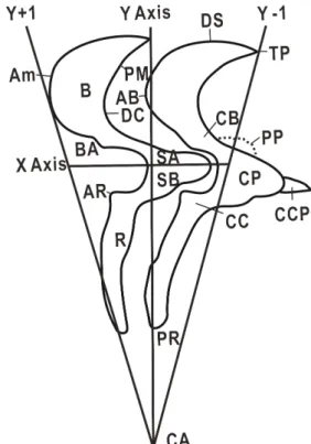
Microsoft Excel 2003. All measurements are presented in micrometres and follow the system proposed by Lom [14]. Minimum and maximum values are given, followed in parentheses by the arithmetic mean and standard deviation. In the case of the denticles and radial pins, the mode is given rather than the arithmetic mean with the number of specimens examined given in parentheses. The span of the denticle is measured from the tip of the blade to the tip of the ray. Body diameter is measured as the adhesive disc plus the border membrane. Detailed descriptions of the denticles are presented in accordance with the method proposed by Van As and Basson [15] (Fig. 2).
Results and discussion
Taxonomic summary
Species: Paratrichodina yangtzeus sp. n. Type host: Silurus asotus Linnaeus, 1758 Fish family: Siluridae
Location: gills
Type locality: Luzhou, Sichuan, China (28°51'20.41"N 105°23'25.43"E)
Etymology: The specific name is coined from the name of the Yangtze River
Reference material: Holotype, slide LZY–664/ 2006 and paratype slides LZY–640/2006, LZY–668/2006, deposited in the Biological Laboratory of Luzhou Vocational Technical College.
Fig. 2. Denticle structure and construction of X and Y axes as fixed references for description of denticles (after Van As and Basson 1989)
Explanations: AB, apex of blade; AM, anterior margin
of blade; AR, apophysis of ray (= thorn); B, blade; BA,
apophysis of blade; CA, central of adhesive disc; CB,
section connecting blade and central art; CC, secton
connecting central part and ray; CCP, central conical
part; CP, central part; DC, deepest point of curve; DS,
distal surface of blade; PM, posterior margin of blade; PP, posterior projection; PR, point of ray; R, ray; SA,
section of central part above x axis; SB, section of
central part below x axis; TP, tangent point
Figs. 3–6. Photomicrographs of the silver nitrate impregnated adhesive discs of Paratrichodina
yangtzeus sp. n. from the gills of Silurus asotus.
Fig.6. Also displays the shape of the whole cell of it. Scale bars=20 µm
Description (Figs. 3–7, Table 1)
Small, freshwater trichodinid with long cylinder or cone shape (Fig. 6). Body diameter 25.8–31.1 (28.1±1.9). Diameter of adhesive disc 20.4–26.3 (23±1.9). Diameter of denticulate ring 12.8–17.8 (14.9±1.6). Centre of disc clear and without any granules. Border membrane 2.0–3.0 (2.5±0.3) in width. Number of denticles 20–22 (21). Number of radial pins (mode) per denticle 5. Span of denticle 5.6–7.8 (6.6±0.7). Length of denticle 2.0–3.5 (2.5±0.4). Denticulate ring constituted by loosely
arranged denticles. Blade length 2.1–2.9 (2.9±0.2). Blade narrow sickle shaped and tangent point tapering or slightly rounded. Apophysis of blade almost close to Y+1 axis. Width of central part 0.5–1.2 (1±0.2). Central part narrow with triangle or bluntly-rounded point, extending less than halfway towards the Y–1 axis. Shape of the central part above and below the X axis almost the same. Ray straight, long and thin, and directed towards the center of adhesive disc. Ray length 1.8–3.8 (2.7±0.6). Adoral ciliary spiral 180–200°.
Table 1. Comparison of Paratrichodina yangtzeus n. sp. with P. erectispina Lom et Haldar, 1977 and P. voikarensis Kashkovsky et Lom, 1979
Trichodinid species P. yangtzeus sp.n. P. erectispina P. voikarensis
Host Silurus asotus Pimephales vigilax Coregonus nasus Coregonus peled
Locality Luzhou, Sichuan, China Vicinity of Detroit USA Russia Russia
Site gills gills gills gills
Reference present study Lom and Haldar (1977) Kashkovsky and Lom (1979)
No. of specimens 20 — — — measured Body diameter 25.8–31.1 (28.1±1.9) 30 (23–36) 34 (29–44) 37 (33–48) Adhesive disc 20.4–26.3 (23±1.9) 25 (20–31) 24 (23–25) 24 (18–26) Denticulate ring 12.8–17.8 (14.9±1.6) 15 (12–19) 15 (15–16) 26 (20–39) Border membrane 2.0–3.0 (2.5±0.3) 2 2–3 1.8–2.4 Denticle number 20–22 (21) 25 (24–27) 23–25 22–25 Radial pins per denticle 5 (5) 4–5 6 6 Denticle span 5.6–7.8 (6.6±0.7) — — — Denticle length 2.0–3.5 (2.5±0.4) — — — Blade length 2.1–2.9 (2.9±0.2) 3–3.5 3–4 3 (2.8–4.6) Central part width 0.5–1.2 (1±0.2) 1.2–1.4 3 (2–4) 1–2.5 Ray length 1.8–3.8 (2.7±0.6) 2–2.5 1–2 0.6–1.3 Adoral ciliary spiral 180–200° 230–280° 240–260° 240–260°
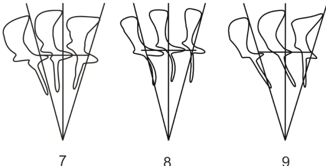
Figs. 7–9. Diagrammatic drawings of the denticles of trichodinid ciliophorans Fig. 7. Paratrichodina yangtzeus sp. n.
Fig. 8. P. erectispina Lom et Haldar, 1977 Fig. 9. P. voikarensis Kashkovsky et Lom, 1979
Remarks (Figs. 7–9, Table 1)
Among all Paratrichodina species, the Paratrichodina yangtzeus sp. n. collected from the gills of Silurus asotus Linnaeus, 1758 in the Yangtze River at Luzhou, China only resembles P. erectispina Lom et Haldar, 1977 obtained from the gills of Pimephales vigilax found in West Birginis, USA [5] and P. voikarensis Kashkovsky et Lom, 1979 obtained from the gills of Coregonus nasus, C. peledamong found in Russia [6].
Paratrichodina yangtzeus sp. n. is clearly distinguished from P. erectispina by the shape of denticle and some other measurements such as morphometric data. The apophysis of blade in the new species is almost close to the Y+1 axis, but that of P. erectispina is far away from the Y+1 axis. The central part of the new species extends less than halfway towards the Y–1 axis, but that of P. erectispina stretches far more than halfway to the Y–1 axis. In the P. erectispina, the rays are curved, the concavity of this arch being directed forwards (i.e., anticlockwise in the aboral view), but in case of the new species the rays are straight and directed towards the center of adhesive disc. In the diagrammatic drawing of the denticles of trichodinid, the ray of the P. erectispina is situated between Y and Y–1 axis, but the new species’ ray is situated between Y and Y+1 axis. Moreover, morphometric data are different. For instance, the new species has fewer denticles (20–22 vs 24–27) and more radial pins per denticle (5 vs 4–5), shorter blade (2.1–2.9 vs 3–3.5) and central part width (0.5–1.2 vs 1.2–1.4). The adoral ciliary spiral is quite different too: 180–200° in the new species, but 230–280° in P. erectispina.
Paratrichodina yangtzeus sp. n. and P. voikarensis also demonstrate dissimilarities. The apophysis of blade in the new species is almost close to the Y+1 axis, but that of P. voikarensis is far away from the Y+1 axis. The central part of the new species is longer than that of the P. voikarensis. It extends less than halfway towards the Y–1 axis, but in P. voikarensis it just stretches across the Y axis. In the new species the ray is straight and parallel to Y axis, while in the P. voikarensis the whole denticle is slanted backward and its ray crosses the Y axis. Some morphometric data are different too. The new species. has smaller body diameter (25.8–31.1 vs 29–48) and denticulate ring (12.8–17.8 vs 15–39), and it has fewer denticles (20–22 vs 22–25), fewer radial pins per denticle (5 vs 6), and shorter blade (2.1–2.9 vs 3–4.6). And the
adoral ciliary spiral is also quite different for both species (180–200° vs 240–260°).
References
[1] Lom J. 1959. On the systematics of the genus
Trichodinella ŠramekHušek (=Brachyspira Ra a
-be). Acta Parasitologica 7: 573–590.
[2] Lom J. 1963. The ciliates of the family Urceolariidae inhabiting gills of fishes (Trichodinella group).
Acta Societatis Zoologicae Bohemoslovenicae 27:
7–19.
[3] Lom J. 1963. Discovery of a Tripartiella in the urinary tract of Phoxinus phoxinus L. Acta Protozoologica 1:1–4.
[4] Lom J., Haldar D.P. 1976. Observations on trichodinids endocommensal in fishes. Transactions
of the American Microscopical Society 95:
527–541.
[5] Lom J., Haldar D.P. 1977. Ciliates of the genera
Trichodinella, Tripartiella and Paratrichodina
(Peritricha, Mobilina) invading fish gills. Folia
Parasitologica 24: 193–210.
[6] Kashkovsky V.V., Lom J. 1979. New species of parasitic infusoria of the family Urceolariidae.
Parazitologiâ 13: 266–268.
[7] Kazubski S.L., El-Tantawy S.A.M. 1986. The ciliate
Paratrichodina africana sp. n. (Peritricha, Trichodinidae) from Tilapia fish (Cichlidae) from Africa. Acta Protozoologica 25: 433–438.
[8] Mitra A.K., Bandyopadhyay P.K. 2006. First record of ectoparasitic African Trichodinids (Ciliophora: Peritrichida) in a cichlid fish Oreochromis
mossambicus (Peters, 1852) from the Churni river
system, West Bengal, India. Animal Biology 56: 323–333.
[9] Asmat G.S.M. 2002. Trichodinid ciliates (Ciliophora: Trichodinidae) from Indian fishes with description of two new species. Bangladesh Journal of Zoology 30: 87–100.
[10] Mitra A.K., Bandyopadhyay P.K. 2006. Trichodina
haldari n. sp. and Paratrichodina bassonae n. sp.
(Ciliophora: Peritrichida) from Indian fresh water fishes. Acta Protozoologica 45: 289–294.
[11] Xu K., Song W. 2000. Parasitic trichodinid ciliates (Ciliophora, Peritrichida) from mariculture fishes II. Genera Paratrichodina, Trichodinella and Dipar
-tiella. Journal of Ocean University of Qingdao 30:
413–417.
[12] Xu K., Song W., Warren A., Choi J.K. 2001. Trichodinid ectoparasites (Ciliophora: Peritrichida) of some marine fishes from coastal regions of the Yellow Sea and Bohai Sea. Systematic Parasitology 50: 69–79.
[13] Klein B.M. 1958. The „dry” silver method and its proper use. Journal of Protozoology 5: 99–103.
[14] Lom J. 1958. A contribution to the systematics and morphology of endoparasitic trichodinids from amphibians of uniform specific characteristics.
Journal of Protozoology. 5: 251–263.
[15] Van As J.G., Basson L. 1989. A further contribution to the taxonomy of Trichodinidae (Ciliophora:
Peritrichia) and a review of the taxonomic status of some fish ectoparasitic trichodinids. Systematic
Parasitology 14: 157–179. Wpłynęło 25 sierpnia 2008