Biologia choroby nowotworowej -
proces transformacji
nowotworowej, angiogeneza,
inwazja i przerzutowanie
ONKOLOGIA - Podstawowe pojęcia
•
onkos (greka) - masa, guz
•
nowotwór (neoplasma) - łagodny/złośliwy
•
rak - nowotwór wywodzący się z tkanek
nabłonkowych,
•
w literaturze anglosaskiej i popularnie synonim
Czym jest rak?
o życiu, śmierci i nieśmiertelności komórki, ewolucji i..
rosyjskiej ruletce
Czym jest rak?
Rak jest wynikiem mutacji (wrodzonych i nabytych lub tylko nabytych) oraz zmian
epigenetycznych.
Mutacje są wrodzone, wywołane przez czynniki szkodliwe (karcynogeny) lub są dziełem
przypadku.
Ryzyko zachorowania na raka jest modyfikowane przez cechy wrodzone, styl życia i
środowisko, jest też kwestią szczęścia....
Transformacja nowotworowa - teoria klasyczna
progresja nowotworu: od
hyperplazji, poprzez dysplazję,
raka in situ do raka
1.000.000 komórek
1.000.000.000 komórek
-granica wykrywalności
klinicznej (historyczna),
ok. 30 podziałów
1.000.000.000.000 -
śmierć organizmu ok.
40 podziałów
wzrost guza - progresja kliniczna
nowotworu
•
komórka (nowotworowa) w wyniku
mutacji somatycznych jest zdolna do
ekspansji klonalnej
Transformacja nowotworowa - teoria klasyczna
Zachorowalność na raka piersi i raka jelita grubego -
teoria vs praktyka
Progresja nowotworu - selekcja klonów i prawa
ewolucji darwinowskiej
Transformacja nowotworowa - wiele klonów czy jeden
klon?
Nowotory lite vs białaczki/chłoniaki
różna biologia, naturalny przebieg i leczenie
•
nowotwory lite
•
raki i mięsaki
•
komórki z których się wywodzą
(nabłonki, mezenchyma) nie są
mobilne
•
(potrzebne dodatkowe mutacje)
•
białaczki/chłoniaki (liquid
tumors)
•
komórki z których się wywodzą są
Wiele mutacji w jednej komórce czy selekcja klonalna -
mikroewolucja w obrębie guza nowotworowego
Transformacja nowotworowa - inicjacja, promocja,
Hodowle komórkowe-
komórki normalne i komórki
nowotworowe
Cechy komórki nowotworowej
•
samowystarczalność w zakresie sygnałów wzrostu
(proliferacji)
•
ignorowanie sygnałów hamowania proliferacji
•
unikanie apoptozy - samobójstwa komórki
•
angiogeneza
•
inwazja tkanek i przerzutowanie
•
nieograniczony potencjał replikacyjny -
krzywa
Gompertza
- guz
normalna
tkanka
Czas
logarytm liczby
komórek
komórki
dzielące się
komórki
macierzyste
komórki
zróżnicowane
komórki
śmierć
Komórka - wzajemnie powiązane
obwody regulacji
•
onkogeny (mutacja powoduje ich aktywację) -
pedał
gazu w samochodzie się zaciął wciśnięty!
•
geny supresorowe (mutacja powoduje
deaktywację) -
hamulce przestały działać!
•
geny stabilizujące (geny opiekuńcze - naprawa
DNA - mutacja powoduje niestabilność genetyczną) -
mechanik nic nie warty!
•
*mutacja żadnego pojedynczego genu nie powoduje raka
Za proces transformacji komórki są
H I S TO R I C A L P E R S P E C T I V E
NATURE MEDICINE VOLUME 10 | NUMBER 8 | AUGUST 2004 791
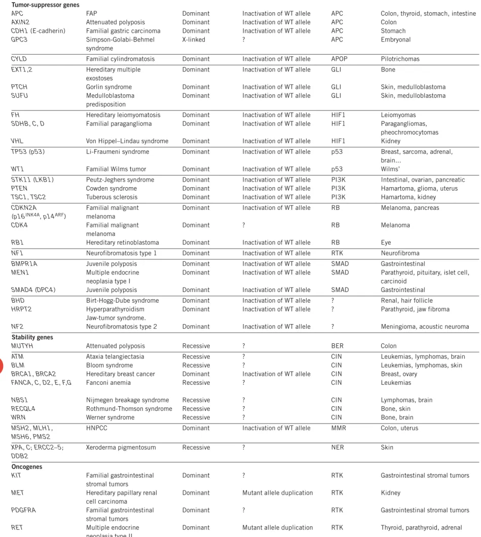
Table 1 Cancer predisposition genes
Gene (synonym(s))a Syndrome Hereditary pattern Second hit Pathwayb Major heredity tumor typesc
Tumor-suppressor genes
APC FAP Dominant Inactivation of WT allele APC Colon, thyroid, stomach, intestine
AXIN2 Attenuated polyposis Dominant Inactivation of WT allele APC Colon
CDH1 (E-cadherin) Familial gastric carcinoma Dominant Inactivation of WT allele APC Stomach
GPC3 Simpson-Golabi-Behmel X-linked ? APC Embryonal
syndrome
CYLD Familial cylindromatosis Dominant Inactivation of WT allele APOP Pilotrichomas
EXT1,2 Hereditary multiple Dominant Inactivation of WT allele GLI Bone exostoses
PTCH Gorlin syndrome Dominant Inactivation of WT allele GLI Skin, medulloblastoma
SUFU Medulloblastoma Dominant Inactivation of WT allele GLI Skin, medulloblastoma
predisposition
FH Hereditary leiomyomatosis Dominant Inactivation of WT allele HIF1 Leiomyomas
SDHB, C, D Familial paraganglioma Dominant Inactivation of WT allele HIF1 Paragangliomas, pheochromocytomas
VHL Von Hippel–Lindau syndrome Dominant Inactivation of WT allele HIF1 Kidney
TP53 (p53) Li-Fraumeni syndrome Dominant Inactivation of WT allele p53 Breast, sarcoma, adrenal, brain...
WT1 Familial Wilms tumor Dominant Inactivation of WT allele p53 Wilms’
STK11 (LKB1) Peutz-Jeghers syndrome Dominant Inactivation of WT allele PI3K Intestinal, ovarian, pancreatic
PTEN Cowden syndrome Dominant Inactivation of WT allele PI3K Hamartoma, glioma, uterus
TSC1, TSC2 Tuberous sclerosis Dominant Inactivation of WT allele PI3K Hamartoma, kidney
CDKN2A Familial malignant Dominant Inactivation of WT allele RB Melanoma, pancreas (p16INK4A, p14ARF) melanoma
CDK4 Familial malignant Dominant ? RB Melanoma
melanoma
RB1 Hereditary retinoblastoma Dominant Inactivation of WT allele RB Eye
NF1 Neurofibromatosis type 1 Dominant Inactivation of WT allele RTK Neurofibroma
BMPR1A Juvenile polyposis Dominant Inactivation of WT allele SMAD Gastrointestinal
MEN1 Multiple endocrine Dominant Inactivation of WT allele SMAD Parathyroid, pituitary, islet cell,
neoplasia type I carcinoid
SMAD4 (DPC4) Juvenile polyposis Dominant Inactivation of WT allele SMAD Gastrointestinal
BHD Birt-Hogg-Dube syndrome Dominant Inactivation of WT allele ? Renal, hair follicle
HRPT2 Hyperparathyroidism Dominant Inactivation of WT allele ? Parathyroid, jaw fibroma Jaw-tumor syndrome.
NF2 Neurofibromatosis type 2 Dominant Inactivation of WT allele ? Meningioma, acoustic neuroma
Stability genes
MUTYH Attenuated polyposis Recessive ? BER Colon
ATM Ataxia telangiectasia Recessive ? CIN Leukemias, lymphomas, brain
BLM Bloom syndrome Recessive ? CIN Leukemias, lymphomas, skin
BRCA1, BRCA2 Hereditary breast cancer Dominant Inactivation of WT allele CIN Breast, ovary
FANCA, C, D2, E, F,G Fanconi anemia Recessive ? CIN Leukemias
NBS1 Nijmegen breakage syndrome Recessive ? CIN Lymphomas, brain
RECQL4 Rothmund-Thomson syndrome Recessive ? CIN Bone, skin
WRN Werner syndrome Recessive ? CIN Bone, brain
MSH2, MLH1, HNPCC Dominant Inactivation of WT allele MMR Colon, uterus
MSH6, PMS2
XPA, C; ERCC2–5; Xeroderma pigmentosum Recessive ? NER Skin
DDB2
Oncogenes
KIT Familial gastrointestinal Dominant ? RTK Gastrointestinal stromal tumors
stromal tumors
MET Hereditary papillary renal Dominant Mutant allele duplication RTK Kidney
cell carcinoma
PDGFRA Familial gastrointestinal Dominant ? RTK Gastrointestinal stromal tumors stromal tumors
RET Multiple endocrine Dominant Mutant allele duplication RTK Thyroid, parathyroid, adrenal
neoplasia type II
WT, wild type. aRepresentative genes of all the major pathways and hereditary cancer predisposition types are listed. For a complete list, see ref. 117. Approved gene symbols are
provided for each entry, with alternative names in parentheses. bIn many cases, the gene has been implicated in several pathways. The single pathway that is listed for each gene
represents a ‘best guess’ (when one can be made) and for the reasons noted in the text and in the legend to Figure 9, should not be regarded as conclusive. APOP, apoptotic
pathway; RTK, receptor tyrosine kinase pathway (see Fig. 1). cIn most cases, the nonfamilial tumor spectrum caused by somatic mutations of the gene includes those occurring in
the familial cases plus additional tumor types. For example, mutations of TP53 and CDKN2A are found in many more tumor types than those to which Li-Fraumeni and familial malignant melanoma patients, respectively, are predisposed.
©
2004
Nature
Pub
lishing
Gr
oup
http://www
.nature
.com/naturemedicine
H I S TO R I C A L P E R S P E C T I V E
most dramatic therapeutic advances have come from agents
targeted against proteins encoded by genes that are mutated in
cancers. These include trastuzumab (Herceptin), an antibody
against the product of the ERBB2 gene amplified in some breast
cancers
87; imatinib (Gleevec), an inhibitor of tyrosine kinases
altered in chronic myelogenous leukemia (CML)
88and
gastroin-testinal stromal tumors (GISTs)
89; and gefitinib (Iressa), an EGFR
kinase inhibitor used to treat lung cancers
90. Though none of these
advances have resulted in cures of many patients with advanced
disease, they can substantially improve and prolong lives. One
important lesson from the use of these agents is that mutations are
more reliable indicators of a good target than is abnormal
expres-sion. For example, virtually all GISTs abnormally express high
lev-els of c-kit, but only those tumors with intragenic mutations of KIT
796 VOLUME 10 | NUMBER 8 | AUGUST 2004 NATURE MEDICINE
Table 2 Genes that are mutated somatically but not inherited in mutant form
Genea(synonym) Somatic mutation typeb Cancers with mutant genec Pathwayd
CTNNB1 (!-catenin) Activating codon change Colon, liver, medulloblastomas APC
BCL2 Translocation Lymphomas APOP
TNFRSF6 (FAS) Activating codon change Lymphomas, testicular germ cell tumors APOP
BAX Inactivating codon change Colon, stomach APOP FBXW7 (CDC4) Inactivating codon change Colon, uterine, ovarian, breast CIN
GLI Amplification, translocation Brain, sarcomas GLI
HPVE6 HPV infection Cervical p53
MDM2 Amplification Sarcomas p53
NOTCH1 Translocation Leukemias p53
AKT2 Amplification Ovarian, breast PI3K
FOXO1A, 3A Translocation Rhabdomyosarcomas, leukemias PI3K
PI3KCA Activating codon change Colon, stomach, brain, breast PI3K
CCND1 (cyclin D1) Amplification, translocation Leukemias, breast RB
HPVE7 HPV infection Cervical RB
TAL1 Translocation Leukemias RB
TFE3 Translocation Kidney, sarcomas RB
ABL1 (ABL) Translocation Chronic myelogenous leukemia RTK
ALK Translocation Anaplastic large cell lymphoma RTK
BRAF Activating codon change Melanoma, colorectal, thyroid RTK
EGFR Amplification, activating codon change Glioblastomas, non–small cell lung cancers RTK
EPHB2 Inactivating codon change Prostate RTK
ERBB2 Amplification Breast, ovarian RTK
FES Activating codon change Colon RTK
FGFR1–3 Translocation Lymphomas, gastric cancers, bladder cancers RTK
FLT3, 4 Activating codon change Leukemias, angiosarcomas RTK
JAK2 Translocation Leukemias RTK
KRAS2, N-RAS Activating codon change Colorectal, pancreatic, non–small cell lung cancer RTK
NTRK1, 3 Translocation, activating codon change Thyroid, secretory breast, colon RTK
PDGFB Translocation Dermatofibrosarcomas and fibroblastomas RTK
PDGFRB Translocation Leukemias RTK
EWSR1 Translocation Ewing’s sarcomas, lymphomas, leukemias SMAD
RUNX1 Translocation Leukemias SMAD
SMAD2 Inactivating codon change Colon, breast SMAD
TGFBR1, TGFBR2 Inactivating codon change Colon, stomach, ovarian SMAD
BCL6 Translocation Lymphomas ?
EVI1 Translocation Leukemias ?
HMGA2 Translocation Lipomas ?
HOXA9, 11, 13; HOXC13, Translocation Leukemias ?
HOXD11, 13; HOX11, HOX11L2
MAP2K4 (MKK4) Inactivating codon change Pancreas, breast, colon ?
MLL Translocation, activating codon change Leukemias ?
MYC, MYCN, MYCL1 Amplification Lymphomas, neuroblastomas, small cell lung cancers ?
PTNP1, 11 Activating codon change Leukemias, colon ?
RARA Translocation Promyelocytic leukemia ?
SS18 Translocation Synovial sarcomas ?
aRepresentative genes of all the major pathways and cancer types are listed. For a complete list, see ref. 76. Approved gene symbols are provided for each entry, with alternative names in
paren-theses. bActivating codon change, intragenic mutation altering one or a small number of base pairs that activates the gene product, indicating that it is an oncogene; inactivating codon change,
any mutation (point mutation, small or large deletion, etc.) that inactivates the gene product, indicating that the gene is a tumor suppressor. Amplifications and translocations generally affect oncogenes, though occasional translocations disrupt a gene rather than activate it (such as has been suggested to occur with RUNX1; ref. 118). cOnly representative types of cancers are listed
when a gene is mutated in many tumor types, Specific types of leukemias and lymphomas are listed only if a given gene is predominantly mutated in a specific subtype. dIn many cases, the gene
has been implicated in several pathways. The single pathway that is listed for each gene represents a ‘best guess’ (when one can be made) and for the reasons noted in the text and in the legend to Figure 9, should not be regarded as conclusive. APOP, apoptotic pathway; RTK, receptor tyrosine kinase pathway (see Fig. 9).