Volume V • No. 2
April –June • 2012 ISSN 1898-6498
MILITARY PHARMACY
AND MEDICINE
QUARTERLY INTERDISCIPLINARY JOURNAL
• PHARMACY
• MEDICINE
• MEDICAL TECHNIQUE
• ENVIRONMENT AND HEALTH
• EDUCATION
The Staff of the Military Center of Pharmacy and Medical Technology in Celestynow – Poland MILIT AR Y PHARMA CY AND MEDICINE • V olume V • No . 2 • 2012
MILITARY PHARMACY
AND MEDICINE
Quarterly Interdisciplinary Journal
of Military Centre of Pharmacy and Medical Technique in Celestynów
on scientific socio-professional
and training issues of Military Pharmacy and Medicine
ISSN 1898-6498
April – June, 2012
MILITARY PHARMACY
AND MEDICINE
SCIENTIFIC BOARD
Anyakora Chimezie, Nigeria Balbaa Mahmoud, Egypt
prof. dr hab. Michał Bartoszcze, Poland prof. dr hab. inż. Stanisław Bielecki, Poland Bisleri Gianluigi, Italy
Blumenfeld Zeev, Israel
dr hab. Kazimiera H. Bodek, Poland Boonstra Tjeerd W, Netherlands Borcic Josipa, Croatia Cappelli Gianni, Italy Yifeng Chai, China Chowdhury Pritish K, India Costa Milton, Brasil
prof. dr hab. inż. Krzysztof Czupryński, Poland Deckert Andreas, Germany
Demeter Pal, Hungary prof. dr hab. Adam Dziki, Poland Ermakova Irina, Russia
prof. dr hab. Zbigniew Fijałek, Poland Florence Sudha Gnanou, India Fontoura Paulo, Portugal dr hab. Ryszard Gajdosz, Poland Ning Gao, China
dr hab. Tomasz Gaszyński, Poland prof. dr hab. Paweł Górski, Poland prof. dr hab. Bożenna Gutkowska, Poland Holko Ivan, Slovakia
Zhenlin Hu, China Huang Feng, USA
dr hab. Czesław Jeśman, Poland prof. dr hab. Wiesław Jędrzejczak, Poland Kaubrys Gintaras, Lithuania
Kashanian Maryam, Iran
prof. dr hab. Andrzej Klimek, Poland dr hab. Józef Knap, Poland Korshunov Andrey, Russia Kusec Sanja, Croatia Shan Lei, China dr hab. Julian Maj, Poland
prof. dr hab. Jerzy Mierzejewski, Poland prof. dr hab. Elżbieta Mikiciuk-Olasik, Poland Newcomb Andrew, Canada
prof. dr hab. Jerzy Z. Nowak, Poland dr hab. Romuald Olszański, Poland prof. dr hab. Daria Orszulak-Michalak, Poland prof. dr hab. Krzysztof Owczarek, Poland prof. dr hab. Marek Paradowski, Poland Perotti Daniela, Italy
Pivac Nela, Croatia Pizzuto Francesco, Italy prof. dr hab. Janusz Pluta, Poland Polat Gurbuz, Turkey
Popescu Irinel, Romania Reddy G. Bhanuprakash, India prof. dr hab. Juliusz Reiss, Poland Rodrigues Gabriel Sunil, India Rossi Roberto, Italy Samarkos Michael, Greece Shen Hui-Liang, China Shevchuk Nikolai, Russia Xianglin Shi, USA Skultetyova Dana, Slovakia Strumylaite Loreta, Lithuania dr Piotr Siermontowski, Poland prof. dr hab. Marek Sosnowski, Poland prof. dr hab. Andrzej Stańczak, Poland prof. dr hab. Zbigniew Lew-Starowicz, Poland dr hab. inż. Marek Strzelczyk, Poland Ding-Feng Su, China
dr hab. Janusz Świniarski, Poland Tchetina Elena, Russia
Tomoum Hoda, Egypt Tufekcioglu Omac, Turkey
prof. dr hab. Jarosław Wysocki, Poland Wang FuZhou, China
Wei-dong Zhang, China Zarkovic Neven, Croatia Ruixin Zhu, China
MILITARY PHARMACY
AND MEDICINE
EDITORIAL BOARD
EDITOR-IN-CHIEF
prof. Piotr Fiedor, Warsaw, Poland
DEPUTY EDITOR
prof. Jarosław Wysocki, Warsaw, Poland
SECTION EDITORS
Biochemistry
dr hab. inż. Marek Strzelczyk, Poland
Bioethics & Medical Law
prof. dr hab. Hieronim Bartel, Poland
Biology
prof. Lidia Chomicz, Poland
Catastrophe Medicine
Adam Pietrzak, Poland
Emergency Medicine
dr hab. Tomasz Gaszyński, Poland
Epidemiology
dr Witold Gnitecki, Poland
Forensic Medicine
dr hab. Paweł Krajewski, Poland
Hematology
prof. dr hab. Wiesław Jędrzejczak, Poland
History of Medicine & Pharmacy
dr Zdzisław Jezierski, Poland
Infectious Diseases
dr hab. Józef Knap, Poland
Linguistic Editor
Mirosław Termar, USA
Maritime & Tropical Medicine
dr hab. Romuald Olszański, Poland
Military Medicine
dr Marek Skalski, Poland
Neurology
prof. dr hab. Andrzej Klimek, Poland
Neurosurgery
prof. dr hab. Jan Podgórski, Poland
Ophthalmology
Piotr Michałowski, Poland
Organization of the Health Care System
prof. dr hab. Tadeusz Mosiniak, Poland
Orthopedics and Traumatology
dr Wojciech Glinkowski, Poland
Patomorfology
dr Piotr Siermontowski, Poland
Pharmacology & Pharmacy
prof. dr hab. Bożenna Gutkowska, Poland
Physiology
prof. dr hab. Józef Kędziora, Poland
Psychiatry
prof. dr hab. Józef Kocur, Poland
Psychology
prof. dr hab. Krzysztof Owczarek, Poland
Radiology
dr hab. Antoni Szymański, Poland
Sexology
prof. dr hab. Zbigniew Lew-Starowicz, Poland
Statistical Editor
dr Janusz Śmigielski, Poland
Stomatology
dr Stanisław Żmuda, Poland
Surgery
prof. dr hab. Adam Dziki, Poland
Toxicology
dr Wotold Kurnatowski, Poland
Urology
MILITARY PHARMACY
AND MEDICINE
EDITORIAL OFFICE
Secretary of the Editorial Office
Krzysztof Barczewski, Poland Remigiusz Radziszewski, Poland
Statistical Editor
dr Janusz Śmigielski, Poland
Technical Editor
Remigiusz Radziszewski, Poland
English Language Professional Service
Miroslaw Termar, USA
Public Relations
Krzysztof Barczewski, Poland
Distribution
Magdalena Ruchniak, Poland
PUBLISHER
Military Centre of Pharmacy and Medical Technique in Celestynów Wojska Polskiego 57 05-430 Celestynow, Poland phone +48 22 689 40 70, fax +48 22 689 40 91 e-mail: wofitm@wp.mil.pl
PUBLISHED BY
International Scientific Literature, Inc
361 Forest Lane,
Smithtown, New York 11787, USA phone +1 516 874 4341 e-mail: office@isl-science.com
Interdisciplinary journal of Military Centre of Pharmacy and Medical Technique in Celestynów, Poland http://military.isl-journals.com/
© MILITARY PHARMACY AND MEDICINE. All rights reserved.
No part of this publication may be reproduced, stored in retrieval system, or transmitted, in any form or by any means, electronic, mechanical, photocopying, recording or otherwise without the prior written permission.
ISSN 1898-6498 quarterly Indexed in: MNiSW, Index Copernicus 140 copies
prof. dr hab. Hieronim Bartel, Poland dr Przemysław Biliński, Poland
dr hab. Romana Bogusławska-Walecka, Poland prof. dr hab. Andrzej Buczyński, Poland prof. dr hab. Marian Brocki, Poland dr hab. Andrzej Chciałowski, Poland dr Wiesław Chudzik, Poland dr Jan Czarnecki, Poland
dr Maria Dziedziczak-Buczyńska, Poland prof. dr hab. Adam Dziki, Poland prof. dr hab. Wojciech Gaszyński, Poland dr hab. Czesław Jeśman, Poland prof. dr hab. Józef Kędziora, Poland prof. dr hab. Józef Kocur, Poland
dr Marek Kołodziejczyk, Poland
prof. dr hab. Krzysztof Kwiatkowski, Poland dr hab. Julian Maj, Poland
prof. dr hab. Eugeniusz Miękoś, Poland prof. dr hab. Tadeusz Mosiniak, Poland dr Dariusz Piotrowski, Poland prof. dr hab. Jan Podgórski, Poland dr hab. Wiesław Raszewski, Poland dr Barbara Sadowska, Poland dr hab. Antoni Szymański, Poland dr Zbigniew Teter, Poland dr Wiesława Trendak, Poland dr hab. Jadwiga Turło, Poland dr Elżbieta Wojtasik, Poland
v
© Military Pharmacy and Medicine • 2012 • 2
Table of Contents
On Roman military doctors and their medical instruments
1
Magdalena Cybulska, Czesław Jeśman, Agnieszka Młudzik, Agnieszka Kula
Analysis of injection systems ampulla-syringe vs. ampulla with respect to application
of parenteral medicinal products on the example of ibadronic sodium
9
Michał Krzysztof Kołodziejczyk
First aid in cases of wounds, fractures, as well as thermal and chemical burns
15
Radosław Ziemba
Consultative problems in the cases of acoustic injuries caused by explosions, according to
documented medical opinions on injuries sustained by Polish soldiers in Afghanistan
25
Zuzanna Raczkowska, Aleksandra Borowska-Solonynko, Krzysztof Krasucki, Paweł Krajewski , Bogdan Ciszek
Microbes indicators of cosmetic preservation efficiency.
Part I – Pseudomonas aeruginosa
32
Jerzy Mierzejewski , Agnieszka Woźniak Kosek
Medical care in the unit of major Henryk Dobrzański aka „Hubal”
42
Donata Syryjczyk
Selected agents of biological warfare
49
Łukasz Szarpak
Psychopathology of combat stress – suicide risks
54
Wiesława Trendak, Józef Kocur
Defense Strategy of the Republic of Poland as foundation of Military Medical Service
58
Adam Wegner, Andrzej Jankowski, Jarosław Wojsa, Marian Dójczyński, Marek Skalski
Criteria of procedures in life-threatening states
62
Radosław Ziemba
Rescue operations in biological hazards
69
Łukasz Szarpak
Characteristics of potential hazards such as chemical, flood and hydrometeorological
to life and health occurring in the area of The Capital City of Warsaw
74
Radosław Ziemba
Periodic fluctuations in the prevalence of epilepsy in adults
97
Łukasz Szarpak, Zbigniew Kopański, Marcin Madziała, Joanna Jadczak, Anna Maria Patynowska, Dariusz Timler
vi
Table of Contents
Population and environmental risk posed by hazardous chemical substances
(HCS) in Warsaw City Centre
103
Radosław Ziemba
Tactical Combat Casualty Care: problem outline,application, rules of proceeding
113
Wiesława Trendak, Jakub Łucki, Maria Bartczak
Estimation of protein-energy and mineral nutritional status of flight engineers and navigators
serving in the Polish Air Force
118
Jerzy Bertrandt, Anna Kłos
Preliminary applications of Ultra High Pressure (UHP) in deactivation
of microflora contaminating cosmetics
122
Jerzy Mierzejewski, Mariola Mendrycka
Biological weapons — uncertainty, frustrations, worries
127
Jerzy Mierzejewski, Agnieszka Woźniak Kosek, Jarosław Kosek
© Military Pharmacy and Medicine • 2012 • 2 • 1 – 8 Magdalena Cybulska at al.: On Roman military doctors and …
History of medicine
On Roman military doctors and their medical instruments
Magdalena Cybulska, Czesław Jeśman, Agnieszka Młudzik, Agnieszka Kula
Department of the History of Science and Military Medicine, 3rd Faculty of Rehabilitation, Medical University of Lodz, Poland
Author’s address:
Magdalena Cybulska, Department of the History of Science and Military Medicine, 3rd Faculty of Rehabilitation, Medical University of Lodz, ul. Zeligowskiego 7/9, 90-643 Lodz, Poland; phone: (+48) 426393270,
e–mail: magdalena.cybulska@umed.lodz.pl
Received: 2012.03.12 • Accepted: 2012.06.08 • Published: 2012.06.28
Summary:
The establishment of a standing Roman army during the reign of Augustus resulted in an increased demand for military doctors. The knowledge about the Roman military medicine comes primarily from the excavations at the valetudinaria. Medical instruments, medicine containers and remains of medici-nal plants found there indicate that the Roman army strived to provide unwell legionaries with excel-lent care. Surgical instruments found in the grave of a Roman doctor from Bingen (2nd c. A.D.) and in the Surgeon’s House in Rimini (2nd c. A.D.) confirm the hypothesis that medicine in the Roman army was at a high level compared to the medical care in other ancient armies. Probes and scalpels are among the medical instruments found most frequently by archaeologists during excavation works. Roman mili-tary doctors also used specialized instruments for specific procedures; those included a trepan called modiolus and a tool used to remove arrowheads. Doctors serving in the army would perform many pro-cedures intuitively, relying on their own experience. Roman military medicine had been heavily influ-enced by the Greek doctors’ views on health and diseases and also by Roman civil medicine.
Key words:
health care in the Roman army, Roman military doctors, ancient medical instruments, history of medicine, valetudinaria.Medical history textbooks usually mention three figures who have had an impact on the development of ancient Roman medicine: two — Asclepiades of Bithynia (2nd-1st c. B.C.) and Galen (2nd c. A.D.) — were physicians; the third one was Cornelius Celsus (1st c. A.D.), an encyclopedist, who wrote the eight books of the medical work On Medicine. They contain descriptions of surgical procedures performed at that time, along with descriptions of the instruments used. Written sources have enabled us to identify the medical instruments in the archaeological material.
In this article, we will focus on the activities of Roman military doctors, together with the medical instruments from the period of Roman influences. In the area of Poland, this period lasted from the beginning of 1st c. A.D. until about 4th c. A.D. [1]. During the modern-day archaeological excavations, carried out within and outside of the area of the Roman Empire, archaeologists encounter medical instruments and medicine containers. These artifacts provide us with the knowledge about the procedures performed by ancient physicians. Such a medical instrumentarium can be found in the graves of Roman doctors, in the wrecks of Roman ships, on the grounds of Roman legionary
hospitals (the so-called valetudinaria), owing to the ancient custom to equip the dead with everyday use items. The tools were made out of bronze and iron, occasionally out of gold and silver, sometimes ivory. They were used by physicians in ancient Rome and in its provinces.
The second objective of this work is to describe how the wounded and unwell legionaries were taken care of. However, we believe that, in discussing the medical care in the Imperial Roman army, some description should also be provided of the civil health care in Rome and its provinces. The beginnings of military surgery can be dated back to ancient times. The development of the art of war was accompanied by an increasing demand for people who knew how to treat injuries and wounds which soldiers were most prone to. Initially, wounded and unwell warriors would be taken care of by their companions. Numerous works of art dating back to antiquity often depict warriors taking care of each other, as well as physicians dealing with injured soldiers. One of the vases recovered from a kurgan in the Crimea (5th c. B.C.) shows Scythians, most likely warriors, undergoing medical procedures. The first scene on the vase relates to extracting a tooth with fingers; the other depicts wound bandaging (Figure 1) [2,3]. One of the scenes on the relief of the Trajan’s Column (early 2nd c. A.D.) portrays attending to a wounded Roman soldier [4]. In the armies of ancient India and Egypt, attempts were made to provide medical care to soldiers. Also among Celtic warriors there were people capable of dealing with injured companions. Medical instruments were identified in the archaeological material, which were most likely used by those individuals. They include probes, hooks and scalpels. However, based on that material it is difficult to conclude whether those people were typical military doctors which appeared in the Imperial Roman army. The type of grave from the La Tène period (the pre-Roman period – the period of Celtic influence in Europe, which lasted in Poland from 400 B.C. until the beginning of 1st c. A.D. [1]), where surgical instruments can be found next to weapons in the grave hole, is described in the literature as a “warrior-surgeon” [5]. These were most probably the graves of warriors who had some medical and surgical skills. Today, it is hard to
identify unambiguously whether they used those surgical tools only for their own needs, or if they treated other warriors.
In the period of the Republic, professional doctors were absent from the army. Assistance was provided by more experienced soldiers who, with time, learned how to handle the wounded. They would often form medical corps with the task to attend to the wounded and to transport them to tents specially prepared for the unwell soldiers. More heavily wounded soldiers requiring a longer treatment would be placed in the homes of private citizens. Affluent Roman families owned slaves, many of them from Greece, who had some folk medical knowledge. Their skills are likely to have been used also in treating other injured soldiers. A permanent, regular army was established during the reign of Augustus (27 B.C. – 14 A.D.) [6, 7]. This had an enormous impact on the direction of the development of Roman militarism as well as military health care. The need was noticed for the presence of educated physicians in the army, who would perform medical procedures and attend to ailing legionaries. The inscriptions: medicus legionis, medicus cohortis, found in the areas where legionary camps were located are proof that every legion, as well as cohort, had its
Figure 1: A vase with scenes from the life of Scythians –
wound bandaging.
Gold der Skythen aus der Leningrader Eremitage, München 1984 – quoted in the article.
© Military Pharmacy and Medicine • 2012 • 2 • 3 – 8 Magdalena Cybulska at al.: On Roman military doctors and …
doctor. Medicus cohortis dealt with sick soldiers in specially designated tents. Those less severely wounded could remain in their quarters. In the Empire period, makeshift hospitals were built within legionary camps, which were composed of tents arranged as a square around an empty yard [6,8,9]. The builders of the first permanent military hospitals are likely to have made an attempt to model them on those early tent hospitals.
Valetudinaria – hospitals, clinics, “places where health is restored” were designed for two groups of people: slaves and soldiers. The former had a character of a care institution and did not enjoy good reputation; the latter aimed to provide care to wounded soldiers [9]. Meanwhile, the residents of Rome would seek healing in the temples of Asclepius and at the iatreia where physicians would attend to their patients. The cult of Asclepius developed in ancient Rome around 291 B.C. During an epidemic which broke out in the city, a statue of the god of medicine was brought from Epidauros in Greece. A temple devoted to Asclepius was erected on the Tiber Island [10,11,12]. Healing in the temples was based on prayers, dream interpretation, and was permeated with mysticism, though the priests probably also performed minor surgical procedures. Physicians with specialist education would visit the sick in their homes; in more severe cases, they would provide medical assistance at the iatreia.
Archaeologists date the early military valetu- dinaria to the beginning of the 1st c. A.D. The Roman army strived to restore a soldier to health as swiftly as possible in order for him to return to active service.
The looks of the valetudinaria, where soldiers would receive treatment, could be reconstructed thanks to the excavation works carried out at military camps within the Roman Empire. Those hospitals were identified in camps located in the strongholds along the Danube: Vindonissa (Vienna), Aquincum (Budapest), Novae (near Svishtov) in Moesia Inferior, as well as in the fortresses along the Rhine: Noviomagus (Nijmegen), Novaesium (Neuss), Vetera (near Xanten), and Bonna (Bonn). In the 1st c. A.D., the rivers Rhine and Danube became the borders of the Roman Empire. Therefore, attempts were made to provide medical assistance to the legionaries stationed there [4,13]. Valetudinaria
were part of almost every legionary camp along the Empire’s border. Based on the excavation data and modern-day reconstructions, we know that the architectural objectives – the arrangement of the buildings around a central, square-shaped or rectangular yard – were similar in all the valetu-dinaria. No two identical hospital buildings have ever been found; they differ in the room count and size as well as their layout inside the buildings. Valetudinaria comprised bathrooms (baths), maintenance rooms, sanitary facilities and operating rooms [4,14,15,16]. The latter are where numerous instruments are often found, which were used by military doctors to perform surgeries. The excavations carried out within the areas of the Roman legionary hospitals have revealed cases which had been used to store those tools. They were mostly made out of wood as well as ivory and bronze. They included smaller compartments for various types of tools [17]. A wide range of diverse surgical instruments can be found in this kind of buildings. Thanks to the use of: scalpels, probes, forceps, needles, scoops and spatulas, performing surgeries on wounded soldiers was made possible. Surgical procedures are likely to have been performed not only in specially designated rooms, but also in rooms where patients were kept [14,16]. According to the archaeological data, the number of patient rooms in a military hospital was about 60; one room comprised 3-8 beds. An average legionary hospital was capable of housing about 200 patients [18,19,20]. Efforts were made to ensure peace and quiet for wounded and sick soldiers. It is worth mentioning that this kind of buildings often encompassed separate places devoted to medicine-related gods: Asclepius and Hygieia. Prayers directed to those deities were likely an important component of the patient treatment process, although medical history textbooks claim that surgery was free from magic and incantations.
Remains of medicinal plants can be often found during the excavation works carried out at the former valetudinaria. At one of them, the following plants, described by ancient physicians, were found: common centaury (used for digestive problems), black henbane (contains alkaloids: atropine, scopolamine, L-hyoscyamine; used as an anesthetic), St. John’s wort (cholagogue, anti-inflammatory and disinfecting properties), ribwort plantain
(anti-inflammatory and expectorant qualities) [21]. They were used for treating wounds and injuries as they would alleviate the pain and accelerate healing. Black henbane, with its poisonous and hallucinogenic properties, might have been used as a sedative and an anesthetic. Another medicine was also used to alleviate the pain, which included the extracts of two plants: black henbane and poppy [22]. It may be emphasized that anesthesia began in the Roman army. Celsus recommended that the diet of sick soldiers should be rich in fruit and vegetables. For this reason, they were fed peas, lentils and figs [21]. In some valetudinaria, sick soldiers’ diet might have been more varied than that of healthy legionaries.
The person responsible for all the issues related to military health care in the fort was praefectus castrorum; he was in charge of hospital administration and educated doctors (medici), while the legionary hospital itself was supervised by optio valetudinarii. Medici capsarii were likely responsible for looking after a chest containing bandages and for bandaging the injured [6,7]. Roman military doctors dealt with surgery, medicine preparation, and used an appropriate diet to more rapidly restore soldiers to health. Hygiene rules were complied with both in the fort and in the military hospital. It was important that the environment and the soldiers’ outfits be clean. The great density of soldiers in a small area was conducive to the spread of infectious diseases. It is likely that all the soldiers were given first aid training. Many of the soldiers serving in the Roman army were of Greek descent.
Roman military doctors were armed with a gladius, a short sword [22].
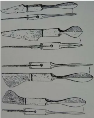
Among the sources of knowledge about the medical instruments used at the valetudinaria are also the burials of Roman military doctors. Two findings related to Roman surgery and military medicine will be described in detail in this article. The first one is the cremation grave of a Roman military doctor from Bingen (Germany). It is dated to 2nd c. A.D. It contained about 60 artifacts, which were described as medical instruments [23]. The most numerous group in the surgical kit is composed of instruments with a spatula probe on one end and a scalpel on the other (Figure 2). The latter have varied blade geometry. Usually only the spatula-shaped handle is found by the
archaeologists, as scalpels were a replaceable part of this medical instrument.
Physicians operating across the Empire also used probes as separate tools. They can be divided into three groups, according to the shape of the working part: spoon probes (the working part was spoon-shaped), spatula probes (the working part was spatula-shaped) and ear probes (with a small head on one end) (Figure 3). Medical instruments used by military doctors included also spatula probes. They could have been used to determine the wound depth and to apply medicament to the wound. The toolkit of the Bingen physician also comprised hook-ended needles as well as hooks used to widen the wound opening. Forceps were used to grab and hold tissues (two of them have toothed edges) and also to remove foreign bodies. It is possible that the kit also included tools for cauterization (burning wounds). Unfortunately, their bad condition prevents their complete identification.
However, we do know that such instruments must have been used by Roman military doctors. The kit also comprised three bronze cupping vessels (one bigger and two smaller ones).
Figure 2: Examples of Roman scalpels with replaceable blades
from the grave of a doctor from Bingen.
© Military Pharmacy and Medicine • 2012 • 2 • 5 – 8 Magdalena Cybulska at al.: On Roman military doctors and …
According to ancient physicians, cupping was performed to restore the balance of bodily fluids, or in headaches and joint pains. Albucasis claimed that large, medium and small vessels were used in treatment, depending on which part of the body they were being used on [14].
One of the tools (modiolus) was used to perform skull trepanation by means of drilling. The auger would be put into rotation by a string attached to its arched handle. Skull fragments removed during the procedure were round in shape [24]. In ancient times, trepanation was performed for magic and medical reasons. It was often thought that an evil spirit, a demon, inhabits the head
and causes the disease; headaches, vertigo, coma, faintings and epileptic seizures were perceived as evidence. Drilling a hole in the skull was an attempt to release the spirit. Cranial cavity would not be opened to perform neurosurgical procedures, though it could have been a method of evacuating hematomas. Skull trepanation was performed in adults; less frequently among children. Various ointments were also used to prevent wound infection.
A range of surgery methods were used. The external table, the diploë and the internal table would be removed; a hole would be cut in the skull using a sharp tool or drilled with an auger. Trepanation was also performed in head injuries, in an attempt to remove a cracked fragment of the skull. Soldiers were particularly prone to head injuries; thus, trepanation surgeries in the Roman army were mostly performed for curative purposes. Special scalpels (blades) can be found in the burials from the La Tène period, with a semicircular cutting edge for performing trepanations by incision [5]. In the Roman army, they were performed by drilling but also by scraping. Trepanation procedures were described by Hippocrates (5th c. B.C.) but also by Roman physicians. The former recommended that trepanation be performed within the first three days after a head injury; he also mentioned complications after the surgery and the way the wound should be dressed afterwards [25]. Galen (2nd c. A.D.) included in his work the techniques for the surgery, listed the indications for its performance and also described the required medical instruments. He thought that trepanation should be performed following skull injuries; the damaged fragment of the bone should then be removed and the sharp wound edges should be smoothed out using a chisel-like tool. Galen provided a description of a method for drilling and scraping bones. The surgery was supposed to bring relief to the patient, alleviate their pain and lower the intracranial pressure [26]. Apart from the modiolus, medical instruments used by Roman physicians for trepanations probably included specific scalpels, chisels and hook-shaped tools for lifting bone fragments. Chisels were also used for the trepanation of the skull of the child whose burial is dated to 1st-2nd c. A.D., which comes from the excavation works carried out in the suburbs of Rome [27]. It is interesting that in Greece, in the period before Hippocrates,
Figure 3: Examples of probes. From the top: ear probe, spoon
probe, spatula probe.
also iron scrapers, scalpel-like knives and iron forceps would be used for trepanation [28]. Throughout centuries, scraping was considered to be the safest method of performing the surgery due to a lesser risk of brain damage.
The physician whose burial was discovered in Bingen is likely to have been educated in Alexandria, proof of which is the figurine of a hippopotamus with a cobra on its back [23]. It was there that he became familiar with Greek and Egyptian medicine. Between 3rd and 2nd c. B.C. Alexandria was the main academic centre of the ancient world; it was the place where Greek medicine, the foundations of which had been laid by Hippocrates, was developing. Herophilus and Erasistratus, two popular ancient physicians, were active there. They put great emphasis on the study of anatomy. Autopsies and vivisections were carried out in Alexandria; the results are likely to have influenced the development of surgery [11]. Plenty of information related to medical tools used by military doctors in ancient times can be obtained by analyzing artifacts from the site in Rimini (Italy). The Surgeon’s House (Domus del Chirurgo), discovered by archaeologists, and dated to 2nd c. A.D., is where about 150 medical instruments were found [29,30]. Most of them had been used for bone surgeries and medical procedures [31]. The house, comprising a surgery, belonged to Eutyches, who was a military doctor. The use of a bow in military operations resulted in the need for physicians to learn the safest way to remove arrowheads from wounds. This kind of wounds was described in the works of the ancient Indian surgeon Susruta (4th c. B.C.) and was also mentioned by Hippocrates. In the Iliad, Homer pointed out how important it was to deal with such injuries correctly. Celsus described a special tool for this kind of operations, named “the spoon of Diocles” (after its inventor, Diocles of Carystus); an example was found in Rimini [32]. In their practice, Roman military doctors had to deal with specific wound types, hence specialist tools were developed to tackle battle wounds. It was also here that instruments were found with a blade on one end and a leaf-shaped probe on the other.
Eutyches also used forceps and tweezers to remove foreign bodies and fractured bone fragments from the body. Among the artifacts related to medicine,
also dental forceps were identified. The surgical toolkit also included chisels and gouges. They might have been used to lift the skull bones in case of some injuries. It is possible that the physician would also perform trepanations.
The proof that Eutyches had links to the Roman army is the votive sculpture of a human hand, also found in the Surgeon’s House. It is related to the cult of Jupiter Dolichenus, which was widespread among Roman soldiers [29,30]. Military doctors serving in the army also prepared medicines for their patients. Proof of the above are not only remains of medicinal plants, found at the valetudinaria, but also small containers, used to store and make medicines, discovered in the Surgeon’s House in Rimini. Dioscorides (1st c. A.D.), who most likely was a military doctor, described, in his numerous works, plant-derived medicaments, their properties and applications [11].
The instruments used by Roman military doctors included general surgery instruments, such as probes, scalpels, hooks, hook-ended needles, as well as specialist tools used for trepanation (modiolus) or to remove arrowheads from wounds (“the spoon of Diocles”). Gynecological instruments (vaginal specula, gynecological probes) have not been found among the medical instruments discovered during excavations at the valetudinaria as well as at cemeteries comprising the graves of Roman military doctors. Vaginal specula were discovered in Pompeii; they are also part of the collections of a number of European museums.
Physicians working at the valetudinaria are likely to have been dealing with a range of wounds: cuts, stabs, contusions; they would remove foreign bodies and arrows from soldiers’ bodies. Improper or incorrect wound handling might have resulted in gangrene and patient death. Outbreaks of infectious diseases were also likely in military camps; therefore, efforts were made to maintain an appropriate level of hygiene.
Greek medicine, the medicine of other ancient armies, along with civil medicine in Rome’s provinces influenced the entire Roman military medicine. When comparing medical instruments used by a “warrior-surgeon” with later tools used by Roman military doctors, one can identify the same types of instruments. However, Roman
© Military Pharmacy and Medicine • 2012 • 2 • 7 – 8 Magdalena Cybulska at al.: On Roman military doctors and …
ones were made with greater precision. Civil physicians with specialist knowledge who worked in Roman provinces used the same medical toolkit as Roman military doctors. Certainly they were not as experienced in dealing with wounds and injuries as the doctors at the valetudinaria. Civil physicians also used special cases or etuis to carry medical instruments. Thanks to that they could perform some surgeries in the homes of their patients [33]. In addition, the cult of Asclepius (called Asklepios in Greece) was widespread among Roman military doctors.
Most trepanations in the Roman army as well as those performed by civil physicians would be carried out for curative purposes or to remove a fractured bone fragment. Such surgeries were often performed intuitively. Lifting and removing the fragment(s) of a fractured bone, as well as smoothing out sharp wound edges may have resulted in an immediate effect in the form of abating neurological irregularities or regaining consciousness due to the fact that the fragments of a damaged bone had been prevented from applying pressure on brain tissue. For this reason, the surgery was repeatedly attempted. Efforts were also often made to remove the cause of pain, which could result from various pathological changes: injuries, related episcleral and subdural hematomas, inflammatory changes, benign and malignant brain tumors. It was not easy to perform such a procedure; it is likely that a physician with relevant expertise and tools was required. Most historical trepanations were episcleral. Related complications would include wound infection, operational bleeding, as well as damage to the meninges and the brain. Those would result from an improper use of tools and poor surgery technique.
Due to the lack of X-rays, the most common instruments used by Roman military and civil doctors were probes. They could be used to identify the wound depth and the location of a foreign body in it. The second largest group of tools were scalpels, used to make cuts of various depths. Incisions of skin, soft tissue and bones were possible.
Health care began and was developing in the Imperial Roman army. It reached a high level compared to medical assistance provided to wounded soldiers in other armies of the ancient world. The demand for professional medical assistance across the Empire was high. Military doctors probably constituted a large part of educated physicians; they worked permanently for the army, but some of them also ran a private practice.
Archaeological artifacts identified as medical instruments, along with the remains of Roman legionary hospitals, enable us to expand our knowledge about military medical service.
Human body was not penetrated. This was due to the lack of knowledge of anesthesia, antiseptics and blood loss replacement. It was only in the second half of the 19th century that all those methods were introduced to surgery. Surgical procedures in the Roman army related mostly to external injuries. However, the diversity of instruments used by Roman military doctors indicates that they were very good practitioners. There was relatively little anatomical knowledge in the ancient period; thus, Roman physicians had to rely rather on their experience, gained during the work in the army.
References:
1. Kaczanowski P, Kozłowski KJ. Wielka historia Polski. Najdawniejsze dzieje ziem polskich. tom 1. Wydawnictwo Fogra; Kraków 1998.
2. Gold der Skythen aus der Leningrader Eremitage. Ausstellung der Staatlichen Antikensammlungen am Königsplatz in München 19. September bis 9. Dezember 1984. Grebner, Aschheim. München 1984. 3. Wojtkowiak S, Talar J, Majewski W, Piotrowski F. Zarys
dziejów wojskowej służby zdrowia. Wydawnictwo Ministerstwa Obrony Narodowej. Warszawa 1974. 4. Davis WR. The roman military medical service.
Saalburg-Jahrbuch. 1998; 49: 76-82.
5. de Navarro MJ. A doctor’s grave of the middle La Téne Period from Bavaria. Proceedings of the Prehistoric Society.1955; 21: 231-248.
6. Byrne HE. Medicine in the roman army. The Classical Journal. 1910; 5: 267-272.
7. Błyskal O. O medycynie, lekarzach i rzymskich lekarzach wojskowych w I-III w. Annales Universitatis Paedagogicae Cracoviensis. Studia Historica. 2009; 8: 37-54.
8. Crislip TA. From monastery to hospital: Christian monasticism & the transformation of health care in late antiquity. University of Mitchigan Press; 2005.
9. Nutton V. Roman medicine 250 BC to 200 AD. In: Lawrence IC, editors. The western medical tradition: 800 B.C-1800 A. D. Cambridge University Press; 1995. 10. Prioreschi P. A History of Medicine: Roman medicine.
Horatius Press; 1996.
11. Brzeziński T. Historia medycyny. PZWL; Warszawa 1988. 12. 12. Elliot SJ. Outlines of greek and roman medicine.
Cretikron Company 1978.
13. Tyszler L. Higiena, pielęgnacja ciała i medycyna – wybrane zagadnienia z europejskich prowincji rzymskich. In: Supady J, editor. Szkice z historii medycyny; 2010 Jun 15-16; Łódź, Polska. 14. Baker P. Archaeological remains as a source of
evidence form Roman Medicine. [cited 2012 Fab 25] Available from: URL:http://www.ucl.ac.uk/~ucgajpd/ medicina%20antiqua/sa_ArchaeologicalRemains.pdf 15. Scarborough J. Roman medicine and the legions:
a reconsideration. Medical History. 1968; 12: 254-261. 16. Nutton V. Medicine and the roman army: a further
reconsideration. Medical History. 1969; 13: 260-270. 17. Künzl E. Medizinische Instrumente aus
Sepulkralfunden der römischen Kaiserzeit. Bonner Jahrbücher. 1982; 182: 1-131.
18. Press L. Odkrycie valetudinarium w Novae. Meander. 1983; 10-11: 385-405.
19. Press L. Szpital legionowy w Novae. Archiwum Historii i Filozofii Medycyny. 1989; 52: 473-483.
20. 20. Press L: Valetudinarium w rzymskiej twierdzy Novae. Balcanica Posnaniensia. 1990; 5: 267-277. 21. Davis WR. Some Roman medicine. Medical History.
1970; 14: 101-106.
22. McCallum EJ. Military medicine: from ancient times to the 21st century. ABC-CLIO; 2008.
23. Como J. Das Grab eines römischen Arztes in Bingen. Germania. 1925; 9: 152-162.
24. Kirkup J.The evolution of cranial related instruments. In: Arnott R, Finger S, editors. Trepanation: History, discovery theory. Swets Zeitlinger Publishers; 2003. 25. Martin G. Craniotomy: the first case histories. Journal
of Clinical Neuroscience. 1999; 6(4): 361-363. 26. Toledo-Pereyra HL. Galen's Contribution to surgery.
Journal of the History of Medicine and Allied Sciences. 1973; 28: 357-375.
27. Mariani-Costantini R, Catalano P, di Gennaro F, di Tota G, Angeletti LR. New light on cranial surgery in ancient Rome. Lancet. 2000; 355: 305-307.
28. Liston AM, Day PL. Is does take a brain surgeon: A successful trepanation from Kavousi, Crete. In: Schepartz AL, Fox CS, Bourtou Ch, editors. New direction in the skeletal biology of Greece. ASCSA; 2009.
29. Gerrie A. Roman ruins cast new light on a trip to doctor. [cited 2012 Fab 25] Available from: URL:http:// www.telegraph.co.uk/news/worldnews/1572030/ Roman-ruins-cast-new-light-on-a-trip-to-doctor.html 30. Ghiretti G. Un ambulatoro medico tico: due libri
recenti sul “Chirurgo di Rimini”. Papyrotheke. 2010; 1: 81-96.
31. Jackson R. Holding on to health? Bone surgery and instrumentation in the Roman Empire. In: King H, editor. Health in Antiquity. London and New York 2005. 32. Karger B, Sudhues H, Brinkmann B. Arrow wounds:
Major stimulus in the history of surgery. World Journal of Surgery. 2001; 25: 1550-1555.
33. Bliquez JL. Two sets of Roman surgical tools from the Holy Land. Saalburg-Jahrbuch. 1998; 49: 83-92.
© Military Pharmacy and Medicine • 2012 • 2 • 9 – 12 Michał K. Kołodziejczykl: Analysis of injection systems ampulla-syringe vs. ampulla …
Pharmacology & Pharmacy
Analysis of injection systems ampulla-syringe vs. ampulla with
respect to application of parenteral medicinal products on
the example of ibadronic sodium
Michał Krzysztof Kołodziejczyk
Department of Drug Formulation, Medical University of Lodz, Poland
Author’s address:
Michał Krzysztof Kołodziejczyk, Department of Drug Formulation Technology, Chair of Applied Pharmacy at the Medical University of Łodz, ul. J. Muszyńskiego 1, 90-151 Łódź, Poland; phone: (+48) 426779240,
e–mail: michal.kolodziejski@umed.lodz.pl
Received: 2012.05.18 • Accepted: 2012.06.08 • Published: 2012.06.28
Summary:
Nowadays, equally to the development of modern pharmaceutical technology and introduction of new biologically active substances to pharmacology we observe a development of techniques with the use of which medicinal products are applied into a living organism. The above regard many types of drug forms, among others, medicinal aerosols, fillers, injections, anal and vaginal forms, etc. Many times the medicinal effect depends on a high technical level of these devices, which are also medical materials. In this work at attempt has been made to analyze the mutual relations between a way of administering an injection in a standard form with the use of a standard syringe and technically higher forms, namely integrated ampulla-syringe systems.
Key words:
ampulla-syringes, injections, fillers, parenteral administration.Introduction
Durability of medicines for injections to a large extent depends on the direct packaging in which the target form of the medicine is stored (a solution, a colloid, an emulsion, etc.) and a way of storing them. Containers to store medicines for injections: ampulla-syringe or ampullas should have the following functions:
•Protect a medicine against the impact of the environment (air, moisture, light);
•Protect against mechanical contaminations;
•Protect against microbiological contaminations [1,2,4];
A direct container having contact with a medicine should be chemically neutral, cannot react with particular substances forming a pharmaceutical form of a medicinal agent, and a biologically active substance itself. Ampulla-syringes and ampullas should be characterized by a proper mechanical durability. They should also have specific thermal durability because of the method of sterilization. Also additional elements such as stoppers, pistons, sealing rings must be chemically and physically neutral, cannot react with the content of the container (a biologically active substance, auxiliary substances), cannot have an impact on any properties of the medicinal product [3,4].
Ampulla
This is a glass container that saves the airtight closing of the particular volume and dosage of the medicinal product after the sealing. An ampulla protects against the air and levels down a problem related to the secondary infection of a sterilized medicine. If a biologically active substance or any of the auxiliary substances is UV sensitive (light, the direct sun light), ampullas made of orange glass are used. However, the intensity of ampulla coloring cannot collide with the visual control of the content assessment. A volume of the containers which serve the purpose of an ampulla is from 1ml up to 20ml, they are mainly designed for solutions [4]. So far in order to open an ampulla one has had to use a file sawing through the structural narrowing of the neck so that the opening of an ampulla was easier.
Currently ampullas are produced in the way which enables the opening by breaking off the top part, without using a file. Such a way of opening the container is risky because tiny mechanical contaminations in the form of glass scraps can get to the solution of a medicine. The opening of an ampulla should take place immediately before injection, however a medicinal product contained in an ampulla after the opening is exposed to the contact with external conditions (moisture, microbiological contaminations!). The content of an ampulla is then taken with the use of a working needle to a syringe. During this process part of the volume of the liquid medicine is lost because it remains either on the bottom of an ampulla or/and in a needle, depending on a suction pressure. The next activity before administering the medicine to a patient is an exchange of a working needle (suction from an ampulla) into a injection needle (introduced to the patient’s tissues). After putting an injection needle the air sucked with a medicinal product into a syringe must be removed externally. Some part of the volume of a medicinal product is also lost resulting from the type of a needle used, its diameter, pressure force into the piston and the amount of the ejected medicine directly after the removed air [5].
The above information indicates that admi-nistration of a sterile medicinal product ion the form of an ampulla has certain procedural inconvenience, and also gives a possibility of
a loss of some part of a medicinal product and/or its potential microbiological contamination. This is more dangerous in a situation when an administered medicinal product contains ‘difficult’ biologically active substances, e.g. of a low index o biological access or of a narrow therapeutic index. The loss of some medicinal product in the aforementioned stages of an injection procedure can lead to dissatisfying treatment results. Because of small sizes and fragility of glass during handling of a medicinal product a total or partial loss of the content of an ampulla as a result of breaking or spilling. Generally, application of a medicinal product in the form of an ampulla must be administered by a qualified personnel who performs these injections [6,7,8].
Ampulla-syringe
A technological reply to the above presented inconveniences related to the use of ampulla-type containers is the application of modern injection forms combining the functions of ampullas, needles and syringes, namely, ampulla-syringes. An ampulla-syringe is a medicinal product container designed for single use (disposable). It guarantees a fast provision of a medicine in a proper, precisely measured dose. The easiness of the use makes patients use it on their own. An ampulla-syringe is sterile, with the use of it the injection is performed without the need to take the preparation from an ampulla or a flask. The whole set prepared in a factory is sterile and ready for direct use. The use of an ampulla-syringe prevents a loss of a biologically active substance and any form of microbiological secondary infection and/ or mechanical contamination [9-12].
The analysis of a loss of an injection
preparation administered with the use of
an ampulla-syringe and a traditional method
with the use of a standard ampulla
.Both in a traditional method and a modern one injection needles are used. In the case of an ampulla-syringe a needle is joined with the set and constitutes it integral part, and during a traditional administration from an ampulla, it is a separate element, exchangeable and exposed to the contact with non-sterile environment. Additionally when exchanging a working needle into an injection one, a medicinal product
© Military Pharmacy and Medicine • 2012 • 2 • 11 – 12 Michał K. Kołodziejczykl: Analysis of injection systems ampulla-syringe vs. ampulla …
contained in an ampulla and in a needle is lost. An injection needle is a pipe set on a cap.
A needle pipe is made of high-class stainless steel. The technological priority is to make needles of the possibly thinnest wall, which leads to the increase of the inner diameter, without the increase of the outer diameter. All needles must have the rigidity and flexibility of a pipe according to the plant-specific and pharmacopeial standards. Razors of needles are modeled in such a way as to ensure a relatively fast and moderately painless penetration of tissues in order to reduce traumatization, as well as minimization of pain.
Depending on a purpose, needles can be cut short (surface of cutting to a needle creates an angle of 16˚-18˚) or needles of long razors (an angle of 11˚-13˚). Injections needles most often used have outer diameters of 0.35-1.4 mm, and preferred lengths of pipes 8-40 mm. a good
solution preventing the remains of a preparation is a system of an integrated needle with a cylinder of a syringe as it is used in ampulla-syringe systems [4,8,13-18].
If we assume that a needle pipe from the point of spatial geometry is a cylinder of the height of h and the radius r, (picture 1 and 2), the a cylinder base and a cylinder upper part is a circle, ad its width is in every place the same. This solid figure in a Cartesian coordinate system is described as a collection of points (x, y, z) meeting the criteria of the inequality
where: r
>0 and is a cylinder radius and h>0
and is its height.
In relations to the aforementioned on the
basis of the following dependence:
we can calculate a figure value of a right
cir-cular cylinder volume, that is, in our case, an
approximate volume of an injection
remai-ning in a needle after operations connected
with performing an injection (table 2) [20].
Calculations of volume of a pipe needle –
V show that when a length and a measured
outer diameter of pipe needle increase, it
volume also increases, and at the same time,
the content of a solution of a medicine with
a biologically active substance. To compare,
needle parameters of an original medicinal
product with ibandronate sodium by Roche
has been listed table 3.
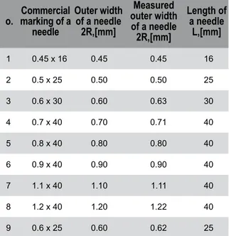
Table 1: Parametric list of most frequently used needles
in a pharmacy trade (No. 1-8) and needles
with ampulla-syringes in an original medicinal
product with ibandronate sodium by Roche (No. 9).
o. marking of a Commercial needle Outer width of a needle 2R,[mm] Measured outer width of a needle 2R,[mm] Length of a needle L,[mm] 1 0.45 x 16 0.45 0.45 16 2 0.5 x 25 0.50 0.50 25 3 0.6 x 30 0.60 0.63 30 4 0.7 x 40 0.70 0.71 40 5 0.8 x 40 0.80 0.80 40 6 0.9 x 40 0.90 0.90 40 7 1.1 x 40 1.10 1.11 40 8 1.2 x 40 1.20 1.22 40 9 0.6 x 25 0.60 0.62 25
Figure 2: A right circular cylinder of the base radius –
r and height – h.
Figure 1: Scheme of a needle pipe with the marked place
Table 2: List of necessary empirical figure values r and h, needed to calculate volume V of examined types
of injection needles
No. Measured diameter of a needle 2r[mm] Radius of a base of a needle pipe r [mm] Square radius of a base of a needle pipe r2 [mm2] Height of a needle pipe h[mm]Volume of a needle pipe V[mm3] V[ml] 1 0.45 0.225 0.050625 16 2.5434 0.0025 2 0.50 0.25 0.0625 25 4.9063 0.0049 3 0.63 0,315 0.099225 30 9.3469 0.0093 4 0.71 0.355 0.126025 40 15.8570 0.0159 5 0.80 0.4 0.16 40 20.0960 0.0201 6 0.90 0.45 0.2025 40 25.4340 0.0254 7 1.11 0.56 0.3136 40 40.9847 0.0410 8 1.22 0,61 0.3721 40 46.7358 0.0467
Table 3: List of necessary empirical figure values r and h, needed to calculate volume V of examined types
of injection needles
No. Measured diam-eter of a needle 2r[mm]
Radius of a base of a
needle pipe r 2 [mm] Square radius of a base of a needle pipe r2 [mm2]
Height of a needle pipe h[mm] Volume of a needle pipe V[mm3] V[mm3] V[ml] 1 0.62 0.31 0.0961 25 7.5439 0.0075
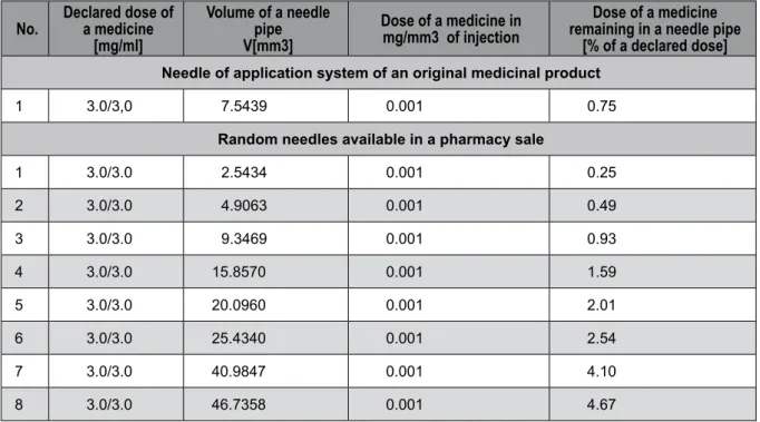
Table 4: Losses of an active substance in relations with the way of administering an injection.
No. Declared dose of a medicine [mg/ml] Volume of a needle pipe V[mm3] Dose of a medicine in mg/mm3 of injection Dose of a medicine remaining in a needle pipe
[% of a declared dose] Needle of application system of an original medicinal product
1 3.0/3,0 7.5439 0.001 0.75
Random needles available in a pharmacy sale
1 3.0/3.0 2.5434 0.001 0.25 2 3.0/3.0 4.9063 0.001 0.49 3 3.0/3.0 9.3469 0.001 0.93 4 3.0/3.0 15.8570 0.001 1.59 5 3.0/3.0 20.0960 0.001 2.01 6 3.0/3.0 25.4340 0.001 2.54 7 3.0/3.0 40.9847 0.001 4.10 8 3.0/3.0 46.7358 0.001 4.67
© Military Pharmacy and Medicine • 2012 • 2 • 13 – 14 Michał K. Kołodziejczykl: Analysis of injection systems ampulla-syringe vs. ampulla …
References:
1. Thurman J.E.: Analysis of Insulin Pen Devices for the Treatment of Diabetes Mellitus, J. Diabetes Sci. Technol. 2008, 5, 2(3), 482–483.
2. Podlewski J., Chwalibogowska-Podlewska A.: Leki Współczesnej Terapii, 2010, vol. 20, Medical Tribune, Warszawa, Polska.
3. Jachowicz R.: Farmacja Praktyczna, 2007, PZWL, Warszawa, vol 1.
4. Farmakopea Polska IX, Urząd Rejestracji Produktów Leczniczych, Wyrobów Medycznych i Produktów Biobójczych, 2011.
5. Jaber A., Bozzato G.B., Vedrine L., Prais W.A., Berube J., Laurent P.E.: A novel needle for subcutaneous injection of interferon beta-1a: effect on pain in
The data presented show that dozing in an
origi-nal mediciorigi-nal product is arranged in such a way
that prevents a loss of a biologically active
sub-stance. A unit packaging of a medicine contains
a needle of specified parameters. Therefore, an
error of medical personnel connected with
cho-osing an improper needle is eliminated. In case
of using a generic medicinal product being a
form of a standard ampulla, a number of
poten-tial errors related to administration and
influ-encing on a reduction of an already small active
substance increases [21,22].
Conclusions
•the conducted loss analysis shows that a loss value increases proportionally to a size of an needle measured by a volume of a pipe needle.
•application of a needle of the sizes bigger than 0.6 x 25mm makes a risk of administering a dose smaller than a recommended one bigger.
•negative phenomena related to API loss in a product increase, if application is from a standard ampulla, with the use of working needles and an injection needle.
•at this stage of research one needs to state that a way of application of an original medicinal product with sodium ibandronate arranged with a set of ampulla-syringe and needle is an optimal guarantee of providing a patient with a full dose of a biologically active substance expected for an effective therapy.
Figure 3: Illustration of a potential loss of an active substance [%] occurring during an injection with the use
of an ampulla-syringe with an originally selected needle and a needle chosen randomly.
volunteers and satisfaction in patients with multiple sclerosis, Neurol. 2008, 8, 38.
6. Chan E., Hubbard A., Sane S., Maa Y.F.: Syringe siliconization process investigation and optimization, J. Pharm. Sci. Technol. 2012, 66, 2, 136-150.
7. Müller R.H., Hildebrand G.E.: Technologia Nowoczesnych Postaci Leków, PZWL, 1998, Warszawa, Polska.
8. Merry A.F., Webster C.S., Hannam J., Mitchell S.J., Henderson R., Reid P., Edwards K.E., Jardim A., Pak N., Cooper J., Hopley L., Frampton Ch., Short T.G. Multimodal system designed to reduce errors in recording and administration of drugs in anaesthesia: prospective randomised clinical evaluation, BMJ, published online 2011, 9, 22.
9. Chan E., Maa Y.F., Overcashier D., Hsu C.C.: Investigating Liquid Leak from Pre-Filled Syringes upon Needle Shield Removal: Effect of Air Bubble Pressure, J Pharm. Sci. Technol. 2011 65, 4, 363-371. 10. Badkar A., Wolf A., Bohack L., Kolhe P.: Development
of biotechnology products in pre-filled syringes: technical considerations and approaches. Pharm. Sci. Tech. 2011 12, 2, 564-572.
11. Maarschalkerweerd A., Wolbink G.J., Stapel S.O., Jiskoot W., Hawe A.: Comparison of analytical methods to detect instability of etanercept during thermal stress testing, Eur. J. Pharm. Biopharm, 2011, 78, 2, 213-221. 12. Kayser O., Müller R.H.: Pharmazeutische
Biotechnologie, WFmbH, 2000, Stuttgart, Germany. 13. Carr J.A., Nalwa K.S., Mahadevapuram R., Chen Y., Anderegg J., Chaudhary S.: Plastic-Syringe Induced Silicone Contamination in Organic Photovoltaic. 14. Mahvash M., Dupont P.E: Mechanics of Dynamic
Needle Insertion into a Biological Material. Trans. Biomed. Eng. 2010, 57, 4, 934–943.
15. Kongsgaard U.E., Andersen A., Øien M., Oswald I.-A.Y., Bruun. L.I.: Experience of unpleasant sensations in the mouth after injection of saline from prefilled syringes, Nurs. 2010, 1, 7.
16. Hansen B., Lilleøre S.K., Ter-Borch G.: Needle with a Novel Attachment Versus Conventional Screw-Thread Needles: A Preference and Usability Test Among Adults with Diabetes and Impaired Manual Dexterity, Diabetes Technol. Ther. 2011, 5, 13(5): 579–585. 17. Paul M., Vieillard V., Roumi E., Cauvin A., Despiau
M.C., Laurent M., Astier A.: Long-term stability of bevacizumab repackaged in 1mL polypropylene syringes for intravitreal administration,
Ann. Pharm. Fr. 2012, 70, 3, 139-154.
18. Alterovitz R., Branicky M., Goldberg K.: Motion Planning Under Uncertainty for Image-guided Medical Needle Steering, Int. J. Rob Res. 2008, 27, 11-12, 1361–1374.
19. Oferta Handlowa dla Aptek Ogólnodostępnych, PZF Cefarm-Kielce, grupa Farmacol, 2012.
20. http://www.wiw.pl/matematyka/geometria.pl 21. http://www.leki.urpl.gov.pl
22. Kołodziejczyk M.K., Zgoda M.M.: Polimery jako substancje pomocnicze stosowane w technologii tabletek powlekanych zawierających pochodne kwasu ibandronowego (bisfosfoniany), 2012, 42, 1, 5-16.
© Military Pharmacy and Medicine • 2012 • 2 • 15 – 24 Radosław Ziemba: First aid in cases of wounds, fractures, as well …
Emergency Medicine
First aid in cases of wounds, fractures, as well as thermal
and chemical burns
Radosław Ziemba
Military Centre of Pharmacy and Medical Technique in Celestynów, Poland
Author’s address:
Radosław Ziemba, Military Centre of Pharmacy and Medical Technique, ul. Wojska Polskiego 57, 05–430 Celestynów, Poland; e–mail: zx11@op.pl
Received: 2012.03.07 • Accepted: 2012.06.08 • Published: 2012.06.28
Summary:
In this work classification and characteristics of wounds are given. Subsequently, different types of treat-ment of patients during different individual stages of medical evacuation are briefly discussed, taking into consideration gas gangrene. Issues concerning the case of tetanus, as well as the rules of surgical aid administration to children, are also discussed.
Key words:
wounds, wounds classification, primary delayed suture, qualified medical aid, gas gangrene, tetanus, surgical aid.1. Wound management procedures
A wound (vulnus) is defined as a break in the body covering. The continuity of skin is broken and the inner tissues are in contact with the surrounding environment, thus a portal of infection is formed, through which pathogenic microorganisms may enter the body. During visual wound examination, special attention is paid to the location, size, shape, margin, channel and bottom of the wound. Wound margin may be smooth, nonlacerated, uneven, lacerated, contused, etc. Wound bottom is a part of the wound located between the margins. The bottom may be even or uneven, lacerated, and may have recesses and pockets. Those two characteristics, i.e. the margin and the bottom, are of high importance for the course of wound healing.Wound, as any injury, induces local and general symptoms, even including circulatory shock. Intensity of pain related to the wound depends
on the object which caused it and innervation of the injured body part. A sharp, fast-acting object induces less pain than a blunt one, e.g. a knife or razor blade cut is less painful than a contused wound caused by a hammer blow. Some areas of the body are particularly sensitive to pain due to high density of nerve endings, e.g. fingertips, eyeballs, surroundings of sexual organs. Breaking the continuity of tissues also causes blood vessel damage. Incised wounds usually induce a more intense bleeding, whereas contused wounds bleed to a lesser extent.
Depending on the type of damaged vessels, arterial, venous and mixed bleeding may be distinguished. Once skin is incised, the contraction of elastic fibres causes wound margins to retract and the wound to open. The intensity of this phenomenon is highly dependent on the direction of elastic fibre incision.
2. Classification of wounds
Depending on the cause and characteristics of the injury, several types of wounds may be listed:
1) Incised wound (vulnus sectum) — caused by
a sharp, cutting object, e.g. knife, razor blade, glass shard. Incised wound margin is smooth and has no recesses or pockets. Haemorrhaging is intense due to wide open blood vessels. Blood leaving the wound mechanically removes any impurities. The wound usually heals well and the risk of infection is low.
2) Slicing wound (vulnus lobatum) — occurs when
the cutting object (e.g. knife) is not applied perpendicularly to the skin, but inclined. The characteristics are similar to those of incised wound.
3) Chopped wound (vulnus caesum) — formed by
a forceful action using a heavy cutting object (e.g. axe, sabre, cleaver). Often results in total amputation of the affected body part.
4) Puncture wound (vulnus ictum s. punctum) — has
similar characteristics to incised wound and is caused by a sharp object with a very small cutting surface, e.g. pin, splinter, nail, fork, dagger, bayonet. Puncture wound often induces internal bleeding, while wound secretion accumulating at the bottom has no appropriate drainage due to the narrow and uneven wound channel, which may facilitate the development of infection. Penetrating puncture wounds of the chest and abdomen are particularly dangerous due to the possibility of heart and lung, or intestine damage, respectively.
5) Contused wound (vulnus contusum) — induced
by a blunt or blunt-edged object (e.g. stone, stick, hammer). Wound margin is contused, crushed, the bottom is uneven and has recesses and pockets. Bleeding is scarce, as crushed blood vessels are not open. Contused tissue undergoes necrosis easily and the necrotic tissue constitutes a base for the development of infection.
6) Crush wound (vulnus conguasatum
s. Guassum) — defined as a particularly vast and deep tissue contusion caused by
a forceful trauma, e.g. in the victims of mine wall collapse, people covered with earth after mine explosion, run over, or crushed between train cars. The lesions are often accompanied by post-traumatic shock. Moreover, advantageous conditions for the development of vulnar infections, e.g. gas gangrene, are created.
7) Lacerated wound (vulnus laceratum) – has
uneven, torn margin, while the bottom has recesses and pockets. The bottom exposes ragged adipose tissue and muscles. Loss of skin and deeper tissues is often experienced, as the blunt object causing the wound operates at a certain angle and tears some tissue off.
8) Bite wound (vulnus morsum) – those caused by
a dog, cat, horse, or human belong to lacerated-contused wounds. Those caused by a horse are particularly dangerous, as the animal's strong mandible often tears off large fragments of soft tissues. The risk of infection of those wounds is high, since the bacterial flora residing in the animal or human oral cavity is abundant and composed of opportunistic strains.
9) Gunshot wound (vulnus sclopetarium) — has
similar characteristics to lacerated and contused wounds, however, lesions originating from the kinetic energy of the bullet also apply in this case and include overpressure and underpressure wave effects, mechanical injuries and resonance inducing tissue and organ breakage around the wound channel. Tissue and organ water content increases the force of the bullet impact, as the mechanical reactions are accompanied by hydrodynamic phenomena: the bullet's energy is transferred to water molecules which tear the organ apart. Such explosive processes take place when the bullet hits a urine-filled bladder or a full stomach. Gunshot wound has the following characteristic features: inlet (small, with a diameter equal to the bullet calibre, even margin, and scorched skin around the inlet with incorporated gunpowder remains), channel (narrow, straight or sinuous), outlet (often not located opposite to the inlet, large surface, considerable bleeding, bottom and margin lacerated with visible bone shards).
© Military Pharmacy and Medicine • 2012 • 2 • 17 – 24 Radosław Ziemba: First aid in cases of wounds, fractures, as well …
3. Wound treatment during
medical evacuation
Treatment of the victims of catastrophes and natural disasters requires clear and detailed injury identification, allowing a precise assessment of the necessary therapeutic actions and evacuation of the wounded. Anatomical identification of the wound type and functional impairment caused by the injury should be thoroughly reported in medical documentation (Evacuation Card). Three basic types of injury-related risks may occur: haemorrhage, potential entrance of pathogenic microorganisms through damaged body covering, as well as general anatomical and functional impairment of tissues and organs. Therefore the essential aims of the therapy cover: stopping the haemorrhage, combating potential infection along with anatomical and functional reconstruction of the affected tissues and organs.
First aid comprises covering the wound with
aseptic bandage and using tourniquets in the case of intense arterial bleeding, impossible to stop by other means. In the case of concurrent bone fracture or massive damage to soft tissues, the damaged limb should be immobilized.
Premedical aid comprises control and, if needed,
correction of bandage covering, tourniquet or limb immobilization.
Medical aid involves changing the bandage only
if it is too loose or too tight, soaked with blood, or if the patient reports strong pain or oedema around the wound. Before placing a fresh bandage, the skin around the wound should be washed with petrol, ether, or alcohol, then sterilized with iodine. Wound surface should be freed from any foreign bodies using forceps and gauze pads damped with hydrogen peroxide solution. Since a longer use of a tourniquet is disallowed, it has to be released and the bleeding vessel along with the surrounding tissue has to be grasped using Kocher's haemostatic forceps or Pean's forceps, or blocked by applying a tamponade.
All the wounded should be administered anatoxin and antitetanus serum, as well as antibiotics, while the wounded potentially at risk of gas
gangrene should be administered antigangrene serum. Serum administration should be noted in the Evacuation Card. Shock prevention therapy may also be introduced (analgesics, Novocaine block, immobilization in the cases of massive damage to soft tissues).
Qualified medical aid involves wound
segregation into two groups: requiring and not requiring surgical intervention.
The latter group comprises:
1) small superficial skin wounds,
2) multiple, blind, superficial wounds caused by small grenade or mine debris,
3) gunshot exit wound with smooth inlet and outlet wounds, no signs of serious tissue damage along the wound channel, and no bone fractures or large blood vessel damage.
Optimal conditions for wound treatment are provided by a single surgery leaving no need for further interventions. However, in the case of massive inflow of victims, medical services will be forced to limit early surgical interventions to deep wounds, complicated by the damage to internal organs, blood vessels, bones and joints. Postponed surgical aid applies to the cases of wounds limited in area and covering tissues well-supplied with blood, mainly those of the limbs. The postponement requires antibiotic and immune serum administration, in order to prevent the development of wound infection.
Surgical wound treatment involves performing
one the following actions: incision, partial excision or complete excision of the wound.
Wound incision is performed in the cases of
infected wounds to facilitate the drainage of purulent secretion. These are particular wounds, as they are caused by debris which produces small wound inlet but seriously damages deeper tissues. The aim of the incision is relieving tissue tension and exposure of its recesses and pockets. The surgery involves cutting through the skin in both directions from wound ends and parallel fascial incision.
Partial excision of fresh wounds is used when
complete excision is impossible. Most of all it regards contused, crush and lacerated wounds,
![Figure 3: Illustration of a potential loss of an active substance [%] occurring during an injection with the use of an ampulla-syringe with an originally selected needle and a needle chosen randomly.](https://thumb-eu.123doks.com/thumbv2/9liborg/3092323.8085/21.892.134.739.124.403/illustration-potential-substance-occurring-injection-originally-selected-randomly.webp)
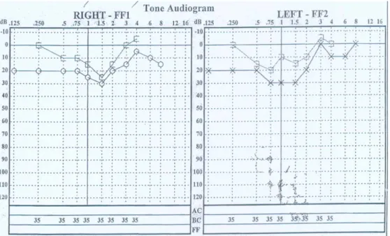
![Figure 2: Pure-tone threshold audiometry – conductive hearing loss x-----x bone, •——• air [2].](https://thumb-eu.123doks.com/thumbv2/9liborg/3092323.8085/35.892.501.747.459.615/figure-pure-tone-threshold-audiometry-conductive-hearing-loss.webp)