TRENDS
in
Sport Sciences
2015; 3(22): 125-132 ISSN 2299-9590Variability of selected hematological
and biochemical markers in marathon runners
JOANNA KAMIŃSKA, TOMASZ PODGÓRSKI, MACIEJ PAWLAK
Abstract
Introduction. The marathon is an increasingly popular form of athletic competition, especially among amateur runners. It seems, therefore, important to determine how this type of exercise affects the runner’s body. Aim of Study. The aim of the study was to identify hematological and biochemical changes in the blood of amateur runners after a marathon run. Material and Methods. The study involved 10 male runners aged from 28 to 42 years. Their blood was collected from the fingertip four times: a day before the competition and one, three and five days after the race, and subjected to hematological and biochemical analysis. Results. The results revealed a statistically significant increase in the number of leukocytes, including monocytes and granulocytes, creatine kinase (CK) and lactate dehydrogenase (LDH) activity, urea concentration, and ferric reducing ability of plasma (FRAP). On the other hand, the values of certain indices, such as the number of erythrocytes, hemoglobin concentration, hematocrit, and red blood cell distribution width, decreased. There were no statistically significant changes in the number of lymphocytes and thrombocytes, the concentration of total protein, albumin, and polyphenols. Conclusions. Prolonged and intense physical eexercise, such as a marathon run, causes multiple significant changes in the body. These changes can lead to health disorders and deterioration of physical fitness. KEYWORDS: long-distance running, biochemistry, effort, catabolism, aerobic exercise.
Received: 2 May 2015 Accepted: 27 June 2015
Corresponding author: pawlak@awf.poznan.pl
University School of Physical Education, Department of Physiology, Biochemistry and Hygiene, Poznań, Poland
What is already known on this topic?
Physical activity has a very beneficial effect on both people’s physical condition and the quality of their lives. Today, long-distance running is an increasingly popular form of recreation, as evidenced by the growing number of marathon participants. However, such prolonged and intense physical effort is a strain for the body and leads to a number of changes in the organism, as demonstrated by the results of hematological and biochemical blood analysis.
Introduction
T
he marathon or other long-distance races are becoming increasingly popular. In the Poznań Marathon alone, the number of participants has increased nearly three times in last ten years, from 2,234 (2004) to 6,426 (2014).Marathon running is considered long and very intensive exercise, and its manifestations include changes of concentrations of certain biochemical indices in the blood and the hematological profile [1]. The reasons for such qualitative and quantitative changes are the direct and indirect consequences of increased physical activity, leading to dehydration [2], depletion of carbohydrate stores in the muscles involved [3], or injury caused by overloading [4].
Participation in long-distance running necessarily involves adaptation of the body to aerobic effort [5].
The main source of energy for the muscles is glucose, which originates from glycogen reserves in the muscles and liver as well as from free fatty acids created as a result of lipolysis of fat tissue [6]. Adequate preparation of athletes for a marathon run requires an appropriate diet with glycogen supercompensation [7] and special training, while any deficiencies in these respects may lead to the occurrence of a phenomenon known as hitting the
wall, which involves severe physical and psychological
weakening of the runner. The metabolic reason for this sensation is the early exhaustion of glycogen accompanied by a delay in energy production from fat [8].
The aim of this study was to describe the variability of certain biochemical and morphological indices in a group of amateur athletes – participants of the 15th Poznań Marathon.
Material and Methods
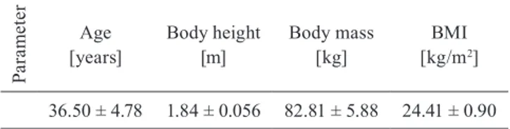
The studied material were samples of capillary blood collected from the fingertips of 10 males aged between 28 to 42, participating in the 15th Poznań Marathon. The athletes were subjected to basic anthropometric measurements (body height and body mass) using a WPT 60/150 OW medical balance (Radwag®, Poland).
Body mass index (BMI) was calculated for each participant (Table 1).
The blood was collected in the morning by qualified professional laboratory staff, after disinfection, using a disposable lancet (Medlance® Red, HTL-Zone,
Germany).
About 600 μl of the capillary blood was collected four times (a day before the marathon and one, three and five days after the run) into a Microvette® CB
300 tube (Sarstedt, Germany) containing K2-EDTA (EDTA dipotassium salt) as an anticoagulant to carry out the hematological analysis using the Mythic®
18-parametric automated hematology analyzer (Orphée, Switzerland); however, in this paper presents only nine parameters are discussed. An additional 300 μl of blood was collected into a Microvette® CB
300 Z (Sarstedt, Germany) to obtain serum.
After hematological analysis, the blood was centrifuged (4000 rpm, 20 min, 4°C), and the extracted plasma and serum were stored at –80°C. Total protein, albumin, creatine kinase (CK), lactate dehydrogenase (LDH), urea, blood antioxidant potential and total polyphenols were determined in this biological material.
The chemical antioxidants present in the plasma were
by Benzie & Strain [9] and modified by Janaszewska & Bartosz [10]. The concentration of total phenol substances (total polyphenols) in the plasma was measured with the method published by Singleton and Rossi [11], which uses the ability to oxidize phenol groups with the Folin-Ciocalteau reagent.
In the remaining blood serum, certain biochemical indices were determined using commercial tests (Cormay, Poland): the activity of creatine kinase (Liquid Cor-CK, catalogue No 1-219) and lactate dehydrogenase
(LDH) (Liquid Cor-LDH), total protein concentration (Liquid Cor-TOTAL PROTEIN), and albumin (Liquid Cor-ALBUMIN), as well as interleukin 1 beta (IL-1b) and interleukin 6 (IL-6) using ELISA tests (DRG Diagnostics, Poland). All of the above biochemical analyses were read on the ELISA Microplate Reader Synergy 2 SIAFRT, BioTek, USA.
The results were statistically analyzed using the STATISTICA 10.0 software package (StatSoft. Inc., USA). The study was approved by the Bioethical Committee of the Poznań University of Medical Sciences in Poland.
Results
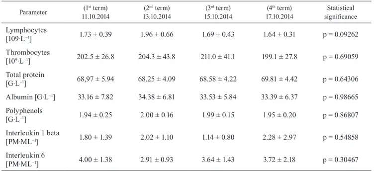
The measurements to check the response of the body to long-lasting and intensive physical activity were carried out during four terms: one before and three after the marathon run. It was found that some hematological markers, such as leukocytes, monocytes, granulocytes, erythrocytes, hemoglobin, hematocrit, and RDW, as well as biochemical values, including CK, LDH, urea, and FRAP, showed statistical differences (p < 0.05) between the tested terms. However, other tested factors, including lymphocyte counts and thrombocyte counts, concentration of total protein, albumin, and polyphenols, were not changed significantly between the four tested days.
The mean white blood cell count in the tested persons during the study ranged from 4.7·109·L–1 to 5.7·109·L–1
(Figure 1A). The substantial increase of leucocytes Table 1. Anthropometric parameters of tested group (mean ± SD)
Pa ra m et er Age
[years] Body height[m] Body mass[kg] [kg/mBMI2] 36.50 ± 4.78 1.84 ± 0.056 82.81 ± 5.88 24.41 ± 0.90
B
C
Figure 1. Changes in the concentration of selected haematological indices in marathon athletes. Terms marked with equal symbols differ significantly (p<0.05)
A
D
E F
the next two tested periods to achieve values similar to those from before the competition by the fifth day. Also, the mean monocyte count changed in the analyzed period, with values from 0.33·109·L–1 up to
0.47·109·L–1 (Figure 1B). As in the case of leucocytes,
a day after the physical eexercise, a clear increase was noted (24%), but two days later the mean monocyte count was comparable with the control data.
The mean value of granulocyte count ranged from 2.6·109·L–1 to 3.3·109·L–1 (Figure 1C). A statistically
significant difference (p < 0.05) was found only between the first and the second measurement term, possibly caused by the very slowly decrease of the curve. The erythrocyte count in the blood of the tested athletes decreased in the studied period from 5.31·1012·L–1 to
4.96·1012·L–1 (Figure 1D), with the lowest value, less
than 7%, the day after the marathon run. Moreover, this index subsequently showed an increasing trend, but did not achieve the control value.
The hemoglobin concentration in the studied period varied from 9.3mmol·L-1 the day before to 8.7mmol·L–1
24 hours after the marathon run. The last test of hemoglobin, five days after the competition, showed the same result as the control value (Figure 1E). The course of the hematocrit curve is similar to the results presented for the number of erythrocytes or hemoglobin concentration. The mean value decreased from 45% at the beginning to 42% a day after the marathon run (Figure 1F). Five days after the competition, the blood of the athletes showed the initial values.
A B
The mean value of red blood cell distribution width (RDW), describing the ability of bone marrow to produce erythrocytes, decreased after the marathon run and ranged between 11.96% and 11.48% (Figure 1G). The mean activity of creatine kinase (CK) in the studied group increased nearly 12 times, from 50.45 U·L–1 one
day before the marathon to 624.94 U·L–1 one day after
the marathon (Figure 2A). The high post-exercise value was significantly different from the results of the remaining test periods.
A course of the curve comparable to that of CK was found in the case of lactate dehydrogenase (LDH). Even though in this case the increase was only two-fold, the level of LDH was 46.7% above the starting value, as measured before the competition, even five days after the marathon (Figure 2B).
The mean concentration of urea in the blood of the tested marathon runners ranged from 1.70 mmol·L–1
to 4.35 mmol·L–1. It has been established that physical
effort required to complete the long-distance run led to a 2.3-fold increase on the first day after the competition. This effect lasted only two days, and on the third day after the marathon the level of urea concentration corresponded to the starting value (Figure 2C).
The last index showing significant differences between the analyzed periods (p < 0.05) was the ferric reducing ability of plasma (FRAP) (Figure 2D). Also in this case, its significant increase was noted only the next
day after the marathon run and then it diminished to an even lower level compared with the starting value. In the case of the other parameters tested (Table 2), no significant differences between the analyzed terms of measurement were found.
Discussion
The high popularity of long-distance running in the USA and Europe, including Poland, is the consequence of a growing interest in physical activity, which is understood as an element of a healthy lifestyle [12]. The results of hematological and biochemical assays of the runners monitored in this study clearly show a statistically significant increase in the majority of tested indices a day after the marathon compared with the starting value. In most cases, the return to the starting values took place five days after completion of the marathon.
Leukocytes, and their fractions (lymphocytes, granulocytes and monocytes) alike, play an important role in sports diagnostics by indicating the deployment of leukocytes to sites of injury, even in the case of micro-injuries [13]. Our results have clearly shown an increase in the leucocytes and all their fractions after a marathon run. Interestingly, the lymphocyte and monocyte counts reached the pre-run values mostly on the third day, but the leucocytes and granulocytes achieved these levels later, even on the fifth day. Results consistent with Table 2. Changes of hematological and biochemical parameters in marathon runners in terms of measurement (mean ± SD)
Parameter 11.10.2014(1st term) 13.10.2014(2nd term) 15.10.2014(3rd term) 17.10.2014(4th term) significanceStatistical
Lymphocytes [109·L–1] 1.73 ± 0.39 1.96 ± 0.66 1.69 ± 0.43 1.64 ± 0.31 p = 0.09262 Thrombocytes [109·L–1] 202.5 ± 26.8 204.3 ± 43.8 211.0 ± 41.1 199.1 ± 27.8 p = 0.69059 Total protein [G·L–1] 68,97 ± 5.94 68.25 ± 4.09 68.58 ± 4.22 69.81 ± 4.42 p = 0.64306 Albumin [G·L–1] 33.16 ± 7.82 34.38 ± 6.81 33.53 ± 5.84 33.39 ± 6.37 p = 0.98665 Polyphenols [G·L–1] 1.94 ± 0.25 2.00 ± 0.16 1.99 ± 0.15 1.95 ± 0.20 p = 0.86807 Interleukin 1 beta [PM·ML–1] 1.80 ± 1.39 2.02 ± 1.10 1.14 ± 0.80 2.28 ± 2.97 p = 0.54858 Interleukin 6 [PM·ML–1] 4.00 ± 1.38 2.91 ± 0.93 3.64 ± 1.43 3.72 ± 2.18 p = 0.30467
the outcomes of our study were obtained by Avloniti et al. [14], who demonstrated that lymphocyte counts depended on the training phase. Also, Wu et al. [15], while studying the effects of 24-hour ultra-marathons, pointed to a similar relationship as regards the leucocyte and monocyte counts; however, in the case of lymphocytes they found an opposite relationship. These observations were confirmed in publications by Del Coso et al. [16] and Lippi et al. [17]; however, the authors of this study found such relationships on the third or fifth day after the marathon run. The erythrocyte count, the concentration of hemoglobin, and hematocrit levels are considered the indices of aerobic performance. Their decrease found in our study is consistent with the majority of scientific articles on this topic [15]. Very few show a decreasing erythrocyte count without any change to the hemoglobin and hematocrit [18].
The mean value of red blood cell distribution width (RDW), describing the quality of the hematopoietic system, shows an increasing trend when the erythrocyte count in the bone marrow grows [19]. This index decreased in our study in all of the tested post-marathon terms. Such an effect is associated with the removal of incorrectly or deficiently built erythrocytes or improper functioning of the bone marrow system, and was also present after intensive physical exercise [15]. In younesian et al. [20], however, these results were not statistically significant. On other hand, a significant increase of RDW was noted by Wu et al. [15] after an ultra-marathon. In the case of extreme physical load, mechanical hemolytic anemia must be taken into account [21].
Our study did not reveal any statistically significant changes in the number of thrombocytes, in spite of its slight increase. The increase in the number of thrombocytes as a possible biological protective mechanism of the body against the impact of small blood extravasation after intense physical exercise was also discussed by other authors [18, 22].
The results of our study show a constant level of total protein and albumin in the blood. The very small number of publications in the last decade makes it difficult to interpret the results obtained by Kupchak et al. [23] who found a diminution in the value of these indices in ultra-marathon runners.
The level of creatine kinase (CK) is a classic marker indicating considerable load on the muscles,
is accompanied, for similar reasons, by an increase of lactate dehydrogenase (LDH) in the blood [24]. The increase in CK and LDH under the influence of increased physical exercise revealed in our study confirms the observations of other authors [18, 25, 26]. Increased blood urea found in our study was also observed during prolonged and intense exercise [24] or in the participants of long-distance runs [27, 28]. The increase in the concentration of this index on the second day after completion of the run may indicate increased degradation of proteins, which is also supported by the increased activity of CK and LDH.
The low ferric reducing ability of plasma and polyphenol content indicate a significant use of antioxidants by the body to eliminate the free radicals formed during exercise [24].
Our results showed an increase in FRAP and polyphenols a day after the marathon. Such a reaction of the organism was observed in the study by Ristow et al. [29], while Pialoux et al. [30] found the opposite relationship, wherein the value of these two parameters decreased after strenuous physical exercise. This inconsistency may be the result of the athletes’ diets and the different intake of antioxidant-rich foods.
Conclusions
Intensive physical exercise, including marathon running, leads to important changes (decrease or increase) in certain hematological and biochemical indices in the blood.
The growing popularity of long-distance running, especially among amateurs, points to the importance of a comprehensive description of the biochemical characteristics of such activity. It would help to define the norm and identify the possible health consequences.
What this paper adds?
The results of the study show how significant the changes in the hematological and biochemical blood profile of amateurs after intense and prolonged physical exercise such as the marathon are. In addition, the study, by presenting the time required for the monitored indices to return to their pre-run value, provides information about the time necessary for the body to regenerate after such strenuous effort.
References
1. Banfi G, Colombini A, Lombardi G, Lubkowska A. Metabolic markers in sports medicine. Adv Clin Chem. 2012; 56: 1-54.
2. Sawka MN, Burke LM, Eichner ER, Maughan RJ, Montain SJ, Stachenfeld NS. American College of Sports Medicine position stand. Exercise and fluid replacement. Med Sci Sports Exerc. 2007; 39: 377-390. 3. Sherman WM, Costill DL, Fink WJ, Hagerman FC,
Armstrong LE, Murray TF. Effect of a 42.2-km footrace and subsequent rest or exercise on muscle glycogen and enzymes. J Appl Physiol. 1983; 55: 1219-1224.
4. Hikida RS, Staron RS, Hagerman FC, Sherman WM, Costill DL. Muscle fiber necrosis associated with human marathon runners. J Neurol Sci. 1983; 59: 185-203. 5. Hagan RD, Smith MG, Gettman LR. Marathon
performance in relation to maximal aerobic power and training indices. Med Sci Sports Exerc. 1981; 13(3): 185-189.
6. Callow M, Morton A, Guppy M. Marathon fatigue: the role of plasma fatty acids, muscle glycogen and blood glucose. Eur J Appl Physiol Occup Physiol. 1986; 55(6): 654-661.
7. Sherman WM, Costill DL. The marathon: dietary manipulation to optimize performance. Am J Sports Med. 1984; 12(1): 44-51.
8. Rapoport BI. Metabolic factors limiting performance in marathon runners. PLoS Comput Biol. 2010; 6(10): e1000960.
9. Benzie IFF, Strain JJ. The ferric reducing ability of plasma (FRAP) as a measure of “antioxidant power”: the FRAP assay. Anal Biochem. 1996; 239(1): 70-76. 10. Janaszewska A, Bartosz G. Assay of total antioxidant
capacity: comparison of four methods as applied to human blood plasma. Scand J Clin Lab Invest. 2002; 62(3): 231-236.
11. Singleton VL, Rossi JA. Colorimetry of total phenolics with phosphomolybdic-phosphotungstic acid reagents. Am J Enol Vitic. 1965; 16(3): 144-158.
12. Smith DD, Schuemann T, Hoogenboom BJ. The role of the sports physical therapist-marathon events. Int J Sports Phys Ther. 2013; 8(4): 531-536.
13. Tidball JG. Inflammatory processes in muscle injury repair. Am J Physiol Regul Integr Comp Physiol. 2005; 288: R345-R353.
14. Avloniti A, Douda H, Tokmakidis S, Tsitskaris G, Chatzinikolaou A, Toubekis A, Kortsaris A. Acute effects of basketball training on white blood cell count. Inquiries Sport Phys Educ. 2007; 5(1): 165-172.
15. Wu HJ, Chen KT, Shee BW, Chang HC, Huang yJ, yang RS. Effects of 24 h ultra-marathon on biochemical and hematological parameters. World J Gastroenterol. 2004; 10(18): 2711-2714.
16. Del Coso J, Fernández D, Abián-Vicen J, Salinero JJ, González-Millán C, et al. Running pace decrease during a marathon is positively related to blood markers of muscle damage. PLoS One. 2013; 8: e57602.
17. Lippi G, Salvagno GL, Danese E, Skafidas S, Tarperi C, Guidi GC, Schena F. Mean Platelet Volume (MPV) Predicts Middle Distance Running Performance. PLoS One. 2014; 9(11): e112892.
18. Žákovská A, Šullová A, Chlíbková D, Tomášková I. Selected immunological and biochemical parametres in ultra runners participating 100 km race. Med Sport Bioh et Slov. 2014; 23(1): 33.
19. Zhuo X, Zheng-Mao M. Variation character of chinese male players’ hemogram parameter before and after the ice hockey training. China Winter Sports. 2008; 30(3): 56-59.
20. younesian A, Mohammadion M, Rahnama N, Cable T. Haemathology of professional soccer players before and after 90 min match. Cell Mol Biol Lett. 2004; 9 (Suppl. 2): 133-136.
21. Jordan J, Kiernan W, Merker HJ, Wenzel M, Beneke R. Red cell membrane skeletal changes in marathon runners. Int J Sports Med. 1998; 9(1): 16-19.
22. Ersöz G, Zergeroglu AM, Fiçicilar H, Ozcan H, Oztekin P, Aytaç S, yavuzer S. Effect of submaximal and incremental upper extremity exercise on platelet function and the role of blood shear stress. Thromb Res. 2002; 108(5-6): 297-301.
23. Kupchak BR, Kraemer WJ, Hoffman MD, Phinney SD, Volek JS. The impact of an ultramarathon on hormonal and biochemicalparameters in men. Wild Env Med. 2014; 25: 278-288.
24. Hyla-Klekot L, Kokot F, Kokot S. Badania laboratoryjne – zakres norm i interpretacja. PZWL, Warszawa 2011. 25. Skenderi KP, Kavouras SA, Anastasiou CA,
yiannakouris N, Matalas A. Exertional Rhabdomyolysis during a 246 km continuous running race. Med Sci Sports Exerc. 2006; 38(6): 1054-1057.
26. Jastrzębski Z, Żychowska M, Radzimiński Ł, Konieczna A, Kortas J. Damage to liver and skeletal muscles in marathon runners during a 100 km run with regard to age running speed. J Hum Kinetics 2015; 45: 93-102.
27. Smith JE, Garbutt G, Lopes P, Tunstall Pedoe D. Effects of prolonged strenuous exercise (marathon running) on biochemical and haematological markers used in the investigation of patients in the emergency department. Br J Sports Med. 2004; 38: 292-294.
28. Lopes RF, Osiecki R, Rama LM. Biochemical markers during and after an olympic triathlon race. J Ex Phys. 2011; 14(4): 87-96.
29. Ristow M, Zarse K, Oberbach A, Klöting N, Birringer M, Kiehntopf M, Stumvoll M, Kahn CR, Blüher M. Antioxidants prevent health-promoting effects of physical exer-cise in humans. Proc Natl Acad Sci USA. 2009; 106(21): 8665-8670.
30. Pialoux V, Mounier R, Rock E, Mazur A, Schmitt L, Richalet J-P, Robach P, Brugniaux J, Coudert J, Fellmann N. Effects of the live high-train low method on prooxidant/antioxidant balance on elite athletes. Eur J Cl Nut. 2009; 63: 756-762.