INVITED REVIEW
TRENDS
in
Sport Sciences
2013; 3(20): 123-134. iSSN 2299-9590Aspects of pain in sport
MAciEJ pAWLAKReceived: 31 August 2013 Accepted: 28 September 2013
corresponding author: pawlak@awf.poznan.pl
University School of Physical Education, Poznań, Department of Biochemistry, Poland
From a physiological point of view, pain in sport has an informative function, indicating the maximum load capacity of the body and especially of those areas which are usually exposed to maximum loads and, consequently, damage or injury. Pain in an athlete’s body usually has a specific cause, predictable duration, and proven methods of treatment. Pain is part of the sporting experience, irrespective of whether the discipline is a contact or a non-contact sport. Interest in the problems of pain in sport has been growing in recent years, as demonstrated by the host of scientific publications referred to in the paper, and in general the number of articles and studies available in thematic databases. The problem of pain in sport will become increasingly important, not least because of the increasingly higher load on athletes in all disciplines, as shown by the successive new world records. Also, the increasing number of amateur and recreational athletes will require appropriate studies. As this group is not sufficiently prepared for the effort, it is very susceptible to injury. Pain in sport can also be expected to continue to gain in importance considering the increasing number of active elderly people, especially in European countries.
The article emphasizes that better knowledge of this area may have practical applications in the training process of athletes as well as persons who are physically active during their working life and after retirement. Furthermore, pain in sport may, due to advances in biological and medical sciences, give rise to new research areas. In this paper, the main trends of scientific problems and research concerning biological aspects of pain in sport are presented.
KEY WORDS: sport, pain, injury, physical exercise, athletes.
Introduction
T
he concept of ‘pain’ is mainly associated with different injuries sustained as a result of accidents, traffic collisions, accidents at work or hospital surgery, rather than with sport. However, in Germany alone about two million sport-related accidents are reported every year [1]. Also in other countries, exercise- and sport-related accidents are an evident health problem; however, the number of injured competitive and recreational athletes can differ depending on the source of information and the criterion used. Therefore, data concerning a single country may vary significantly; for example, in the United States, the number of people who get injured each year while participating in a range of sports, exercise and recreational activities is estimated to be 3 to 5 million, close to 7 million or even 25 million [2, 3, 4], while other authors estimate that sports injuries in high schools alone affect 2 million students each year [5].The growing number of studies dealing with the issue of pain in sport published every year clearly indicates increased expectations in this regard. A search in the National Library of Medicine (pub Med) database browser using the keywords “pAiN and SpoRT” in August 2013 resulted in 12,618 papers published in the last 40 years, from 1973 to 2013. However, over two-thirds of all analyzed studies concerning “pain and sport” were published in the last ten years. A more detailed analysis of the papers concerning “pain in sport” shows that the majority of them is related to a particular discipline of sport (football, hockey, martial arts, etc.) or concerns a specific injury, overuse of tissue or physical discomfort (joint pain, back pain, headache,
muscle pain, etc.). The remaining works on “pain and sport” deal with other aspects in this area, and it is worth emphasizing that the issue of pain and injury in sport is also extensively covered in literature on sport psychology, pain experience as well as consequences of injury [6, 7, 8].
Interestingly, in the scientific literature, some aspects of pain in sport remain unexplored or have been investigated only to a small degree, especially those relating to the effects of exercise on pain reception, the ability to tolerate pain during extreme physical stress, painkiller abuse – also by amateurs – pathophysiology of ischemia, individual sensory perception or the placebo effect. Also, the synergic effects of physical, pharmacological and psychophysiological factors modulating the variability in pain perception or affecting pain tolerance are not fully understood.
Better knowledge of this area, in addition to research impact, may have practical applications in the training process of athletes as well as persons who wish to be physically active during their working life and after retirement. Pain in sport and physical fitness can be expected to continue to gain in importance, given, in particular, the increasing number of active elderly people, especially in European countries.
pain in sport may, due to advances in biological and medical sciences, become a new research area. This paper mentions the main trends in scientific research concerning biological aspects of pain in sport.
Pain, what does it mean?
The human body is able to perceive most environmental stimuli, including electromagnetic waves (light), sound, temperature, smell, taste or pressure. All those stimuli are usually of chemical, thermal or mechanical origin. in the last case, their wide spectrum ranges from delicate touch, trough pressure to vibration, tickle or even pain. interestingly, all of those sensations are registered, through special nerve structures, mainly corpuscular endings of peripheral nerve fibres, known as sensors or receptors. They are characterized by clearly defined anatomical structure and physiological function [9, 10, 11, 12]. Receptors are concentrated in the sense organs such as eyes or ears, or can be distributed around the surface of the body, as in the case of skin sensors specialized in thermoreception to recognize and distinguish cold and warm or in proprioception to perceive the body’s current position and movements of joints and muscles. Many of them are localized in internal organs and have a clear
physiological function, such as the pressure sensors, also known as baroreceptors.
Unlike other sensory modalities, pain offers little help in the recognition of our surroundings. its special function is to inform us about the risk of injury or damage to our body originating both externally and internally. interestingly, almost all sensory impressions, such as smell, taste, vision or hearing, can generate feelings of pleasure or displeasure, depending on the concentration, conditions or other circumstances. in this respect, pain seems to be an exception because of the almost always unpleasant emotions associated with it.
The definition of ‘pain’ was published in 1979 in the scientific journal Pain – an official body of the inter national Association for the Study of pain (iASp). This sensory phenomenon was characterized as “unpleasant sensory and emotional experience associated with actual or potential tissue damage, or described in terms of such damage” [13]. it means that pain must be considered in more terms than just pure sensation. in addition to a sensory-discriminative component, informing about the site, intensity and duration of nociceptive stimuli, the consequences of reception, conduction and processing of noxious signals include also other components, which are classified as affective, autonomic, and motor. Usually, all four of them appear together, but their intensity varies almost in each case.
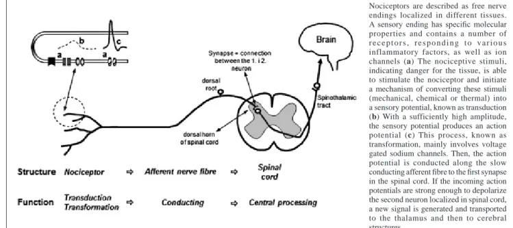
Apart from the term ‘pain’ described above, ‘nociception’ is another closely associated term. Nociception (from the Latin word nocere, which means “to injure”) is defined as objective processes in which the nervous system receives and processes noxious stimuli or stimuli that have the potential to damage tissue. Thus, the term refers to the peripheral recording, transmission and processing of noxious signals in the central nervous system (cNS). The structures in which this process takes place are described as the nociceptive system consisting of peripheral and central elements (Fig. 1).
Peripheral and central mechanisms of pain
The phenomenon of pain will be signalized or have its origin at the site of injury, and is created in the brain. The first element of the nociceptive system is the nociceptive neuron, where, in its ending part, the converting process of the mechanical, chemical or thermal stimuli into action potentials takes place. Such structures, termed nociceptors by Sherington [14], constitute the initial element of the nociceptive system [9, 10, 12]. They are
a kind of “sensing devices” performing only one, but important informative function – warning of danger. Nociceptive endings possess a number of receptors and channels, whose activation causes a change of the electric charge of the sensor’s membrane and, consequently, typical physiological depolarization [11, 15, 16]. Through those receptors and channels in the cell membrane, nociceptors are also targeted by algesic substances, particularly those known as inflammatory mediators, including substance p, bradykinin or histamine, leukotrienes and prostaglandins. When such substances, produced mainly in inflamed tissue, are released, the threshold of nociceptors is lowered, in some cases to such an extent that even normal non-noxious stimuli are able to excite the nociceptors [10, 17, 18]. This phenomenon is called sensitization. it is known that the action and behaviour of nociceptors may be modulated by the gene expression of ion channels or receptors in response to tissue damage or/and inf lammatory processes [11, 12, 16, 18].
Built in sensors, the nociceptive stimuli in the form of action potentials (not pain signals) are transported along the first neuron to the spinal cord, and more specifically the dorsal horn, where they make synaptic contacts with interneurons or with second neurons. This anatomical area is the site of primary integration
of segmental pain information. Much of the molecular aspects of those processes are already known and are extensively discussed in neuroscience literature [12, 19, 20]. The second neuron leads the pain signal through
tractus spinothalamicus to the thalamus and then to the
cortex, where the information is completed by emotional aspects (Fig. 1).
This physiological role of pain, which focuses on warning the organism against potential tissue damage may be modified and/or disturbed peripherally by the aforementioned sensitization of nociceptors or pathological impulse generation in nociceptive fibres known as spontaneous activity. increased spontaneous activity of nerve fibres is the result of functional or/and structural changes to neurons, and was found mainly after exposure of nerve endings to inf lammatory mediators or as a result of severing or rupturing the nerve [21]. This physiological mode of pain can also be changed centrally. in this case, functional disturbances or defects in the spinal cord and the supraspinal nociceptive system can lead to enhanced excitability and even spontaneous activity in those structures.
in addition to warning about the risk of tissue-damaging stimuli, the nociceptive system may also participate in inhibiting the flow of those stimuli to the structures of the central nociceptive system. in particular, the Nociceptors are described as free nerve endings localized in different tissues. A sensory ending has specific molecular properties and contains a number of r e c e p t o r s , r e s p o n d i n g t o v a r i o u s inflammatory factors, as well as ion channels (a) The nociceptive stimuli, indicating danger for the tissue, is able to stimulate the nociceptor and initiate a mechanism of converting these stimuli (mechanical, chemical or thermal) into a sensory potential, known as transduction (b) With a sufficiently high amplitude, the sensory potential produces an action potential (c) This process, known as transformation, mainly involves voltage gated sodium channels. Then, the action potential is conducted along the slow conducting afferent fibre to the first synapse in the spinal cord. if the incoming action potentials are strong enough to depolarize the second neuron localized in spinal cord, a new signal is generated and transported to the thalamus and then to cerebral structures.
Figure 1. Structure and function of the nociceptive system. The peripheral elements of this system involve nociceptive neurons
converting stimuli, in their ending parts, into action potentials and axons conducting them further up to the central ending, where the synapse is built. central elements of the nociceptive system include neurons of the spinal cord and of the brain
nociceptive neurons of the spinal cord are constantly controlled by descending inhibitory systems [12].
Diversity of pain
The sensation of pain can be categorized in relation to the site of origin as somatic, visceral, superficial or deep, in terms of its duration as acute and chronic, and even with respect to the site of perception as local or generalized. Figure 2 provides partial insight into this complex subject.
The physiological form of pain occurs as a consequence of excitation of nociceptors. However, pain may also be produced non-physiologically as a result of pathological events, mainly by direct excitation of elements of the nociceptive system, such as in the case of mechanical pressure, which generates discharges in a nerve, or by damage to the nerve, respectively. Similarly, continuous irritation of a dorsal root leads to pathological impulse generation in nociceptive fibres, not nociceptors [22].
interestingly, the lacking input of nociceptive and sensory stimuli from a part of body that is no longer present after amputation can also induce pain. This phenomenon, known as phantom pain, is seen as a consequence of plastic changes in primary somatosensory cortex [23, 24]. it has been discovered recently that also an itch is a quality of cutaneous sensation closely related to pain [25].
Feeling of pain and factors that alter this sensation in sport
pain is a very subjective sensation that begins with the action of noxious stimuli affecting nociceptors. The gentle electrical changes of membrane potential induced this way lead to local depolarization, generate sensor potentials, and then action potentials (b and c in Fig. 1A, respectively). Functionally, an effective stimulus exceeds the sensitivity threshold of peripherally localized “pain sensors”. The threshold of the nociceptors to noxious stimuli is neither uniform among all nociceptors nor constant in a given nociceptor. As a consequence, pain threshold, defined as the current amplitude that evokes pain in 50% of stimuli [26] is subjective and differs from person to person. in practice, the assessment of pain threshold is more complicated than that of other sensory thresholds because of the subjective nature of pain. other useful terms which help describe the concept of pain include “pain experience” and “pain tolerance”, which is understood as the amount of pain applied under experimental conditions in the form of defined stimuli which a person is able to withstand before suffering from an emotional or physical breakdown.
There are several factors that can influence pain threshold values, including age [27], gender [28], stress [29], supporting persons [30], the subject’s pain experience history, and, obviously, physical activity [25].
Also, regular physical activity is supposed to be associated with specific alterations in pain perception [31]. There are numerous papers indicating that athletes have higher pain tolerance than non-athletes [32, 33]. Furthermore, the author’s own research demonstrates that even slight physical effort may shift the threshold
Figure 2. classification of pain according to different criteria (site,
perception, origin, and duration). Acute pain indicates impending actual tissue damage, and is thus a clear signal with a warning function. Chronic pain lasting some months does not fulfill this function. in terms of the area in which pain is perceived, it can be described localized or generalized. The origin of pain may be physiological, when the nociceptors are directly activated by a stimulus, or pathological, when pain is produced by excitation of the structures of nociceptive systems at more proximal sites
of perception of both sensory stimuli and nociceptive stimuli [12, 34]. it has also been shown that strong aerobic exercise during cycling inhibited the perception of vibration and myofascial pain [35]. interestingly, also professional ballet dancers, similarly to sports professionals, were found to have a higher pain threshold and pain tolerance threshold compared with matched controls in the cold pressor Test [36]. Some authors, such as Ryan and Kovacic [37], found many years ago that athletes participating in contact sports rated their pain as less severe and had greater pain tolerance time than non-contact sports athletes. Those authors also found no significant differences between the tested groups as regards pain threshold, indicating, however, that a high pain threshold can coexist with a low level of pain tolerance. Also, pain perception by 30 competitive swimmers and 28 non-competitive athletes showed that while pain thresholds differed little between the groups, pain tolerances were considerably different [38]. Raudenbush et al. [39] confirmed such observations, finding additionally that physical contact of athletes is a factor that may desensitize them to pain. They also noticed that athletes with a history of injuries were more willing to accept a high level of pain before discontinuing to play the game than athletes without such a history.
in addition, pain tolerance has been found to vary in different sport disciplines, with possible divisions emerging between individual and team sports, and contact and non-contact sports [36]. interestingly, Raudenbush [40] found that pain tolerance was the greatest during playing video sports and fighting games. Thus, certain games produce greater distraction, which may have implications for the medical field as an adjunct to pain management.
pain experience in sport has been the subject of increasing research in recent years. While sports professionals have generally been found to have higher pain thresholds than control subjects, the reasons for this are not entirely clear [36, 41]. The causes of the differences in somatosensory processing in athletes in comparison with controls may be linked to the probably less responsive endogenous pain inhibitory system [41]. This finding may explain the paradoxical tendency of athletes to develop chronic widespread pain.
Some objective factors mentioned earlier, such as age, gender, or even ethnic group or religious affiliation, are able not only to influence the sensory threshold but also to modulate the perception of pain. Moreover, the
same injury in two different people will be experienced in two completely different ways, depending upon the context, past experience, levels of anxiety, and other circumstances. it is a well-known fact that soldiers wounded in action need far less treatment with painkillers than would be required for comparable injuries in their civilian life [42]. Also in sport, especially in some disciplines such as boxing or martial arts, athletes experience pain during competitions or sparring fights. This pain is, however, experienced in a different way. it does not cause anxiety and is tolerated by the athlete, and a punch from the opponent triggers an additional desire to fight and gives a motivation to win. Also, muscle pain after exercise can be perceived as positive, suggesting, sometimes incorrectly, that the training was good and intensive [12].
in the literature, evidence for the inf luence of sex and menstrual phase on pain perception was already presented in the 1930s [43]. Bergstrom et al. [44] found that among adolescents aged 15-19 attending a ski high school, female students suffered more knee pain (88%) than males (57%). on the other hand, there are no clear results to indicate that gender could be a factor affecting the willingness to play through pain and injury [4]. Furthermore, under experimental conditions, the relationship between reported pain intensity and the peripherally applied pain stimulus also depends on the emotional status, which can be influenced by many subjective factors. Weinberg et al. [4] found in their study of male and female basketball players that individuals with a higher athletic identity had more positive attitudes and reported a higher level of willingness to play through pain than those with a lower athletic identity.
pain is a phenomenon that becomes a stronger and more explicit element in the later periods of life. There is also a noticeable tendency that with age the frequency of cases of acute pain decreases, while the number of patients complaining about chronic pain increases [45]. changes in pain perception in the elderly should be looked at in a wider context. As we age, both functional and structural changes occur in the nervous system [46, 47], including reduction in the density of opioid and serotonin receptors [48]. Neurophysiological observations in animal models also indicate the possibility of a more intense pain response in older animals [27].
What is special about “pain in sport”?
in sports, during training or a match, one must accept the possibility of injuries or micro-injuries that are not
perceived explicitly as noxious stimuli. However, the aggregation of such events is particularly dangerous as it may lead to chronic pathological processes in the musculoskeletal system, disorders and dysfunction, and pain [49]. Usually, pain in an athlete’s body has a specific cause, predictable duration, and can be managed with proven methods. Generally, pain has an informative function with a clear protective role, aiming to minimize the damage of tissue at the time of injury. on the other hand, from the clinical point of view, pain has a very important diagnostic role, forcing one to consult a physician. The diagnostic function of pain, manifesting itself as a decreased, the same or an increased level of its intensity, is also useful in the assessment of the therapy applied. in athletes, pain plays an additional function to indicate the maximal load of the organism, and particularly of the area of the body that gets injured the most.
pain and sport also show another interesting relationship. Sport activity can induce and even intensify pain. on the other hand, sporting activities can reduce pain intensity and improve the comfort of living, as found out by many patients suffering from pain.
pain in sport is mainly associated with acute injury during training or competition. However, it is suggested that overuse injuries and pain problems such low back pain and jumper’s knee are often chronic, with periods of reemission and exacerbation, and may represent as much of a problem in many sports as do acute injuries [50]. one should not forget that pain is a prevalent factor not only in competitive sport, but also in other physical performance domains such as dance, military operations, and outdoor adventure activities.
pain must be seen not only as a pathological symptom but also as a useful and necessary adjunct to inhibit sports participation when injury is involved [51]. A number of studies conducted to date suggest that many athletes play through injuries. As much as 90% of collegiate athletes continued to play through injury and pain at some points during their sporting career [52]. Such an attitude to pain may have different underlying causes, but it often involves the athlete’s image in the eyes of the coach, the team, the fans or even the media. Athletes experience, endure, tolerate, and accept pain as part of their daily lives [53]. it is also important for the athlete’s success to be able to play while hurt.
interestingly, even recreational athletes are ready to play with pain and injury [4]. in a variety of sports, athletes maintain their ability and willingness to play through
pain as a demonstration of character building, as a way of gaining respect from others, and as a way of attaining success [52, 54].
Also, ballet dancers reported to dance through pain, which can lead to serious injury and may impose a limitation on training intensity [55, 56, 57]. interestingly, male and female Standard and Latin style dancers also experience pain, with males experiencing pain primarily in the hips and calves and females in the toe region (44.9%); however, back pain affects both female and male dancers [58]. Because Standard and Latin style dancers practice and compete together, the partner’s absence from training may adversely affect the performance of the couple during a competition, which could lead to lower ranking and possible partner replacement [59].
pain as a consequence of athletic injury may result from acute trauma or overuse; however, previous injuries and their sequelae may also play a role in generating pain [60]. Therefore, the number of pain events in sport can be assessed either directly or indirectly. Mostly, accordingly to sport, muscle pain, joint pain, headache as well as back pain will be discussed.
Muscle pain
Muscles are served by nociceptive nerve fibres, which can be a source of pain that manifest itself in the form of functional limitations. Muscle nociceptors do not respond to natural stimuli generated during normal muscle activity, hence minor muscle deformations, spasms or st retching within the physiological limits are not adequate stimuli for nociceptors [61]. A typical muscle nociceptor, however, responds to both noxious mechanical stimuli (local pressure, such as pinching) and chemical stimuli. The latter may include reduction in tissue pH, or, in other words, increased acidity, which is usually associated with hypoxia or muscle inflammation or production of pain-inducing and sensitizing substances, such as bradykinin, prostaglandins, substance p and others, which also affects pain receptors within the muscle [62, 63, 64]. it is also worth mentioning that previously lactic acid was considered to be the cause of exercise-induced muscle pain. Although the concentration of this compound increases up to 10 times during exercise [65], it then normalizes within 1-1.5 hours, even after intense effort accompanied by muscle pain.
Another cause of muscle pain induced by chemical stimuli is the release of adenosine triphosphate (ATp),
usually from damaged muscle cells, and its binding by special purinergic receptors p2X2 and p2X3 [66]. Muscle soreness can occur after a single training session of high intensity. Muscle pain onsets within 8 to 24 hours after the load, peaks after 1 or 2 days, and disappears completely within the next few days. Therefore, this condition is known as delayed onset muscle soreness (DoMS). Histological tests indicate damage to the contractile and/or structural elements of sarcomeres. The period of noticeable changes in those structures coincided with the length of muscle pain. The process of removal of mechanical micro-damage to sarcomeres is accompanied by the release of endogenous substances that irritate the nociceptors located in the muscle [67, 68]. This type of muscle soreness can affect physical activity by limiting the mobility of joints, causing muscle weakness, and resulting in problems with coordination between the individual motor units during muscle contraction. Although there are different markers to evaluate muscle lesions [69, 70], no efficient biochemical methods to assess muscle pain are known.
An interesting but still unexplored issue is muscle pain caused by local hypoxia. Hypoxic pain, although associated mostly with the heart muscle, or angina pectoris, applies, however, directly or indirectly, also to other types of muscle tissue, including the skeletal muscles. A hypoxic and working muscle will soon manifest its discomfort with pain [71, 72].
Joint pain
pain in the joints, particularly the knee joint, may have different etiology [73, 74, 75, 76, 77], but their overloading is a serious clinical problem for many athletes, including top athletes [78].
if the structure of the joint is damaged, pain and swelling, which sometimes spread beyond the site of injury, onset within a short period of time after the event. pain that intensifies during movement in the joint’s functional range is the result of sensitization of nociceptors, which, in healthy tissue, are activated only to indicate the possibility of damage when the joint is reaching its limit position. it is worth noting that nociceptors are found substantially in all structures of the joint (joint capsule, ligaments and tendons), with the exception of the articular cartilage, which is not innervated [73, 74]. Other factors may also contribute to intensified activity of nociceptors in this region, such as an increase in pressure in the joint capsule observed in the course of an inflammatory process.
Hahn and Foldspang [79] suppose that knee pain is a common symptom in athletes; however, its prevalence is associated with the type, amount and duration of sports participation. in Denmark, they found that among sports club members aged 14-35 the prevalence of knee pain within the preceding 12 months, constant or recurrent knee pain, absence from sport, and absence from work due to knee pain was 54%, 34%, 19% and 4%, respectively. Also, 73% of students of a ski high school reported activity-related pain of the knee [44, 80].
Headache
A classification by the International Headache Society (iHS) lists more than 250 types of headaches, which are divided into primary headaches, which are themselves a disease and a primary condition, such as migraine, and secondary headaches, and which are the result of damage or injury to the head or an infection or poisoning. Damage to the head and face is very common in athletes, especially in team sports or martial arts. Moreover, headaches after exercise (also known as exertional headaches or headaches on exertion) are very common in modern athletes and people who exercise [81]. Analysis of the literature indicates that the causes of headache in people who do sport or exercise are not homogeneous. A study of 440 football players showed that 85% of them reported the occurrence of headaches associated with their discipline, and 21% of the players experienced headaches during the game [82]. A similar incidence of headache was also observed in Australian professional football players. of the 160 persons surveyed, 80% declared the presence of that condition. interestingly, fewer respondents experienced headache during a match than during training, 49% and 60%, respectively [83]. on the other hand, the probability of head injury in female hockey players was six times higher during a match than during training [84]. it was also demonstrated that the headache can be triggered by other events, such as fast ice skating [85] or mountaineering [86], where the cause may be the cold air leading to the contraction of blood vessels and/or stimulation of cold receptors.
pain in sport is an area which is usually investigated in terms of injuries, trauma or medical treatment and recovery. There are, however, aspects of pain in sport which remain rather unexplored, such as the effect of exercise on pain reception, the ability to tolerate pain during extreme physical stress, painkiller abuse (also by amateurs), pathophysiology of ischemia, individual
sensory perception or the placebo effect. A fuller knowledge of this area may have practical applications in the training process.
Another specific and relatively unexplored problem relating to the issue of pain in sport is the reduced perception of pain by athletes who are constantly and intensely affected by minor or major injuries. it is common knowledge that generally pain accompanies every injury. However, repetitive strain injuries in athletes, especially in those who train contact sports or martial arts, are, over time, perceived as increasingly weaker and less burdensome. The mechanism of this phenomenon is still not clear. Several possibilities have been discussed. it is possible that during the healing process, connective tissue replacement occurs instead of regeneration. As a consequence, the structure might become weaker and inflexible, which, especially in the case of tendons, may result in joint contractures or in an inability to adapt to external and internal stress very well [87]. Also, a bone is able to adapt to altered physical stimuli or injury. Exercise can thus affect bones in the same limb in different ways [88].
Alleviation of pain – playing through pain
injuries and pain were already treated by athletes in Ancient Greece [89]. The intensity of pain as well as the perception of this sensation can be modulated by three important processes: pharmacological procedures aimed at either preventing the reception and conduction of noxious signals or inhibiting central processing and diminishing affective participation in the pain event; physical procedures used in physiotherapy, which employ extremely diverse routes to counteract pain; and a broad spectrum of psychological methods [42]. in sport, particularly in last two decades, self-administration of drugs, mainly non-steroidal anti-inflammatory drugs and opioids used to manage athletic injuries, has significantly increased [90, 91]. The same growing trend has been observed as regards non-medical use of prescription opioids among adolescents, including athletes, in recent years. Veliz et al. [5] found that adolescent participants in high-injury sports, especially football players and wrestlers, had 50% higher odds of non-medical use of prescription opioids than adolescents who did not participate in those types of sports. The authors suppose that the greater odds may be related to the fact that football players and wrestlers have the highest severe injury rate among high school athletes [92]. A dangerous trend has been observed in
participants of different types of mass sporting events, such as marathon running. According to Brune [91], even more than half of those people may be taking analgesics before the event to relieve existing pain or pain anticipated during the run.
When discussing the possibilities of pain alleviation in athletes, one must mention the placebo effect. placebo, which can activate the same brain structures which modulate pain as opioids [93], is a special aspect of pain in sport. This phenomenon is clearly more complex in the case of sport than in the case of a specific disease. It has been demonstrated that athletes convinced that they had been administered anabolics [94, 95], caffeine [96] or a hypothetical “super substance” [97] or who trained with the assistance of a respiratory device [98], performed better than the baseline or control group. Furthermore, Benedetti et al., [99] showed also that opioid-mediated placebo applied after repeated administration of morphine in the pre-competition training phase is able to boost pain endurance and physical performance to a similar extent.
Speaking of placebo in sport, additional questions arise, for example, about events that can be classified as a placebo effect. During sporting rivalry, placebo effects may, in fact, be modulated or superseded by motivational factors [100]. on the other hand, it is possible that changes in motivation are part of the placebo effect.
Conclusions
interest in the problems of pain in sport has been growing in recent years, as demonstrated, for example, by the host of scientific publications referred to in the paper, and in particular the number of articles and studies available in databases. This is a consequence of both the increasing loads on professional athletes’ bodies and the growing number of amateur and recreational athletes who are not sufficiently prepared for the effort. It is worth noting that the research to date has not focused or has focused to a very limited extent on several important aspects of pain, both in professional and amateur sport, such as the ability to tolerate pain during extreme physical stress, painkiller abuse, including by amateurs, pathophysiology of ischemia, individual sensory perception of stimuli, including pain stimuli, the synergic effects of physical, pharmacological and psychophysiological factors that modulate variability in pain perception or the placebo effect. New studies on the issue of pain in sport should also deal with topics concerning the ageing generation
of people who wish to remain fit or actively participate in a sport discipline of their choice.
References
1. Gläser H, Henke T. Sportunfälle – Häufigkeit, Kosten. prävention. informationsblatt. Ruhr-Universität Bochum, Lehrstuhl für Sportmedizin, Arag Versicherungs-AG Hrsg. 2002.
2. Ball DR. The psychology of sport and exercise injury. iDEA Health & Fitness inc. 2002.
3. conn JM, Annest JL, Gilchrist J. Sports and recreation related injury episodes in the US population, 1997-1999. inj prev. 2003; 9(2): 117-123.
4. Weinberg R, Vernau D, Horn T. playing through pain and injury: psychological considerations. J clin psychol. 2013; 7: 41-59.
5. Veliz pT, Boyd c, Mccabe SE. playing through pain: sports participation and nonmedical use of opioid medications among adolescents. Am J public Health. 2013; published online ahead of print March 14: e1-3. 6. Jirásek i, Hurych i. pain and suffering in sport. Human
Movement. 2012; 13(2): 185-189.
7. Gaz DV, Smith A M. psychosocial benef its and implications of exercise. pM R. 2012; 4(11): 812-817. 8. Nippert AH, Smith AM. psychologic stress related to
injury and impact on sport performance. phys Med Rehabil clin N Am. 2008; 19(2): 399-418.
9. Messlinger K, pawlak M, Steinbach H, et al. A new combination of methods for the localization, identification, and three-dimensional reconstruction of the sensory endings of articular afferents characterized by electrophysiology. cell Tissue Res. 1995; 281(2): 283-294.
10. Messlinger K. What is nociceptor? Schmerz. 1997; 11(5): 353-366.
11. Pawlak M. Praktyczne aspekty sensorycznej i modulującej funkcji nocyceptorów (practical aspects of the sensory and modulatory function of nociceptors). Fizjoterapia polska. 2008; 2: 115-127.
12. pawlak M. Biologiczne uwarunkowania bólu (Biological determinants of pain). Podręcznik. Poznań: AWF. Podręczniki nr 62; 2010.
13. Bonica JJ. The need of a taxonomy. pain. 1979; 6: 247-252.
14. Sherrington CS. Qualitative differences of spinal reflex corresponding with qualitative difference of cutaneous stimulus. J physiol. 1903; 30: 39-46.
15. Binshtok AM. Mechanisms of nociceptive transduction and transmission: a machinery for pain sensation and tools for selective analgesia. int Rev Neurobiol. 2011; 97: 143-177.
16. petho G, Reeh pW. Sensory and signaling mechanisms of bradykinin, eicosanoids, platelet-activating factor, and nitric oxide in peripheral nociceptors. physiol Rev. 2012; 92(4): 1699-1775.
17. Schmidt RF, Schaible H-G, Messlinger K, et al. Silent and active nociceptors: structure, functions and clinical implications. in: Gebhart DL, Hammond DL, Jensen TS, eds. progress in pain research and management, Vol. 2, iASp press, Seattle; 213-250; 1994.
18. Mizumura K, Sugiura T, Katanosaka K, et al. Excitation and sensitization of nociceptors by bradykinin: What do we know? Exp Brain Res. 2009; 196(1): 53-65.
19. Woolf cJ, costigan M. Transcriptional and post-translational plasticity and the generation of inflammatory pain. proc Natl Acad Sci USA. 1999; 96(14): 7723-7730.
20. Salter MW. cellular signalling pathways of spinal pain neuroplasticity as targets for analgesic development. curr Top Med chem. 2005; 5(6): 557-567.
What this paper adds?
This paper stresses that the available results of studies published to date show clearly that some aspects of pain in sport remain insufficiently explored. Especially studies regarding the effect of exercise on pain reception, the ability to tolerate pain during extreme physical stress, painkiller abuse – also by amateurs – pathophysiology of ischemic pain as well as individual sensory perception or the placebo effect seem to be neglected. Similarly, the problems of synergic effects of physical, pharmacological and psychophysiological factors modulating the variability in pain perception or affecting pain tolerance in athletes are not fully understood. There are growing indications that the topic of sports injuries and the associated pain requires closer cooperation between specialists in the field of sports medicine, sport science, and psychology. Furthermore, the intense physical activity is not only limited to areas traditionally defined by sports disciplines, but it is also important in other physical performance domains such as dance, outdoor adventure activities or even military operations.
Pain in sport and physical fitness can be also expected to continue to gain in importance, given, in particular, the increasing number of active elderly people, especially in European countries. on the other hand, the wide spectrum of problems concerning pain in sport may, due to advances in biology and medicine, become a new attractive area of scientific research.
21. Pawlak M, Trawiński B. Aspekty aktywności spontanicznej dośrodkowych włókien nerwowych (Aspects of spontaneous activity of afferent nerve fibers). Fizjoterapia polska. 2011; 11, 3(4): 186-197.
22. Schaible H-G. Nozizeption und Schmerz. in: Schmidt RF, Lang F, Heckmann M, eds. physiologie des Menschen. Springer. 2010.
23. Flor H, Elbert T, Knecht S, et al. phantom-limb as a perceptual correlate of cortical reorganization following arm amputation. Nature. 1995; 375: 482-484.
24. Flor H. phantom-limb pain: as a perceptual correlate of cortical reorganization following arm amputation. Lancet Neurol. 2002; 1: 182-189.
25. Schmelz M. itch and pain. Neurosci Biobehav Rev. 2010; 34(2): 171-176.
26. Schmidt RF, Willis DW. Encyclopedia of pain. Springer. 2007.
27. McDougall JJ. pain and oA. J Musculoskelet Neuronal interact. 2006; 6: 385-386.
28. palmeira cc, Ashmawi HA, posso ide p. Sex and pain perception and analgesia. Rev Bras Anestesiol. 2011; 61(6): 814-828.
29. Vachon-presseau E, Martel Mo, Roy M, et al. Acute stress contributes to individual differences in pain and pain-related brain activity in healthy and chronic pain patients. J Neurosci. 2013; 33(16): 6826-6833.
30. Breitenstein c, Flor H, Birbaumer N. Kommunikations- u n d p r o b l e m l ö s e v e r h a l t e n v o n c h r o n i s c h e n Schmerzpatienten und ihren partnern. Zeitschrift für Klinische psychologie. 1994; 23(2): 105-116.
31. Tesarz J, Schuster AK, Hartmann M, et al. pain perception in athletes compared to normally active controls: a systematic review with meta-analysis. pain. 2012; 153(6): 1253-1262.
32. Janal MN, Glusman M, Kuhl Jp, et al. Are runners stoical? An examination of pain sensitivity in habitual runners and normally active controls. pain. 1994; 58, 109-116.
33. Walker J. pain distraction in athletes and non-athletes. percept Mot Skills. 1971; 33(3): 1187-1189.
34. Pawlak M. Trawiński B. Aspekty bólu w sporcie: pomiędzy teorią a praktyką (Aspects of pain in sport: between theory and practice). Medycyna Sportowa. 2012; 28(S1): 34.
35. Ristic D, Baltatu L, Aparecida c, et al. Reduced perception of vibration and myofascial pain during strong aerobic exercise in man. Acta physiologica. 2009; 195, Suppl. 669: o107.
36. Tajet-Foxell B, Rose FD. pain and pain tolerance in professional ballet dancers. Br J Sports Med. 1995; 29 (1): 31-34.
37. Ryan ED, Kovacic cR. pain tolerance and athletic participation. perceptual and Motor Skills. 1966; 22:
38. Scott V, Gijsbers K. pain perception in competitive swimmers. Br Med J (clin Res Ed). 1981; 283(6284): 91-93.
39. Raudenbush B, canter RJ, corley N, et al. pain threshold and tolerance differences among intercollegiate athletes: implication of past sports injuries and willingness to compete among sports teams. North Am J psychol. 2012; 14(1): 85-94.
40. Raudenbush B, Koon J, cessna T, et al. Effects of playing video games on pain response during a cold pressor task. percept Mot Skills. 2009; 108(2): 439-448.
41. Tesarz J, Gerhardt A, Schommer K, et al. Alterations in endogenous pain modulation in endurance athletes: an experimental study using quantitative sensory testing and the cold-pressor task. pain. 2013; 154(7): 1022-1029. 42. Birbaumer N, Schmidt RF. Biologische psychologie.
Springer; 1999.
43. Goolkasian p. phase and sex effects in pain perception: a critical review. psychol Women Q. 1985; 9(1): 15-28. 44. Bergstrøm KA, Brandseth K, Fretheim S, et al.
Activity-related knee injuries and pain in athletic adolescents. Knee Surg Sports Traumatol Arthrosc. 2001; 9(3): 146-150. 45. Helme RD, Gibson SJ. The epidemiology of pain in
elderly people. clin Ger Med, 2001; 17(3): 417-431. 46. Jacobs JM, Love S. Qualitative and quantitative
morphology of human sural nerve at different ages. Brain. 1985; 108: 897-924.
47. Rolke R, Baron R, Maier c, et al. Quantitative sensory testing in the German Research Network on Neuropathic pain (DFNS): Standardized protocol and reference values. pain. 2006; 123: 231-243.
48. Amenta F, Zacchero D, collier WL. Neurotransmitters, neuroreceptors and aging. Mech Aging Dev, 1991; 61: 249-273.
49. Dziak A, Tayara S. Urazy i uszkodzenia w sporcie (Traumas and injuries in sport). Wydawnictwo Kasper, Kraków. 2000.
50. Bahr R. No injuries, but plenty of pain? on the methodology for recording overuse symptoms in sports. Br J Sports Med. 2009; 43(13): 966-972.
51. Prokop L. The significance of pain in sport. Kinesiology. 2000; 32: 77-84.
52. Nixon Hi. Accepting the risks of pain and injury in sport. Mediated cultural influences on playing hurt. Sociol Sport J. 1993; 10: 183-196.
53. Addison T, Kremer J, Bell R. Understanding the psychology of pain in sport. irish J psychol. 1998; 19(4): 486-503.
54. Messner MA. power of play. Sports and the problem of masculinity. Boston: Beacon press. 1992.
55. Koutedakis Y, Jamurtas A. The dancer as a performing athlete: physiological considerations. Sports Med. 2004; 34(10): 651-661.
56. Macintyre J, Joy E. Foot and ankle injuries in dance. clin Sports Med. 2000; 19(2): 351-368.
57. Jacobs cL, Hincapié cA, cassidy JD. Musculoskeletal injuries and pain in dancers: a systematic review update. J Dance Med Sci. 2012; 16(2): 74-84.
58. Ambegaonkar Jp, Shultz SJ, perrin DH, et al. Anterior cruciate ligament injury in collegiate female dancers. Athletic Therapy Today. 2009; 4: 13-16.
59. Miletic A. pai n prevalence among competitive international dancers. international J Athletic Ther Train. 2011; 1: 13-16.
60. Brolinson pG, Sampson M. pathophysiology of pain in sports. curr Sports Med Rep. 2003; 2(6): 310-314. 61. Graven-Nielsen T, Mense S, Arendt-Nielsen L. painful
and non-painful pressure sensations from human skeletal muscle. Exp Brain Res. 2004; 159(3): 273-283.
62. Hoheisel U, Unger T, Mense S. Excitatory and modulatory effects of inflammatory cytokines and neurotrophins on mechanosensitive group iV muscle afferents in the rat. pain. 2005; 114(1-2): 168-176.
63. Mense S. considerations concerning the neurobiological basis of muscle pain. can J physiol pharmacol. 1991; 69(5): 610-606.
64. Mense S. Nociception from skeletal muscle in relation to clinical muscle pain. pain. 1993; 54(3): 241-289. 65. Podgórski T, Kryściak J, Domaszewska K, et al. Influence
of maximal exercise on organism’s antioxidant potential in field hockey players. Med Sport. 2006; 10(4): 102-104. 66. Burnstock G. physiology and pathophysiology of
purinergic neurotransmission. physiol Rev. 2007; 87: 659-797.
67. cheung K, Hume p, Maxwell L. Delayed onset muscle soreness: treatment strategies and performance factors. Sports Med. 2003; 33(2): 145-164.
68. Lewis pB, Ruby D, Bush-Joseph cA. Muscle soreness and delayed-onset muscle soreness. clin Sports Med. 2012; 31(2): 255-262.
69. Guerrero M, Guiu-comadevall M, cadefau JA, et al. Fast and slow myosins as markers of muscle injury. Br J Sports Med. 2008; 42(7): 581-584.
70. Brancaccio p, Lippi G, Maffulli N. Biochemical markers of muscular damage. clin chem Lab Med. 2010; 48(6): 757-767.
71. Mense S, Stahnke M. Responses in muscle afferent fibres of slow conduction velocity to contractions and ischaemia in the cat. J physiol. 1983; 342: 383-397.
72. issberner U, Reeh pW, Steen KH. pain due to tissue acidosis: a mechanism for inflammatory and ischemic myalgia? Neurosci Lett. 1996; 208(3): 191-194.
73. Heppelmann B. Anatomy and histology of joint innervation. J peripher Nerv Syst. 1997; 2(1): 5-16. 74. Kodali p, islam A, Andrish J. Anterior knee pain in the
young athlete: diagnosis and treatment. Sports Med Arthrosc. 2011; 19(1): 27-33.
75. Haversath M, Hanke J, Landgraeber S, et al. The distribution of nociceptive innervation in the painful hip: a histological investigation. Bone Joint J. 2013; 95-B(6): 770-776.
76. Mithoefer K, Scopp JM, Mandelbaum BR. Articular cartilage repair in athletes instr course Lect. 2007; 56: 457-468.
77. Biedert RM, Sanchis-Alfonso V. Sources of anterior knee pain. clin Sports Med. 2002; 21(3): 335-347.
78. Hoch AZ, pepper M, Akuthota V. Stress fractures and knee injuries in runners. phys Med Rehabil clin N Am. 2005; 16(3): 749-777.
79. Hahn T, Foldspang A. prevalent knee pain and sport. Scan J Soc Med. 1998; 26(1): 44-52.
80. Jonasson p, Halldin K, Karlsson J, et al. prevalence of joint-related pain in the extremities and spine in five groups of top athletes. Knee Surg Sports Traumatol Arthrosc. 2011; 19(9): 1540-1546.
81. Turner J. Exercise-related headache. curr Sports Med Rep. 2003; 2(1): 15-17.
82. Sallis RE, Jones K. prevalence of headache in football players. Med Sci Sports Exerc. 2000; 32: 1820-1824. 83. Mccrory p, Heywood J, coffey c. prevalence of headache
in Australia footballers. Br J Sports Med. 2005; 39(2): 1-2.
84. Dick R, Hootman J, Jennifer M, et al. Descriptive epidemiology of collegiate women’s field hockey injuries: national collegiate athletic association injury surveillance system, 1988-1989 through 2002-2003. J Athl Train. 2007; 42: 211-220.
85. Jankelowitz SK, Zagami AS. cold-stimulus headache. cephalalgia. 2001; 21: 230-235.
86. Serrano-Dueňas M. High altitude headache. A prospective study of its clinical characteristics. cephalalgia. 2005; 25: 1110-1116.
87. Sandrey MA. Effects of acute and chronic pathomechanics on the normal histology and biomechanics of tendons: A review. J Sport Rehabil. 2000; 9: 339-352.
88. Li Kc, Zernicke RF, Barnard RJ, et al. Differential response of rat limb bones to strenuous exercise. J Appl physiol. 1991; 70(2): 554-560.
89. Bartels EM, Swaddling J, Harrison Ap. An ancient Greek pain remedy for athletes. pain pract. 2006; 6(3): 212-218.
90. Houglum JE. pharmacologic considerations in the treatment of injured athletes with nonsteroidal anti-inflammatory drugs. J Athl Train. 1998; 33(3): 259-263. 91. Brune K, Niederweis U, Kaufmann A, et al. Jeder
Zweite nimmt vor dem Start ein Schmerzmittel. MMW- Fortschr. Med. 2009; 40: 39-42.
92. Darrow cJ, collins cL, Yard EE, et al. Epidemiology of severe injuries among United States high school athletes 2005-2007. Am J Sports Med. 2009; 37(9): 1798-1805.
93. petrovic p, Kalso E, petersson KM, et al. placebo and opioid analgesia – imaging a shared neuronal network. Science. 2002; 295: 1737-1740.
94. Ariel G, Saville W. Anabolic steroids: the physiological effects of placebos. Medicine and Science in Sport and Exercise. 1972; 4: 124-126.
95. Maganaris cN, collins D, Sharp M. Expectancy effects and strength training: do steroids make a difference? Sport psychologist. 2000; 14: 272-278..
96. Beedie cJ, Stuart EM, coleman DA, et al. placebo effects of caffeine on cycling performance. Med Sci Sports Exerc. 2006; 38(12): 2159-2164.
97. Foster c, Felker H, porcari Jp, et al. The placebo effect on exercise performance. Medicine and Science in Sport and Exercise. 2004; 36, Suppl. S171.
98. Sonetti DA, Wetter T, pegelow DF, et al. Effects of respiratory muscle training versus placebo on endurance exercise performance. Respir physiol. 2001; 127 (2-3): 185-199.
99. Benedetti F, pollo A, colloca L. opioid-mediated placebo responses boost pain endurance and physical performance: is it doping in sport competitions? J Neurosci. 2007; 27(44): 11934-11939.
100. Beedie cJ. placebo effects in competitive sport: Qualitative data. J Sports Sci Med. 2007; 6: 21-28.