Received: 1 August 2019 Accepted: 30 August 2019
Corresponding author: gronek@awf.poznan.pl
1 Poznan University of Physical Education, Department of
Dance and Fitness, Poznań, Poland
2 Poznan University of Physical Education, Department of
Physiology, Poznań, Poland
3 Coventry University, Faculty of Health and Life Sciences,
Coventry, United Kingdom
PIOTR GRONEK1, JAKUB KRYŚCIAK2, CAIN C.T. CLARK3, WERONIKA STROIŃSKA1
Exercise for endurance and strength: always separate?
TRENDS
in
Sport Sciences
2019; 3(26): 107-112 ISSN 2299-9590 DOI: 10.23829/TSS.2019.26.3-1 AbstractPhysical training can be classified into three main types: endurance, resistance, and patterned movements. The first two of them have a significant impact on muscle phenotype and metabolism while patterned movement exercises concern changes in a motor program in the central nervous system and result in only slight changes in muscle tissue. Adaptation to endurance versus resistance training in most aspects is extremely different. Due to the mutually opposite nature, in classical training systems, endurance and resistance exercises are very often separated. Nowadays, in sport as well as recreation and rehabilitation it is postulated to combine both types of exercises. Because of this, the very important question arises as to how combined workouts including strength and endurance exercises will affect the body. An even more important question concerns the proportions of both types of exercises, their intensity and duration. Therefore, defining safe and effective training systems can be beneficial not only for athletes but also for the prevention of civilization-related diseases and aging effect.
KEYWORDS: endurance training, resistance training, combined training, aging.
Introduction
A
ccording to Baar exercise can be classified into three subclasses: endurance, resistance, and patterned movements, where resistance and endurance exercises have a significant influence on muscle phenotype while patterned movement exercises concern mainly a motor program in the central nervous system and result in relatively small or very small biochemical changes in the muscles [2]. Moreover, as the first, Hickson observed that resistance exercises were negatively affected by concurrent endurance exercise [16].In present, when the mechanisms underlying muscle adaptation to exercise are better understood, it allowed Baar to propose a model explaining the concurrent training effect [2]. The main variables of physical activity (PA) are intensity and duration of exercise, and their reciprocal relations (increase/decrease of one variable) resulting in quite different adaptation within the myocytes.
The results of high-resistance training relate to RNA content [40], protein content [40], fast-twitch fibers cross-sectional area [35, 36], wet mass [3], and the capacity to generate force [9]. In opposite, increasing the duration parallel to decreasing the intensity of exercise results in quite different adaptations within the myocytes including decreased glycolytic enzymes [30], increased oxidative enzymes [20], increased slow contractile and regulatory proteins, increased mitochondrial mass [17], increased capillarization [23] and decreased fast-twitch fiber area [34].
Combined training (endurance and resistance in one single bout of exercise) results in decreased strength
gains [16] however, as it will be further described, in certain cases and circumstances such combining seem adequate and thus recommended, not only as recovery or rehabilitation.
In adaptation to exercise two serine/threonine protein kinases play a particularly dominant role.
1. Protein kinase B/Akt (PKB) activates protein synthesis (and decrease protein breakdown), which leads to hypertrophy phenotype.
2. AMP-activated protein kinase increases the amount of mitochondrial protein, glucose transport, as well as a number of other paths leading to an endurance phenotype.
3. Moreover, AMPK and PKB block each other’s downstream signaling and, as the consequence of this competition between enzymes, is a direct molecular blockade handicapping the development of the concurrent exercise phenotype [2].
Endurance exercise
The endurance training generally refers to training the aerobic metabolic systems to energy production. Endurance exercises induce many physiological
adaptations causing increased oxygen delivery to the muscles and ability of the muscles to consume oxygen. Most of these endurance training-dependent adaptations affect the cardiovascular system, respiratory system and skeletal muscle tissue [4, 8, 18, 20, 32].
During the exercise of long duration, two essential intracellular signals trigger muscle adaptation: (i) progressive increase in the AMP/ATP ratio and (ii) increase of free Ca2+ in myocytes [18].
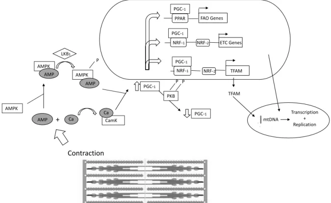
The simplified intracellular algorithm (Figure 1) of signaling during aerobic exercise is as follows: the rise in the AMP/ATP ratio → increased AMP bound to AMPK [14] → a conformational change in AMPK → AMPK becomes a more desired substrate for upstream kinase LKB1 → an increase in AMPK activity after aerobic exercise [39]. After the rise of free Ca2+ appearance
intracellular signaling algorithm is as follows: activating Ca2+ sensitive signaling molecules just like protein kinase
(CamK) [12] → phosphorylation histone deacetylases (HDAC) and binding myocyte-enhancing factor (MEF) 2 to the promoter of PGC-1alpha [10] → increased expression of mitochondrial regulating gene → the expression of respiratory genes, GLUT4, the fatty acid–
CamK Contraction AMP + Ca AMPK AMPK AMP Ca AMPK AMP LKB1 P PGC-1 PKB P P PGC-1 PGC-1 NRF-1 NRF-2 TFAM TFAM PGC-1 NRF-1 NRF-2 ETC Genes PGC-1
PPAR FAO Genes
mtDNA
Transcription + Replication
Figure 1. The molecular pathways activated by endurance exercise
Note: AMP – adenosine monophosphate; AMPK – AMP-activated protein kinase; Ca – calcium; CamK – calcium-calmodulin kinase; LKB1 – serine-threonine kinase; NRF-1 and NRF-2 – nuclear respiratory factors; PPAR – peroxisome proliferator activating receptor; PGC-1 – PPARF coactivator; PKB – protein kinase B/akt
oxidation enzymes, mitochondrial transcription factor A, [10] → increased PGC-1alpha mRNA and protein [1, 22]. In this way, AMPK is involved in mitochondrial biogenesis causing increased mitochondrial capacity [5], increased muscle glycogen storage and hexokinase activity [21], translocation of GLUT4 to the myolemma for glucose uptake [15] and glycolysis stimulation [25]. The listed mechanisms allow the contracting myocytes to adapt to the increased energy expenditure during exercise. Most of these adaptations can be caused by training-induced ischemia. Not only biochemical adaptations of skeletal muscles that take place during prolonged endurance training are mediated by AMPK. Recent research shows a crucial AMPK role in stimulating both vasculogenesis and angiogenesis, and though increasing blood supply to exercising muscles [28]. Increased muscles aerobic capacity after endurance training allows performing prolonged exercises with low or moderate intensity and – what was very often opposed – generating higher force in short bouts of exercises.
Resistance exercise
Resistance exercise may be described as any form of exercise that causes the muscles contraction in the circumstances of external resistance. Its subtypes may
be distinguished as static, dynamic, concentric, eccentric exercises and others. After resistance training, muscle hypertrophy and increased bone density are expected. Yet, these phenotypes may only be expected as the result of systematic and progressive activity of adequate frequency, duration, and intensity, causing the body’s adaptation according to the overload principle.
Whereas in endurance exercise the primary acute response is increased level of oxidative enzymes [19], increased mitochondrial mass [17], increased slow contractile and regulatory proteins [30], decreased glycolytic enzymes [30], and decreased fast-twitch fiber area [34], in case of resistance exercise an increased protein synthesis is the primary acute response. After a single bout of resistance exercises, a 50% increase of protein synthesis at 4 h and 115% at 24 h was observed [2]. Hamosh et al. [13] clarified that the increased intracellular protein synthesis in myocytes after resistance exercise is the result of an increase in the amount of protein synthesized per molecule of RNA since no changes in the RNA content have been observed [13].
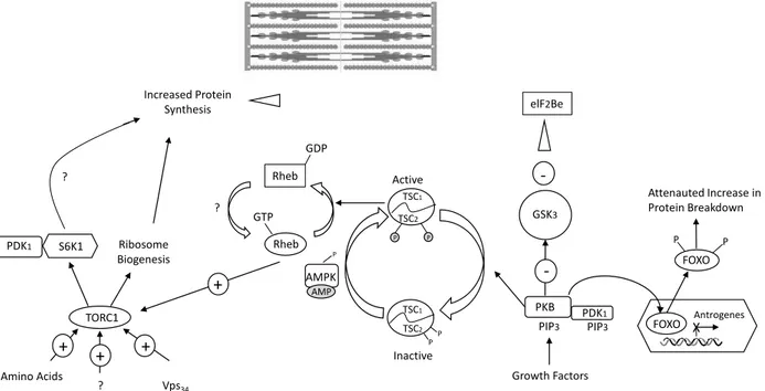
In enhanced protein synthesis after resistance exercise mTOR plays a key role (Figure 2). The mTOR pathway is a specific response of skeletal muscles, to physical,
Increased Protein Synthesis TORC1 + + + Amino Acids ? Vps34 S6K1 PDK1 Ribosome Biogenesis ? Rheb Rheb ? GDP GTP + Active TSC1 TSC2 P P TSC1 TSC2 P P AMPK P AMP Inactive GSK3 -elF2Be PKB -PDK1 PIP3 PIP3 Growth Factors FOXO XAntrogenes FOXO P P Attenauted Increase in Protein Breakdown
Figure 2. The molecular pathways activated by resistance exercise
Note: AMP – adenosine monophosphate; AMPK – AMP-activated protein kinase; eIF2B – eukaryotic initiation factor 2B; FOXO – forkhead transcription factor; GSK3 – glycogen synthase kinase; TORC1 – mammalian target of rapamycin complex 1; PDK1 – 3-phosphoinositide- dependent protein kinase; PKB – protein kinase B/akt; Rheb – ras-like protein enriched in brain; S6K – ribosomal S6 protein kinase; TSC1 – tuberculosis sclerosis 1; TSC2 – tuberculosis sclerosis 2
nutritional or environmental stimuli. The associated mTOR kinase is the enzyme responsible for activating the pathway. An important aspect is also activation through systematic and adequate intensity of resistance exercise. It is a key factor stimulating intracellular muscle protein synthesis and adaptation of the body to changing environmental conditions. mTOR and/or 3-phosphoinositide-dependent protein kinase-1 (PDK1) is required for the primary controls of protein synthesis initiation including eukaryotic initiation factor 2 (eIF2), 4E binding proteins (4E-BP), and the 70-kDa S6 protein kinase (S6K1). mTOR consist of two structurally distinct complexes: mTORC1 and mTORC2 [41] and both subunits localize to different intracellular compartments [6].
Three different mechanisms have been identified for the activation of TORC1 and its downstream targets: growth factor stimulation, nutrient deprivation and refeeding of amino acids, class III PI 3-kinase Vps34 overexpression [7]. Moreover, Barr et al. as well as others have shown that resistance exercise or muscle strain can transiently activate mTOR, S6K, PKB, increase eIF2B activity and inactivate GSK3A [3, 11, 24, 26, 29].
Combined exercise
In the light of knowledge and arguments mentioned above, it seems obvious that exercise applied to highly specialized elite athletes require highly specific either endurance or resistance exercise, rather than their combined version. However, there are numerous individuals, including people above 50 years old, who practicing physical activity expect rather health profits and well-being instead of simple competing and sports awards [33]. Thus, also a different approach to the body’s response to exercise could be expected. Molecular interactions after exercise are complex but recently it became more clear how we can compare and possibly combine endurance and resistance training. Some questions still remain unanswered (especially those concerning interrelation of volume, intensity, and frequency) and some answers are still modified over the years, according to new data appearance and increasing health condition of modern man. Especially for older adults, above 65 y. of age, the combined exercise training should be recommended, since it results in greater improvements in postural control, balance and walking performance [33].
Recommendations on physical activity for health
Moderate-intensity physical activity refers to the physical activity that is performed at 3.0-5.9 times the intensity
of rest (3-6 MET’s, where 1 MET equal to the oxygen cost of sitting quietly, equivalent to 3.5 ml/kg/min or 1 kcal/kg/hour [31]. Recommended by WHO levels of PA for health distinguish (i) at least 60 minutes of moderate to vigorous-intensity physical activity daily (5-17 years old), (ii) at least 75 minutes of vigorous-intensity aerobic physical activity throughout the week (18-64 y.), (iii) at least 75 minutes of vigorous-intensity aerobic physical activity throughout the week, or an equivalent combination of moderate- and vigorous-intensity activity (adults aged 65 years and above) [38]. Given by WHO recommendations seem somewhat conservative and dictated by the principle primum
non nocere, since most international physical activity
guidelines recommend the incorporation of moderate-intensity physical activity on all days of the week. Moreover, WHO recommendations seem rather to maintain health than to development/progress health and fitness performance.
Older adults, above 65 years of age, comprise ca. 13% of the population. By the year 2030 the number of people considered old is expected to increase up to 20% [27]. Moreover, worldwide the number of people over 60 years is expected to nearly triple in 2050 (760 million in 2010 to 2 billion in 2050) [37]. Thus, it is inevitably necessary to face the changing situation of human demography and its consequences.
There is an absolutely common approach that the aging process is irrevocably associated with decreasing fitness performance and fitness functionality. Nonetheless, there are data showing that it can be slowed down since individual being above 60 years old is able to improve his or her fitness performance and even fitness functionality. Maintaining physical fitness in older age requires much more intensity, frequency and duration compared to WHO recommendations. For a narrow group of healthy individuals above 60 years of age, it seems adequate to recommend even higher values of intensity, loads, frequency and duration of exercise allowing progress in functional fitness and fitness performance. Although in old age irreversible pathological changes appear (concerning cardio-metabolic health, muscle-tendon health, sarcopenia, pathologic changes of joints and vessels, glands, etc.), the muscles and bone density are still under influence of individual well-programmed exercise. Intensive exercise and as a consequence better functional fitness allows better general quality of life.
References
1. Akimoto T, Pohnert SC, Li P, Zhang M, Gumbs C, Rosenberg PB, Williams RS, et al. Exercise stimulates
Pgc-1α transcription in skeletal muscle through activation of the p38 MAPK pathway. J Biol Chem. 2005 May 20; 280(20): 19587-19593.
2. Baar K. Training for endurance and strength: lessons from cell signaling. Med Sci Sports Exerc. 2006 Nov; 38(11): 1939-1944.
3. Baar K, Esser K. Phosphorylation of p70(S6k) correlates with increased skeletal muscle mass following resistance exercise. Am J Physiol. 1999 Jan 1; 276(1): C120-C127. 4. Benzi G, Panceri P, De Bernardi M, Villa R, Arcelli E,
D’angelo L, et al. Mitochondrial enzymatic adaptation of skeletal muscle to endurance training. Appl Physiol. 1975 Apr 1; 38(4): 565-569.
5. Bergeron R, Ren JM, Cadman KS, Moore IK, Perret P, Pypaert M, et al. Chronic activation of AMP kinase results in NRF-1 activation and mitochondrial biogenesis. Am J Physiol Endocrinol Metab. 2001 Dec 1; 281(6): E1340-E1346.
6. Betz C, Hall MN. Where is mTOR and what is it doing there? J Cell Biol. 2013 Nov 25; 203(4): 563-574. 7. Byfield MP, Murray JT, Backer JM. hVps34 is a
nutrient-regulated lipid kinase required for activation of p70 S6 kinase. J Biol Chem. 2005 Sep 23; 280(38): 33076-33082. 8. Clarke DH. Adaptations in strength and muscular
endurance resulting from exercise. Exerc Sport Sci Rev. 1973 Jan 1; 1(1): 73-107.
9. Colliander EB, Tesch PA. Effects of eccentric and concentric muscle actions in resistance training. Acta Physiol Scand. 1990 Sep; 140(1): 31-39.
10. Czubryt MP, McAnally J, Fishman GI, Olson EN. Regulation of peroxisome proliferator-activated receptor γ coactivator 1α (PGC-1α) and mitochondrial function by MEF2 and HDAC5. Proc Natl Acad Sci U S A. 2003 Feb 18; 100(4): 1711-1716.
11. Farrell PA, Fedele MJ, Vary TC, Kimball SR, Lang CH, Jefferson LS. Regulation of protein synthesis after acute resistance exercise in diabetic rats. Am J Physiol. 1999 Apr 1; 276(4): E721-E727.
12. Fluck M, Waxham MN, Hamilton MT, Booth FW. Skeletal muscle Ca(2+)-independent kinase activity increases during either hypertrophy or running. J Appl Physiol (1985). 2000 Jan; 88(1): 352-358.
13. Hamosh M, Lesch M, Baron J, Kaufman S. Enhanced protein synthesis in a cell-free system from hypertrophied skeletal muscle. Science. 1967 Aug 25; 157(3791): 935- -937.
14. Hardie DG, Sakamoto K. AMPK: a key sensor of fuel and energy status in skeletal muscle. Physiology. 2006 Feb; 21(1): 48-60.
15. Hayashi T, Hirshman MF, Kurth EJ, Winder WW, Goodyear LJ. Evidence for 5’ AMP-activated protein
kinase mediation of the effect of muscle contraction on glucose transport. Diabetes. 1998 Aug 1; 47(8): 1369- -1373.
16. Hickson RC. Interference of strength development by simultaneously training for strength and endurance. Eur J Appl Physiol Occup Physiol. 1980 Dec 1; 45(2-3): 255- -263.
17. Holloszy JO. Biochemical adaptations in muscle effects of exercise on mitochondrial oxygen uptake and respiratory enzyme activity in skeletal muscle. J Biol Chem. 1967 May 10; 242(9): 2278-2282.
18. Holloszy JO. Exercise-induced increase in muscle insulin sensitivity. J Appl Physiol. 2005 Jul; 99(1): 338-343. 19. Holloszy JO, Oscai LB, Don IJ, Mole PA. Mitochondrial
citric acid cycle and related enzymes: adaptive response to exercise. Biochem Biophys Res Commun. 1970 Sep 30; 40(6): 1368-1373.
20. Holloszy JO, Rennie MJ, Hickson RC, Conlee RK, Hagberg JM. Physiological consequences of the biochemical adaptations to endurance exercise. Ann N Y Acad Sci. 1977 Oct; 301(1): 440-450.
21. Holmes BF, Kurth-Kraczek EJ, Winder WW. Chronic activation of 5’-AMP-activated protein kinase increases GLUT-4, hexokinase, and glycogen in muscle. J Appl Physiol (1985). 1999 Nov; 87(5): 1990-1995.
22. Irrcher I, Adhihetty PJ, Sheehan T, Joseph AM, Hood DA. PPARγ coactivator-1α expression during thyroid hormone-and contractile activity-induced mitochondrial adaptations. Am J Physiol Cell Physiol. 2003 Jun 1; 284(6): C1669-C1677.
23. Klausen K, Andersen LB, Pelle I. Adaptive changes in work capacity, skeletal muscle capillarization and enzyme levels during training and detraining. Acta Physiol Scand. 1981 Sep; 113(1): 9-16.
24. Kubica N, Bolster DR, Farrell PA, Kimball SR, Jefferson LS. Resistance exercise increases muscle protein synthesis and translation of eukaryotic initiation factor 2Bϵ mRNA in a mammalian target of rapamycin-dependent manner. J Biol Chem. 2005 Mar 4; 280(9): 7570-7580.
25. Marsin AS, Bertrand L, Rider MH, Deprez J, Beauloye C, Vincent MF, et al. Phosphorylation and activation of heart PFK-2 by AMPK has a role in the stimulation of glycolysis during ischaemia. Curr Biol. 2000 Oct 14; 10(20): 1247-1255.
26. Nader GA, Esser KA. Intracellular signaling specificity in skeletal muscle in response to different modes of exercise. J Appl Physiol (1985). 2001 May; 90(5): 1936-1942. 27. Nied RJ, Franklin B. Promoting and prescribing exercise
for the elderly. Am Fam Physician. 2002 Feb 1; 65(3): 419-426.
28. Ouchi N, Shibata R, Walsh K. AMP-activated protein kinase signaling stimulates VEGF expression and angiogenesis in skeletal muscle. Circ Res. 2005 Apr 29; 96(8): 838-846.
29. Parkington JD, Siebert AP, LeBrasseur NK, Fielding RA. Differential activation of mTOR signaling by contractile activity in skeletal muscle. Am J Physiol Regul Integr Comp Physiol. 2003 Nov; 285(5): R1086-R1090. 30. Pette D, Heilmann C. Transformation of morphological,
functional and metabolic properties of fast-twitch muscle as induced by long-term electrical stimulation. Basic Res Cardiol. 1977 Mar-Jun; 72(2-3): 247-253.
31. Physical Activity Guidelines for Americans. Office of Disease Prevention & Health Promotion, US Department of Health and Human Services, October 2008. Retrieved 11 January 2010 from: www.health.gov/paguidelines. 32. Sharkey B. Intensity and duration of training and the
development of cardiorespiratory endurance. Med Sci Sports. 1970 Winter; 2(4): 197-202.
33. Sousa N, Mendes R, Silva A, Oliveira J. Combined exercise is more effective than aerobic exercise in the improvement of fall risk factors: a randomized controlled trial in community-dwelling older men. Clin Rehabil. 2017 Apr; 31(4): 478-486.
34. Staron RS, Hikida RS, Hagerman FC, Dudley GA, Murray TF. Human skeletal muscle fiber type adaptability to various workloads. J Histochem Cytochem. 1984 Feb; 32(2): 146-152.
35. Tesch PA. Skeletal muscle adaptations consequent to long-term heavy resistance exercise. Med Sci Sports Exerc. 1988 Oct; 20(5 Suppl): S132-S134.
36. Tesch PA, Karlsson J. Muscle fiber types and size in trained and untrained muscles of elite athletes. J Appl Physiol (1985). 1985 Dec; 59(6): 1716-1720.
37. United Nations. World Population Prospects: The 2010 Revision. Department of Economic and Social Affairs, New York 2011.
38. WHO. Global recommendation on physical activity for health. 2010.
39. Winder WW, Hardie DG. Inactivation of acetyl-CoA carboxylase and activation of AMP-activated protein kinase in muscle during exercise. Am J Physiol. 1996 Feb; 270(2 Pt 1): E299-E304.
40. Wong TS, Booth FW. Skeletal muscle enlargement with weight-lifting exercise by rats. J Appl Physiol (1985). 1988 Aug; 65(2): 950-954.
41. Wullschleger S, Loewith R, Hall MN. TOR signaling in growth and metabolism. Cell. 2006 Feb 10; 124(3): 471-484.