October – December • 2012
MILITARY PHARMACY
AND MEDICINE
QUARTERLY INTERDISCIPLINARY JOURNAL
• PHARMACY
• MEDICINE
• MEDICAL TECHNIQUE
• ENVIRONMENT AND HEALTH
• EDUCATION
The Staff of the Military Center of Pharmacy and Medical Technology in Celestynow – Poland MILIT AR Y PHARMA CY AND MEDICINE • V olume V • No . 4 • 2012
Dr
uga s
tro
na o
kład
ki
Mr Radosław Sikorski
Minister of Foreign Affairs of the Republic of Poland
on the death of his Father
Col Radosław Ziemba, M.D., Ph.D.
Commander of the Military Center for Pharmacy
and Medical Technique in Celestynów
MILITARY PHARMACY
AND MEDICINE
Quarterly Interdisciplinary Journal
of Military Centre of Pharmacy and Medical Technique in Celestynów
on scientific socio-professional
and training issues of Military Pharmacy and Medicine
MILITARY PHARMACY
AND MEDICINE
SCIENTIFIC BOARD
Anyakora Chimezie, NigeriaBalbaa Mahmoud, Egypt
prof. dr hab. Michał Bartoszcze, Poland prof. dr hab. inż. Stanisław Bielecki, Poland Bisleri Gianluigi, Italy
Blumenfeld Zeev, Israel
dr hab. Kazimiera H. Bodek, Poland Boonstra Tjeerd W, Netherlands Borcic Josipa, Croatia Cappelli Gianni, Italy Yifeng Chai, China Chowdhury Pritish K, India Costa Milton, Brasil
prof. dr hab. inż. Krzysztof Czupryński, Poland Deckert Andreas, Germany
Demeter Pal, Hungary prof. dr hab. Adam Dziki, Poland Ermakova Irina, Russia
prof. dr hab. Zbigniew Fijałek, Poland Florence Sudha Gnanou, India Fontoura Paulo, Portugal dr hab. Ryszard Gajdosz, Poland Ning Gao, China
dr hab. Tomasz Gaszyński, Poland prof. dr hab. Paweł Górski, Poland prof. dr hab. Bożenna Gutkowska, Poland Holko Ivan, Slovakia
Zhenlin Hu, China Huang Feng, USA
dr hab. Czesław Jeśman, Poland prof. dr hab. Wiesław Jędrzejczak, Poland Kaubrys Gintaras, Lithuania
Kashanian Maryam, Iran
prof. dr hab. Andrzej Klimek, Poland dr hab. Józef Knap, Poland Korshunov Andrey, Russia Kusec Sanja, Croatia Shan Lei, China dr hab. Julian Maj, Poland
prof. dr hab. Jerzy Mierzejewski, Poland prof. dr hab. Elżbieta Mikiciuk-Olasik, Poland Newcomb Andrew, Canada
prof. dr hab. Jerzy Z. Nowak, Poland dr hab. Romuald Olszański, Poland prof. dr hab. Daria Orszulak-Michalak, Poland prof. dr hab. Krzysztof Owczarek, Poland prof. dr hab. Marek Paradowski, Poland Perotti Daniela, Italy
Pivac Nela, Croatia Pizzuto Francesco, Italy prof. dr hab. Janusz Pluta, Poland Polat Gurbuz, Turkey
Popescu Irinel, Romania Reddy G. Bhanuprakash, India prof. dr hab. Juliusz Reiss, Poland Rodrigues Gabriel Sunil, India Rossi Roberto, Italy Samarkos Michael, Greece Shen Hui-Liang, China Shevchuk Nikolai, Russia Xianglin Shi, USA Skultetyova Dana, Slovakia Strumylaite Loreta, Lithuania dr Piotr Siermontowski, Poland prof. dr hab. Marek Sosnowski, Poland prof. dr hab. Andrzej Stańczak, Poland prof. dr hab. Zbigniew Lew-Starowicz, Poland dr hab. inż. Marek Strzelczyk, Poland Ding-Feng Su, China
dr hab. Janusz Świniarski, Poland Tchetina Elena, Russia
Tomoum Hoda, Egypt Tufekcioglu Omac, Turkey
prof. dr hab. Jarosław Wysocki, Poland Wang FuZhou, China
Wei-dong Zhang, China Zarkovic Neven, Croatia Ruixin Zhu, China
MILITARY PHARMACY
AND MEDICINE
EDITORIAL BOARD
EDITOR-IN-CHIEF
prof. Piotr Fiedor, Warsaw, PolandDEPUTY EDITOR
prof. Jarosław Wysocki, Warsaw, PolandSECTION EDITORS
Biochemistry
dr hab. inż. Marek Strzelczyk, Poland
Bioethics & Medical Law
prof. dr hab. Hieronim Bartel, Poland
Biology
prof. Lidia Chomicz, Poland
Catastrophe Medicine
Adam Pietrzak, Poland
Emergency Medicine
dr hab. Tomasz Gaszyński, Poland
Epidemiology
dr Witold Gnitecki, Poland
Forensic Medicine
dr hab. Paweł Krajewski, Poland
Hematology
prof. dr hab. Wiesław Jędrzejczak, Poland
History of Medicine & Pharmacy
dr Zdzisław Jezierski, Poland
Infectious Diseases
dr hab. Józef Knap, Poland
Linguistic Editor
Mirosław Termar, USA
Maritime & Tropical Medicine
dr hab. Romuald Olszański, Poland
Military Medicine
dr Marek Skalski, Poland
Neurology
prof. dr hab. Andrzej Klimek, Poland
Neurosurgery
prof. dr hab. Jan Podgórski, Poland
Ophthalmology
Piotr Michałowski, Poland
Orthopedics and Traumatology
dr Wojciech Glinkowski, Poland
Patomorfology
dr Piotr Siermontowski, Poland
Pharmaceuticlal Science
prof. dr hab. Aleksander P. Mazurek, Poland
Pharmacology & Pharmacy
prof. dr hab. Bożenna Gutkowska, Poland
Physiology
prof. dr hab. Józef Kędziora, Poland
Psychiatry
prof. dr hab. Józef Kocur, Poland
Psychology
prof. dr hab. Krzysztof Owczarek, Poland
Sexology
prof. dr hab. Zbigniew Lew-Starowicz, Poland
Statistical Editor
dr Janusz Śmigielski, Poland
Stomatology
dr Stanisław Żmuda, Poland
Surgery
prof. dr hab. Adam Dziki, Poland
Toxicology
dr Wotold Kurnatowski, Poland
Urology
MILITARY PHARMACY
AND MEDICINE
EDITORIAL OFFICE
Secretary of the Editorial Office
Krzysztof Barczewski, Poland Remigiusz Radziszewski, Poland
Statistical Editor
dr Janusz Śmigielski, Poland
Technical Editor
Remigiusz Radziszewski, Poland
English Language Professional Service
Miroslaw Termar, USA
Public Relations
Krzysztof Barczewski, Poland
PUBLISHER
Military Centre of Pharmacy and Medical Technique in Celestynów Wojska Polskiego 57 05-430 Celestynow, Poland phone +48 22 689 40 70, fax +48 22 689 40 91 e-mail: http://military.isl-journals.com/
PUBLISHED BY
International Scientific Literature, Inc
361 Forest Lane,
Smithtown, New York 11787, USA phone +1 516 874 4341 e-mail: office@isl-science.com
Interdisciplinary journal of Military Centre of Pharmacy and Medical Technique in Celestynów, Poland http://military.isl-journals.com/
© MILITARY PHARMACY AND MEDICINE. All rights reserved.
No part of this publication may be reproduced, stored in retrieval system, or transmitted, in any form or by any means, electronic, mechanical, photocopying, recording or otherwise without the prior written permission.
ISSN 1898-6498 quarterly Indexed in: MNiSW, Index Copernicus 130 copies
prof. dr hab. Hieronim Bartel, Poland dr Przemysław Biliński, Poland
dr hab. Romana Bogusławska-Walecka, Poland prof. dr hab. Andrzej Buczyński, Poland prof. dr hab. Marian Brocki, Poland Patrick Cadet, USA
dr hab. Andrzej Chciałowski, Poland dr Wiesław Chudzik, Poland dr Jan Czarnecki, Poland
dr Maria Dziedziczak-Buczyńska, Poland prof. dr hab. Adam Dziki, Poland prof. dr hab. Wojciech Gaszyński, Poland dr hab. Czesław Jeśman, Poland prof. dr hab. Józef Kędziora, Poland prof. dr hab. Józef Kocur, Poland dr Marek Kołodziejczyk, Poland
Richard Kream, USA
prof. dr hab. Krzysztof Kwiatkowski, Poland dr hab. Julian Maj, Poland
Kirk Mantione, USA
prof. dr hab. Eugeniusz Miękoś, Poland dr Dariusz Piotrowski, Poland prof. dr hab. Jan Podgórski, Poland dr hab. Wiesław Raszewski, Poland dr Barbara Sadowska, Poland George B. Stefano, USA dr hab. Antoni Szymański, Poland dr Zbigniew Teter, Poland dr Wiesława Trendak, Poland dr hab. Jadwiga Turło, Poland dr Elżbieta Wojtasik, Poland
REVIEWERS
v
Table of Contents
Age-related macular degeneration (AMD): a critical appraisal of diet and dietary
supplements as therapeutic modalities
1
Jerzy Z. Nowak
Assessment of the energy expenditure of soldiers of the Representative Battalion of the Polish Army
during three days of drill training as part of preparations for the celebration of the National
Independence Day of November 11
th 17Jerzy Bertrandt, Anna Kłos, Roman Łakomy
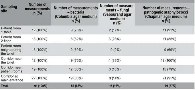
Assessment of microbial quality of drinks not included in the hospital diet as consumed by
patients during hospitalization and the assessment of microbial contamination of hospital air
21
Jaśmina Żwirska, Wanda Jabłońska, Paweł Jagielski, Stanisław Stępniewski, Małgorzata Schlegel Zawadzka
Cholesterol oxyethyleneation products as modifiers of the absorption base in
anti-inflammatory ointments
27
Justyna Kołodziejska, Marian Mikołaj Zgoda,
Katarzyna Ejzenchart, Magdalena Piechota-Urbańska
Non-steroidal anti-inflammatory drugs (NSAIDs) in ophthalmology:
pharmacological and clinical characteristics
33
Jerzy Z. Nowak
The role of paramedics in British emergency aid system
51
Dorota Kołodziej, Radosław Ziemba
Energy expenditure as the basis for determination of nutritional demand in soldiers
57
Jerzy Bertrandt, Anna Kłos, Bartosz Bertrandt
Sodium and potassium content of daily food rations of students of
the Main School of Fire Service in Warsaw
61
Anna Kłos, Maryla Długaszek, Jerzy Bertrandt, Wiesława Szymańska
Cardiac arrest under special circumstances. Part II:
poisoning, …, anaphylactic reaction, …, traumatic injuries
67
Radosław Ziemba
Prevalence of atrial fibrillation in emergency medicine practice
77
Łukasz Szarpak, Dariusz Timler
Autopsy marks on anthropological material contributing
to research on history of anatomy in Poland
81
Magdalena Cybulska, Agnieszka Adamczyk, Agnieszka Młudzik, Czesław Jesman
vi
Table of Contents
Infusion solutions supply in critical circumstances and disasters
87
Katrzyna Parzuchowska, Radosław Ziemba, Jan Hołyński, Adam Ziemba, Jarosław Hołyński, Ewa Ziemba
Scope of knowledge regarding administration ofoxygen therapy among firefighter
rescue teams from Volunteer Fire Departments (OSP)
91
Łukasz Szarpak
Sustainable development in light of international cooperation
97
Radosław Ziemba
Anthropological and archeological sources in medical historian’s studies
105
Magdalena Cybulska, Agnieszka Adamczyk, Agnieszka Młudzik, Czesław Jesman
Selected aspects of communication between emergency services
and the mass media in crisis situations
113
Łukasz Szarpak, Marcin Madziała
Bioterrorism — nature of the problem
119
Radosław Ziemba
Anti-drown ring — a patented drowning protection device (patent no. 197623)
123
Jerzy Mierzejewski
Speech made by Commandant of the Military Centre for Pharmacy and
Medical Technology in Celestynów, Colonel DMSc Radosław Ziemba
127
Radosław Ziemba
Ophthalmology
Age-related macular degeneration (AMD): a critical appraisal of
diet and dietary supplements as therapeutic modalities
Jerzy Z. Nowak
Department of Pharmacology, Chair of Pharmacology and Clinical Pharmacology of Medical University of Lodz, Poland
Author’s address:
Jerzy Z. Nowak, Department of Pharmacology, Medical University, ul. Żeligowskiego 7/9, 90-752 Lodz, Poland; phone: ( + 48) 42 6393270, e–mail: jerzy.nowak@umed.lodz.pl
Received: 2012.10.12 • Accepted: 2012.11.22 • Published: 2012.12.08
Summary:
Age-related macular degeneration (AMD) is a vision-threatening ocular disease, affecting the central region of the retina — the macula — and manifesting in the elderly. Its pathogenesis is multifactorial, molecularly complex and only poorly recognized. AMD is a degenerative disease, and the degeneration affects primarily the retinal pigment epithelial (RPE) cells and secondarily the photoreceptors, which leads to disturbances or partial loss of central vision and legal blindness. Principal processes contributing to the development of the disease include: oxidative stress, lipofuscinogenesis, drusogenesis and local inflammation (so-called para-inflammation). A severe complication of clinically recognized dry form AMD (geographic atrophy) is the aggressive neovascularization originating from choriocapillary system (CNV; wet form AMD). At present, there are no therapeutic agents capable of slowing or inhibiting degeneration process in the photoreceptors-RPE complex, therefore preventive rather than therapeutic modalities are under consideration; they include properly adjusted everyday diet and intake of dietary supplements, both rich in antioxidants of which macu-lar pigments (lutein, zeaxanthin, meso-zeaxanthin) are of particumacu-lar importance. Long-chain unsaturated omega-3 fatty acids (PUFA-ω3) are also recommended to people with already progressing AMD and at an increased risk of the disease development. Despite wide commercial offer of “ophthalmic” antioxidative and PUFA-ω3-rich preparations, no convincing evidence is available to date to support their protective activ-ity in AMD patients. AREDS-2 clinical trials that actually approach completion may likely provide more conclusive answer whether macular pigments (lutein, zeaxanthin) and PUFA-ω3 (EPA, DHA) can really be useful for AMD patients. The aim of this article is twofold: 1. it presents molecular mechanisms underlying the early stages of AMD pathogenesis, which form a platform for the disease development, and 2. it provides a critical appraisal of the prophylactic/therapeutic value of properly profiled diet and the intake of “ophthal-mic” dietary supplements rich in macular pigments and omega-3 PUFAs.
Key words:
Age-related macular degeneration, AMD, oxidative stress, omega-3 fatty acids, macular pigments, prevention, diet, dietary supplements.Introduction
This article refers to an earlier article by the same author, entitled Dry form AMD — do we know
how to treat it?, published in 2011 in issue 5(1)
of Magazyn Lekarza Okulisty [1] — a Polish-language journal specialized in ophthalmology and widely distributed among ophthalmologists. There are no drugs specific to this condition; the
therapeutic approach focuses on prophylaxis in the form of properly profiled diet and the intake of dietary supplements. The number of these sup-plements is constantly growing, and all of them are freely available, i.e. without prescription. The available supplements differ in both qualitative and quantitative composition. A detailed analy-sis of supplements available at Polish market as described by the author in 2010 in an article entitled Ophthalmic antioxidant preparations:
a survey and supportive arguments for their use in AMD [2], included 73 products — all of them
contained the macular pigment — lutein, some contained also zeaxanthin, and only one con-tained three macular pigments, namely lutein, zeaxanthin and meso-zeaxanthin.
Currently nearly 100 ophthalmic antioxidant preparations are available only at Polish mar-ket. This does not include numerous prepara-tions containing polyunsaturated fatty acids of the omega-3 series (PUFA-ω3), discussed by the author in his earlier study entitled Omega-3
polyunsaturated fatty acids in retina and medical practice — pros and cons [3]. Ophthalmologists,
as well as patients, may have and, according to author’s knowledge, indeed have problems with choosing an appropriate preparation. However, resorting to dietary supplement may be per-haps unnecessary if appropriate choice of food products is made for one’s everyday diet. The author hopes that this study, confronting the dietary and supplementation aspects of prophy-laxis will contribute to appreciation of the role of everyday diet rich in selected products in AMD prophylaxis and acknowledging the value of such diet as not inferior to that of pharmaceutical supplementation.
AMD – origin and development
of the pathology
Age-related macular degeneration (AMD) devel-ops inconspicuously over many years. Despite numerous studies conducted worldwide, the pathogenesis of the disease is not fully under-stood. Due to the plurality of factors, both endog-enous (e.g. genetic predispositions) and exog-enous (history of exposure to light) or behavioral (e.g. smoking, improper diet), which might pre-dispose, or even contribute to the development of the disease, the critical physiological processes responsible for the vision process (e.g. the visual
excitation cascade) move beyond the limits of homeostasis, creating a biochemical platform for the future pathology [4–9]. This involves inten-sified lipofuscinogenesis in retinal pigment epi-thelium (RPE) cells, formation of drusen and pseudodrusen — the former occurring under the RPE monolayer (i.e. in the direction of Bruch’s membrane), the latter above the RPE monolayer (i.e. towards photoreceptors) and next, a chronic inflammatory process known as para-inflam-mation. All these phenomena contribute to the development of AMD. Clinically, AMD can be divided into the dry, or atrophic, form (known as geographic atrophy), and wet — exudative, or neovascular form; the latter can be considered a complication of the atrophic form manifesting as choroidal neovascularization (CNV) [1,4,5]. Aging favors the development of AMD, particu-larly in predisposed individuals (due to concomi-tant presence of the risk factors listed above); the term “age-related” in the disease name – AMD is fully justified, as the age is the major (albeit indi-vidual, i.e. specific to each subject) and unavoid-able determinant of various dysfunctions at the cellular and organ level, including these associ-ated with the deficiency of necessary microele-ments/nutrients or with the loss of function in the aging cells — particularly post-mitotic, i.e. non-regenerable cells. While the lacking micro-elements/nutrients may be supplied from the outside, thus attempting to compensate for their deficiency in particular cells/tissues, we are una-ble until now to stop the systemic aging process. Our bodies contain many non-regenerable (post-mitotic) cells, particularly within the central nervous system. These include the photorecep-tors (only the external segments that contain photopigments are subject to regeneration) and retinal pigment epithelium (RPE) cells. In the pathogenesis of AMD, RPE cells are the first cells that become metabolically inefficient and thus undergo degeneration; dysfunction and atrophy of photoreceptors is secondary, as they are una-ble to function and survive without functionally efficient RPE cells, and therefore also undergo degeneration. The pathological process involves mostly a small region of the retina, known as the macula, where cone photoreceptors, respon-sible for acute and color vision, are predomi-nant. Therefore, first clinical symptoms of AMD include blurred vision, defects of central vision of
various intensity or gradual loss of color vision, “warping” of perceived images [10,11]. Early form of AMD is often referred to as age-related macu-lopathy (ARM).
When the eyes are open, photoreceptors are continuously working, absorbing light photons and “recording” the image of the environment. Similarly to TV cameras being turned on, eyes record this image in an automatic fashion, gener-ating the first signal of a complex, multisynaptic vision process. Photoreceptors’ outer segments (POS), filled with visual pigment molecules, are characterized by significant functional dynam-ics; they wear off upon continuous function and, as a consequence, the apical fragments are con-stantly shed and captured by neighboring RPE cells. At the same time, POS is being rebuilt in order to maintain appropriate size (which is an important parameter determining the efficacy and survival of photoreceptors!). Regeneration proceeds from the perikaryon, i.e. the photore-ceptor inner segment (PIS) and requires numer-ous building blocks, including docosahexaenoic acid (DHA). These building blocks are supplied by the RPE cells and originate partly from the captured POS fragments and partly from circu-lation (consumed food) [9].
One of the many important roles played by the RPE cells is “digestion” of the absorbed (and con-tinuously being absorbed) photoreceptor mate-rial stored in phagolysosomes. Despite the fact that phagocytosis and enzymatic degradation occurring as a result of the activity of numerous lysosomal enzymes are physiological processes that had developed over thousands of years in creatures dwelling on Earth and making use of the visual organ system (retinal processes that govern visual perception in many vertebrates, including humans, are generally very similar), they seem to be of limited efficacy, at least in humans. This claim is supported by systematic accumulation of lipofuscin, known as the age pigment, in RPE cells.
Lipofuscin and the process of its formation, i.e. lipofuscinogenesis,are not the attributes of RPE cells and connections between photoreceptors and RPE, as they are also present in other non-renewable cells, such as neurons, cardiomyocytes or skeletal muscle cells. However, it is in this region of the eye, or more precisely, of the retina,
i.e. in the photoreceptors/RPE cells region, where a unique characteristics of lipofuscin accumu-lated in RPE cells becomes evident: lipofuscin contains retinoids (vitamin A derivatives) origi-nating from the visual cycle, particularly bis-retinoids — products of spontaneous fusion of two molecules of all-trans-retinal, generated via photoreaction (i.e. by absorption of photons) by 11-cis-retinal, a cofactor of the visual pigment. The cis → trans retinal transformation is the cru-cial first step of visual cycle, initiating further conformational transformations of opsin (i.e. the visual pigment protein) into its active forms (e.g. meta-rhodopsin II), capable of progressing the visual cycle with the final effect consisting in the closure of cGMP-dependent cation chan-nel in the cellular membrane of photoreceptors and quenching the so-called darkness current. In this time, the cellular membrane of photore-ceptors is hyperpolarized only to regain the state of being ready to absorb another photon, i.e. the depolarization state; the active pigment, capable of absorbing photons, is a molecule, e.g. rho-dopsin, that contains a light sensitive co-factor, 11-cis-retinal [5,6].
The all-trans-retinal formed following photon absorption is completely dissociated from the vis-ual pigment and undergoes further physiological transformations in the retinoid cycle that takes place in both photoreceptors and RPE cells. How-ever, part of all-trans-retinal that does not bind the ABCA4 (ATP-binding cassette [transporter] A4 type, also known as ABCR) transporting the retinoid into the sites with all-trans-retinal dehydroghenase activity, “falls out” the cycle and spontaneously dimerizes (using ethanolamine as a “linker”) into a highly phototoxic bisretin-oid known as A2E. There may be more similar and toxic bisretinoids; however, A2E, which has been studied in more detail, seems to represent an established stress-inducing product. Furano – and peroxy –mtabolites of A2E have signifi-cant ability to activate the complement system (an alternative pathway) which represents the innate immunity, capable of efficiently and auto-matically acting in system’s defense, including destruction of “own” cells [5,12].
In addition, the photoreceptor cell membranes contain exceptionally high amounts of polyun-saturated fatty acids (PUFAs), particularly the
most-unsaturated docosahexaenoic acid (DHA). It is this high unsaturation (six double C=C bonds in the hydrocarbon chain) makes DHA particularly susceptible to spontaneous peroxi-dation and fragmentation into cytotoxic com-pounds, including 4-hydroxy-2-hexenal (HHE) or 4-hydroxy-7-oxyhept-5-enoic acid (HOHA); the latter may additionally form immunogenic carboxyethylpyrrole-protein (e.g. CEP-albu-min) adducts. More details regarding formation of the products of peroxidation of long-chain PUFAs and the properties of carboxyethylpyr-role-protein adducts are available in other arti-cles by the same author [2,5].
Accumulation of lipofuscin deposits in RPE cells is a manifestation of metabolic inefficacy of their lysosomal compartment, character-ized by reduced autophagy. The reason for this inefficacy remains unknown, although consid-ering molecular complexity of autophagic pro-cesses, the reasons may be multiple, including hypofunction or insufficient quantity/activity of lysosomal enzymes – cathepsins being most predominant in normal conditions. Lipofuscin deposits accumulate with age, and the adverse effect of accumulating oxidative stress that accompanies lipofuscinogenesis is intensified. Local inflammatory reaction that develops at certain moment and is manifested by an atypi-cal process referred to as para-inflammation, as well as drusogenesis (drusens, pseudodrusens), become the driving forces of the developing AMD pathology [4-7, 13].
One should not forget that the supply of oxygen (and microelements/nutrients) via the choriocap-illary system into the photoreceptors-RPE cells complex is one of the largest in primates. Tak-ing into account the functional specificity of the retina, particularly of photoreceptors (photosen-sitivity, extensive metabolism, high partial pres-sure of oxygen being the substrate for the forma-tion of oxygen radicals), one may suspect that retina is particularly well predisposed for forma-tion of reactive oxygen species (ROS) [14].
The nature must have predicted the potentially adverse, propathogenic processes, such as oxida-tive stress or lipofuscinogenesis, in the retina, as the tissue, and particularly the macular region, very important for acute and color vision in primates, has been equipped with an array of
antioxidative defense systems, including specific macular pigments (MPs) – lutein and zeaxanthin and meso-zeaxanthin produced from lutein [15].
It should also be mentioned that the classical antioxi-dative systems present within the body are classified as either enzymatic or non-enzymatic. Enzymatic systems include three basic enzymes: superoxide dys-mutase (SOD), catalase and glutathione peroxidase, which are dependent on metal ions, such as zinc, copper, manganium or selenium. The non-enzy-matic system consists of carotenoids (including the aforementioned macular pigments), vitamins E and C and glutathione. The enzymatic system is endog-enous; however, the metal ions that are required for its proper function are exogenous, i.e. must be sup-plied with food. As far as the non-enzymatic system is concerned, only glutathione is an endogenous anti-oxidant. The remaining elements of the system, i.e. carotenoids and vitamins E and C are exogenous fac-tors supplied with food or appropriate dietary supple-ments. The role of carotenoids is to neutralize singlet oxygen and reactive free radicals. What’s interesting, the mechanisms underlying these two activities of carotenoids are different and neutralization of free radicals may, in certain conditions (e.g. those when excessive amounts of radicals are present), change the antioxidative activity of these compounds into prooxidative activity.
Proper functioning of retinal antioxidation defense systems is believed to avert potential early propathogenic changes that may lead to AMD. These propathogenic changes are in fact physiological reactions that become function-ally (chemicfunction-ally) impaired due to their intensity and the accompanying overproduction of reac-tive oxygen species (ROS), crucial transforma-tions of “visual” retinoids and peroxidation and fragmentation of long-chain PUFAs. This may be generally due to indisposition of the aging body – its organs, tissues and cells, and hypofunction of the aforementioned antioxidative defense sys-tems, both enzymatic and non-enzymatic. Since these systems are dependent on the supply of exogenous nutrients, thus their activity is diet-dependent [2,14–17].
A diversified diet, containing vegetables rich in carotenoids, or — more precisely — oxycarotenoids
or xanthophylls (mainly lutein and zeaxanthin;
meso-zeaxanthin is not of dietary origin, being
first recommendation for patients in whome AMD development is suspected on the basis of gener-ally discrete symptoms, albeit disturbing visual sensations.
Diet
Specific dietary recommendations are crucial in many ailments and diseases. For instance, in diabetic patients, diet is an integral element of therapy, as no therapeutic success can be achieved in these patients without an appropri-ately profiled diet. Gluten-free diet is necessary in individuals, particularly children, with gluten intolerance. Patients with arterial hypertension should avoid salt, i.e. sodium chloride, in their diet. Excess sodium ions retain water in the body via a kidney-based mechanism; thus the daily dietary content of sodium should be well below 85 mmoles (less than 5 g NaCl) — in practice, one should follow the rule “the less salt, the better”. Many examples may be provided, but how about a diet for AMD patients?
Indeed, appropriate dietary recommendations for AMD patients are well justified, although one should expect no miraculous effects of such diets. Individuals who rigorously follow dietary recom-mendations (abundance of selected vegetables rich in lutein, marine fish rich in PUFA-ω3, etc.) also suffer from AMD. This is the nature of the disease, as it develops inconspicuously over many years and becomes symptomatic after the age of 50, most frequently after the age of 60. Its back-ground is multifactorial, including both environ-mental/genetic and inflammatory/immune fac-tors; in general opinion, there is no way to avoid it, as reported by the author in numerous stud-ies [1, 2, 4, 5, 18]. Nonetheless, appropriate diet is important in AMD patients.
Considering the threat of AMD or active AMD, one should recommend vegetables that con-tain high amounts of macular pigments (MPs), i.e. lutein and zeaxanthin [19]. Recommended vegetables include spinach, corn, pumpkin, red grapes (particularly seedless grapes), broccoli etc. (a more detailed list is presented in Table 1), as well as egg yolk which contains high amount of zeaxanthin in addition to lutein [15, 20]. It was calculated that the average Western diet content of lutein and zeaxanthin is between 1.3 and 3 mg, with lutein to zeaxanthin ratio of 7:1 [21, 22]. A
properly profiled diet may contain more macu-lar pigments, and therefore be more beneficial for individuals with AMD. Macular pigments are guards of metabolic order in the macular region and play a dual role of a filter, as they absorb blue light which is most dangerous for eyes and of a scavenger of free radicals, as they neutralize sin-glet oxygen and continuously generated free oxy-gen radicals.
Table 1: The content of lutein and zeaxanthin in widely
used vegetables and fruits, given in mol %.
Products zeaxanthinLutein and Lutein Zeaxanthin
Egg yolk 89 54 35 Corn 86 60 25 Kiwi fruit 54 54 0 Red seed-less grapes 53 43 10 Pumpkin 49 49 0 Spinach 47 47 0 Cucumber 42 38 4 Celery 34 3 2 White grapes 31 25 7 Brussels sprouts 29 27 2 Green peas 25 22 3 Broccoli 22 22 0 Mango 18 2 16 Lettuce 15 15 0
Properly selected daily rations of selected veg-etables provide the body not only with macular pigments, but also with microelements (includ-ing zinc, copper, manganese, selenium, vitamins E and C) required for proper functioning of anti-oxidative enzymes (superoxide dysmutase, cata-lase, glutathione peroxidase).
One should not forget other important dietary ele-ments, i.e. unsaturated fatty acids of the omega-3 series, particularly DHA[1], abundantly present in
marine fish[2]. Two or three fish-based meals per
[1] DHA is an example of a compound of dual nature, both beneficial and adverse. On one hand, DHA is required for the function of many cells, tissues and organs; on the other hand, due to its unsaturation, it is associated with the risk of adverse phenomena; at the same time, its functional indispensability is associated with structural unsaturation. The pros and cons of DHA were discussed in more detail in [3]. A disadvantage of DHA, as well as of other long-chain polyunsaturated fats, including the omega-3 eicosapentaenoic acid (EPA), omega-6 arachidonic acid (ARA or AA), or docosapentaenoic acid (DPA) is that in the presence of oxygen they easily undergo peroxidation and cleavage leading to propagation of oxidative stress. Despite the dual nature of the omega-3 fatty acids (DHA, EPA, DPA), one should not avoid their presence in food, particularly in the context of AMD prevention. The presence of vitamin E in food/supplement protects long chain polyunsaturated fats from oxidation. [2] Marine fish contain high amounts of long-chain polyunsaturated fatty acids (PUFAs), including acids of the omega-3 series (PUFA-ω3). Of tested and commonly eaten fish
week (e.g. salmon, herring, mackerel) provide the body with these fatty acids in amounts sufficient not only for needs of the organs of vision, but also of the entire system.
Polyunsaturated fatty acids of the omega-6 series (PUFA-ω6), also essential for human body, are usually consumed in high amounts, as they are present in commonly used vegetable oils – e.g. linoleic acid (C18:2ω6 or C18:2n-6, which is the first acid in the omega-6 series) is abundant in grape seed oil and sunflower oil (63–66%), as well as in corn oil and soybean oil (55–56%); other oils have much lower contents of linoleic acid (rape oil, linen oil, olive oil — 10–21%). In case of PUFA-ω3, the case is more difficult, as EPA and DHA are virtually absent in vegetable oils, while alpha-linolenic acid (ALA; C18:3ω3 or C18:3n-3, being the first acid in the omega-3 series) is present only in moderate amounts in rape oil and soybean oil (7–11%), as well as in large amounts (!) in linen oil (55%)[3]. For the
sake of comparison, the overall PUFA-ω3 (ALA, EPA and DHA) content in fish oil (herring oil) is > 28%, while the linoleic acid (ω6) content is slightly above 12%. Cold-pressed linen oil is available in an increasingly broad commercial offer. Although not everyone finds it tasty, linen oil is a recommended addition to the diet, not only of AMD patients. However, one should remember that conversion of ALA into EPA or DHA in human body is very low, and therefore the supply of ALA may not substitute for direct supply of DHA and DPA (which are abundant in marine fish).
species, highest amounts of DHA and EPA (expressed overall in g/100 g of weight) can be found in Atlantic salmon (> 2; farm-raised and wild caught) and herring (mainly Atlantic herring ≈ 2), then in mackerel and tuna (1.2–1.5); the popular canned tuna contains much smaller amounts (0,3–0,8); less DHA + EPA can be found in halibut and cod (0,2–0,6), while a higher content can be found in trout (farm-raised and wild caught) (≈ 1). As far as DHA is concerned, the highest content can be found in salmon, herring and tuna (excluding canned tuna in brine) and trout. Although fish contain particularly high amounts of EPA, DHA and DPA, other natural sources of these acids are human milk, farm-grown marine algae, marine mammals and krill; as mentioned in the article, some vegetable oils, including linen oil, do not contain EPA and DHA, although they may contain large amounts of alpha-linolenic acid (PUFA-ω3).
[3] The percentage composition of selected fatty acids in the linen oil is as follows:
alpha-linonenic acid (18:3ω3) – 54.5%, oleic acid (18:1ω9) – 19.7%, linoleic acid (18:2ω6)
– 16.2%, palmitic acid (16:0) – 5.1%, stearic acid (18:0) – 3.7%, other acids – 0,8%. The linen oil contains no EPA or DHA. The ω6/ω3 ratio is 0.3. For the sake of comparison, the percentage composition of fish oil (herring oil) is as follows: eicosapentaenoic acid (EPA; 20:5ω3) – 17.2%, palmitic acid (16:0) – 13.9%, palmitoleic acid (16:1ω7) – 13.1%, linoleic acid (18:2ω6) – 12.4%, oleic acid (cis-Δ9-octadecenoic acid; 18:1ω9) – 11.6%, docosahexaenoic acid (DHA; 22:6ω3) – 9%, myristic acid (tetradecanoic acid; 14:0) – 7.4%, stearic acid (octadecanoic acid; 18:0) – 2.7%, linolenic acid (18:3ω3) – 2.1%, elaidic acid (trans-Δ9-octadecenoic acid; 18:1ω9) – 2%, gadoleic acid (cis-Δ11-icosenoic acid; 20:1ω9) – 1.5%; ω6/ω3 ratio = 0.4.
Modern diet, particularly the Western diet, is rich in the fatty acids of the omega-6 series, and the ratio of these acids to the omega-3 (ω3) acids may be as high as 20:1, or even higher! The proper ratio should be about 4:1, with a trend towards balanced supply of both types of acids; this leads to the natural need for omega-3 acids supplemen-tation (EPA, DHA, ALA and docosapentaenoic acid — DPA-ω3); in case of AMD, DHA is of the highest importance. Despite its disadvantages (easy peroxidation and fragmentation), DHA is absolutely necessary for regeneration of pho-toreceptor outer segments worn off in the pro-cess of vision as well as to maintain appropriate plasticity/susceptibility of the cell membrane in rods and cones. In addition, EPA and DHA are substrates for production of anti-inflammatory resolvins and maresins (the latter are formed only from DHA) which are very important for the photoreceptors-RPE cells complex. DHA is also a substrate for production of neuroprotec-tin, which is involved in many protective, anti-inflammatory and cytoprotective mechanisms [8]. More information on the pros and cons of DHA may be found in a recent article by the same author [3].
The advantage of thus-profiled diet (as mentioned above) is that the elements valuable, among oth-ers, for intraocular metabolism, are delivered to the organism in natural, purely physiological fashion, which guarantees optimum gastrointes-tinal absorption and transport to target tissues/ cells as long as a diversified and well-balanced diet is maintained. One should remember that these microelements, being so important not only for AMD patients, are absorbed into circu-lation from the gastrointestinal tract in a diverse manner, as they represent different types of chemical structures and molecular mass ranges. Well-balanced diet containing diverse proteins, carbohydrates and all types of fats (long – and short-chain, saturated and unsaturated) estab-lishes within the stomach and the intestines a natural chemical environment that favors pas-sive or active absorption of microelements sup-plied with food.
This natural, physiological situation is very much different from situation taking place in the stom-ach (oftentimes an empty one) after ingestion of dietary supplement tablets/capsules followed by a glass of water! Microelements contained in the
supplements, albeit selected, have no optimum conditions for absorption. This accounts for the superiority of comprehensive natural nutrition over the intake of selected substances contained in dietary supplements, which do not have to be (and, in fact are not) fully absorbed from the gastrointestinal tract. A separate issue relates to the substances being delivered just where they are needed, which is a problem not less impor-tant from the standpoint of efficacy and expected results, and providently passed over in silence by the producers or suppliers of dietary supplements. However, some individuals may, for various rea-sons, including the fact that preparation of appro-priate meal requires time and effort, while swal-lowing a capsule “solves the problem”, may prefer ready-made dietary supplements. Therefore, the dietary supplements must also be included in our considerations.
Dietary supplements and the efficacy
of AMD prophylaxis/treatment
From the medical standpoint, the issue as stated in the heading is of primary importance for a wide group of mature population at risk of dis-comforts associated with the developing or potential AMD. As mentioned before, pathogen-esis of the disease is still unknown, which makes it impossible to both early diagnose the devel-oping pathology and to efficiently treat it using appropriate medications.
This does not pertain to the neovascular form of AMD, originating from choroidal neovascularization and considered by many researchers, including the author of this article, to be a serious complication of advanced AMD. This form, or more precisely, the dynamic neo-vascularization that accompanies AMD, may cur-rently be treated pharmacologically using agents that neutralize the main proangiogenic factor, i.e. the vascular endothelial growth factor (VEGF). Follow-ing agents are available: monoclonal anti-VEGF-A antibodies (Avastin – bevacizumab, Lucentis – ranibi-zumab), the recently registered soluble decoy receptor for the factors of the VEGF-A family, VEGF-B and placenta growth factor – PlGF (Eylea – aflibercept), as well as the less commonly at present used modified pegylated aptamer, an oligonucleotide strongly and selectively binding the VEGF-A165 protein, thus inhib-iting its activity (Macugen – pegaptanib sodium) – the
first registered drug for the treatment of neovascular AMD [23].
However, when starting the treatment of CNV, one should keep in mind, that:
1) the use of anti-VEGF medications will be efficacious only in VEGF-dependent neovascularization (luckily, in a large group of patients CNV is started from a VEGF-dependent process; however, a blockade, particularly a prolonged blockade of this angiogenic pathway may lead to spontaneous switch to another angiogenic pathway, which may depend on PDGF, FGF, CEP, or other factors; also possible is that a non-VEGF-dependent mechanism of neovascularization is activated first – in this case, the CNV process will be refractory to anti-VEGF medications); and
2) fighting neovascularization is a symptomatic treatment , as the AMD pathology continues to develop despite pharmacological inhibition of neovascularization and/or elimination of already formed pathological vessels using verteporfin-based photodynamic therapy (PDT).
How should one therefore manage AMD, par-ticularly the dry (atrophic) form of AMD? There are no appropriate drugs or reliable diagnostic methods for early stages of the disease. What remains is only physician’s intuition and knowl-edge, and prophylactic rather than therapeutic actions. Since the diet has been discussed above, let’s focus on dietary supplements.
The dietary supplements or, more precisely, “oph-thalmic” supplements, as their trade names often refer, either explicitly or implicitly, to the eye or the retina, include ophthalmic antioxidant prep-arations (OAPs) and prepprep-arations containing PUFA-ω3. Recently, a trend is observed to combine the active ingredients so that one capsule/tablet con-tains both macular pigments (lutein ± zeaxanthin), microelements, and PUFA-ω3. Thus, the capsules become ever bigger in size, making them hard to swallow without plenty of water.
This, however, is not as important as the qualita-tive and quantitaqualita-tive composition of the offered products/ingredients. This may be completely arbitrary, including specific substances (such as lutein and zeaxanthin, vitamins, metal salts) and less precisely defined ingredients such as plant extracts with presumed antioxidative
or cytoprotective properties or, as in the case of PUFA-ω3 – extracts from the livers of e.g. shark, cod or other fish, without any quantita-tive specification with regard to DHA, EPA and other fatty acids possibly included in the extract. Many producers seem to adhere to the motto: “the more, the better”, and therefore the dosage of the active ingredients is higher than in com-peting products: some products contain as much as 20 or more milligrams of lutein per capsule or, besides plant extracts, contain substances which have no chance of arriving at ocular tissues in untransformed, active form (e.g. the tripeptide glutathion) — the goal of all these endeavors is to differentiate the product from other products available at the market and thus promote the purchase.
However, the question whether the therapeutic efficacy of dietary supplements, including OAPs, is an established fact or wishful thinking remains unanswered. What complicates the problem are varied clinical data being published: according to some studies, macular pigments (lutein, zeax-anthin) may have effects protecting against the developing AMD [24–28], while other authors suggest that no such effects can be observed [29– 33]. The former suggest that the use of OAPs in AMD (both in prevention and treatment) is justi-fied, while the latter would preclude such conclu-sion or suggest to think over the deciconclu-sion before pharmaceutical supplementation.
As mentioned earlier, macular pigments are chemical substances present in large quanti-ties in the retina, particularly in the macular region. Exogenous supply of these compounds (e.g. as part of the diet or as dietary supplements) is assumed to lead to the increase in their reti-nal levels/concentrations. Indeed, the intake of lutein-containing dietary supplements leads to an increase in both the serum lutein level and the macular pigment optical density (MPOD) – both parameters are increased in parallel [34]. The lat-ter value, i.e. the increased MPOD, is ascribed to be associated with the positive effect of supple-mentation on the improvement of vision; how-ever, there are still no clinical data that would justify this opinion!
Many researchers measure the MPOD – some-times in addition to clinical evaluation of vision in patients developing AMD, and sometimes as the only
measurement ever made. One should keep in mind that reliable MPOD readout is subject to certain limi-tations stemming from the measurement method, as endogenous lipofuscin also emits light, which might interfere with the measurement. For instance, lipofus-cin may be excited by light in the wavelength range of 400-580 nm to emit fluorescent light in the spectral range of 500-800 nm; on the other hand, the macular pigments absorb blue light at wavelengths shorter than 550 nm, with the absorbance peak at 460 nm [35]. In order for the obtained MPOD readout to be reliable, i.e. free of interferences from (auto)fluorescence of lipofuscin, a special methodological approach should be made, such as the use of dual wavelengths to excite the macular pigments, e.g. 488 nm (well absorbed by MPs) and 514 (minimally absorbed by the MPs) [33], and conclusions should be drawn from the differences in measured values. Another factor very important for interpretation of the MPOD results, particularly in AMD patients, is that macular pigments are usu-ally characterized by very slow turnover rates. This means, that in patients taking dietary supplements increased levels of macular pigments in serum and macula (MPOD) would persist for long periods after the dietary supplements, e.g. lutein-rich supplements, are discontinued. Sometimes, such periods may last several months or longer.
In the context of increasing popularity of PUFA-ω3 dietary supplements being recommended to AMD patients, Delyfer et al. [37] studied the potential rela-tionship between MPOD and serum PUFA-ω3 levels in 107 healthy volunteers (PIMAVOSA study). The authors confirmed the correlation between MPOD and serum lutein/zeaxanthin, and additionally iden-tified high correlation between MPOD and serum PUFA-ω3 (overall levels). When considering the main 3 acids of the omega-3 series separately, observations regarding correlations between the acid levels and MPOD were as follows: DPA —high correlation, EPA – intermediate correlation, DHA – no statistically sig-nificant correlation. Thus, the French authors pointed out another factor that might affect the MPOD value, i.e. the PUFA-ω3, levels/concentrations of which (e.g. in serum) increase after the intake of meals or dietary supplements rich in these acids.
Among recently published data on macular pigments supplementation and its influence on quality of vision in AMD patients, the author would like to take as an example one article, which appeared in PubMed database on August 1, 2012, and was published only in November
issue of Ophthalmology [38] — it presents data on the effect of supplementation with lutein (10 and 20 mg per day) and zeaxanthin (10 mg per day) for 48 weeks on MPOD and best corrected visual acuity (BCVA) in 108 patients with early AMD. The results are similar to those pub-lished earlier by other authors, and therefore it is worth to present the conclusions drawn from these results (as the conclusions may as well pertain to the earlier works): „Among patients
with early AMD, supplementation with lutein and zeaxanthin improved macular pigment, which played a causative role in boosting visual function and might prevent [highlighted by the
author] the progression of AMD. Future studies
are required to evaluate the effect of these carot-enoids on the incidence of late AMD”. In other
words, lutein and zeaxanthin, as expected, increased the value of MPOD (i.e. increased the pigment levels), while their effect on vision was comparatively small, and the preventive effect with regard to AMD was debatable.
Recent Cochrane review, based on randomized, controlled studies in more than 62,500 subjects also reports the lack of effects of some widely recommended antioxidant supplements [39]. The author’s conclusions are as follows: “There
is accumulating evidence that taking vitamin E or beta-carotene supplements will not prevent or delay the onset of AMD. There is no evidence with respect to other antioxidant supplements, such as vitamin C, lutein and zeaxanthin, or any of the commonly marketed multivitamin combinations. Although generally regarded as safe, vitamin supplements may have harmful effects and clear evidence of benefit is needed before they can be recommended.”
Ophthalmologists are divided in their practical observations in AMD patients receiving OAPs. Many of them ask the well-justified question: Can OAPs cure AMD, delay its progression, or better yet, prevent its development? Considering the marketing activity of OAP manufacturers, as well as the ever-increasing number of commer-cially available “antioxidant” preparations with varied qualitative and quantitative compositions, one may have serious doubts regarding the actual therapeutic value of OAPs. The most impor-tant factors affecting the “aye” or “nay” decision for ophthalmic antioxidant preparations and PUFA-ω3 preparations are presented below.
Dietary supplements vs. drugs
Contrary to drugs, i.e. registered medications with precise and constant composition of active substance(s) and excipient(s) and documented therapeutic potential, dietary supplements may have variable compositions and information leaflets, if included, are often laconic or lacking detail. Self-respectful manufacturers present composition, and even intended use/indications for their products, sometimes referring to pub-lished results of research suggesting potentially beneficial effects of the components of these components. And this is pretty much all. Conclu-sion: There is no equals sign between a dietary
sup-plement and drug, although many manufacturers
would like their products being treated as drugs. Dietary supplements are sold freely, and there-fore everyone may purchase them and use them, either recommended by the doctor or at one’s own discretion, oftentimes prompted by loud, albeit imprecise and sometimes groundless information/messages regarding the efficacy of these supplements presented by the mass media. Dietary supplements must not contain ingredi-ents which, when used in excess (i.e. in a reason-able excess, as nearly everything consumed in appropriate excess might have detrimental effects on health) might have any adverse effects. Die-tary supplements should therefore be absolutely safe, as manufacturers want to avoid medical problems resulting from the intake of their prod-uct and potentially leading to elimination of such product from the market.
Many physicians, while recommending dietary supplements, including OAPs, to their patients, do not fully believe in therapeutic effects thereof. Some even say that such preparations do no harm while they might actually help (!) etc., so that patients may take them safely hoping they would really help. However, the despaired patients expect the purchased product(s) to have ben-eficial effects — they trust their physicians and believe, or try to believe, that the product will be efficacious. Patients perceive the paeans pre-sented by the mass media to be reliable informa-tion, not marketing tricks, which, unfortunately, is not always true.
Let’s assume that a patient purchased a packaging with a month’s supply of the dietary supplement
recommended to him/her by his/her physician and that he/she started the treatment. How long should the patient take the recommended prod-uct? And, what’s more important, when should the patient expect to experience an improvement in vision? There are no satisfactory answers to these well justified (as the patient does not get the products for free — he/she buys them) questions, as the beneficial effect will surely not be observed
after the first month, or even after the first sev-eral months of using the dietary supplement(s); in some patients, the effect may be observed after a long-term use that lasts many months, and even years. What’s more, there may be no effect at all!
Patients may feel disappointed with such “supple-mentation therapy”, while the manufacturer of the supplement holds no responsibility whatso-ever, as they may always bring up what is already well known, i.e. that the product is a dietary sup-plement, and not a drug! A question/statement often raised in such cases by the supporters of “supplementation therapy”, i.e. what if the sup-plements had not been taken, is purely rhetorical one. Indeed, there is no answer to this question. Patients disappointed with their hitherto ineffec-tive therapy, i.e. patients with conditions refractory to the established pharmaceuticals or patients with conditions with no pharmacological treatment available are particularly susceptible to the use of dietary supplements. Cases where no specific drugs are available — as in the case of AMD — favors all kinds of therapeutic speculations and para-medical activities, opening a lucrative area for the manufacturers and suppliers of dietary ments — appropriately promoted dietary supple-ments may even be perceived as drugs!
The abundant offer of dietary supplements avail-able at Polish market and targeted at individuals suffering from AMD or at risk of AMD includes nearly 100 products of the OPA type. In a recent study, published in 2010 [2], the author analyzed 73 ophthalmic antioxidant preparations, i.e. all products commercially available in Poland at that time. The call sign of all OAPs is the presence of the macular pigment–lutein; some products con-tain also another macular pigment, i.e. zeaxan-thin (e.g., in alphabetical order, Klarin Perfekt, MaxiVision Total, Nutrof Total, Ocuvite Lutein Forte, Vislea), and one product (Macushield) contains three pigments–lutein, zeaxanthin and
meso-zeaxanthin in one capsule. The analysis led
to the following conclusions:
•All 73 preparations contained lutein in the
amounts of 0.125/6–10/50 mg (the range of 6–10 mg was most common), with 7 prepara-tions not stating the precise dose;
•30 preparations contained also zeaxanthin
in the amounts of 0.12–2.4 mg, with no data regarding the dose provided for 3 preparations;
•1 preparation contained meso-zeaxanthin in
addition to the two above pigments;
•52 products contained vitamin E;
•42 products contained zinc and/or selenium; •9 products contained PUFA-ω3;
•3 products contained glutathione •39 products contained vitamin A or
β-carotene.
As mentioned above, patients may currently choose from about 100 preparations; however, let us restrict our considerations to the 73 prepara-tions that were analyzed in detail by the author. When purchasing a product, the patient must make a choice, and thus, what factors should the patient be guided by when making this choice? Using the simplest exclusion criteria, such as presence of vitamin A or β-carotene, symbolic dose (< 1 mg) of lutein, lack of details regarding the source/quantity of macular pigments, or the presence of ineffective compounds, such as glu-tathione, at most 20 out of the total of 73 prepara-tions may be selected. When applying additional criteria (e.g. the seniority and importance of the manufacturer in the drug and/or dietary supple-ment market), the number of products that could be recommended to patients would not exceed 10. However, for an individual patient who would like to purchase an inexpensive but “good” prod-uct, 10 products is simply 9 too many!
The trend to enrich the OAP-type supplements with PUFA-ω3 has already been mentioned. Manufacturers of such “combination” supple-ments argue that they provide AMD patients with everything they need in a single capsule! Another, more convincing arguments (com-pared to the “everything you need in one” mes-sage, as mentioned above) are arriving from the nearly completed Age-Related Eye Disease Study 2 (AREDS-2), which, contrary to the AREDS-1 (or simply AREDS) study, test the use of macular
pigments and PUFA-ω3 in AMD patients. It should be mentioned that the AREDS study is a multicenter, 5-year clinical study conducted in more than 3,000 patients. The objective of the study was to assess the effect of high doses of antioxidants (these included vitamin C, vitamin E and β-carotene) and zinc on the progression of AMD. As shown by the published reports, the tested AREDS formula did not inhibit vision loss, although it had some beneficial effects reducing the risk of further development of AMD. Authors of the promising article titled “New approaches
and potential treatments for dry age-related mac-ular degeneration”, published in 2012, make
ref-erence to the AREDS study reports of 2001 and the Erratum of 2008 — report as follows: “the
AREDS formula does not prevent GA [geographic atrophy] from forming or progressing” [40].
AREDS-2 (ClinicalTrials.gov; Identifier NTC 00345176; sponsored by National Eye Institute (NEI) with collaboration from National Heart, Lung, and Blood Institute – NHLBI), is, similarly to AREDS, a 5-to-6-year (2007–December 2012), multicenter (82 clinical centers in the United States), randomized clinical study to evaluate the effect of oral supplementation with macular xan-thophylls (lutein, zeaxanthin) and omega-3 poly-unsaturated acids (PUFA-ω3) (DHA, EPA) on AMD progression. Below is the list of agents tested in AREDS (AREDS-1) and AREDS-2 studies:
AREDS-1: vitamin C — 500 mg, vitamin E — 400
IU, beta-carotene — 15 mg, zinc (as zinc oxide) — 25 and 80 mg, copper (as cupric oxide) — 2 mg;
AREDS-2: lutein — 10 mg, zeaxanthin — 2 mg, vitamin
C — 500 mg, vitamin E — 400 IU, copper (as cupric oxide) — 2 mg, EPA – 650 mg, DHA – 350 mg. Smaller studies (Secondary Randomization
Agents — AREDS-Type Supplement) were also
conducted to examine the effects of zinc (as zinc oxide) at doses of 25 and 80 mg, with par-ticular focus on the lower dose, and elimina-tion of β-carotene form the AREDS formula.
Of note is the lack of beta-carotene and zinc in the Primary Randomization Agents of AREDS-2 study. As shown by the alpha-tocopherol, carotene cancer prevention study (ATBC), beta-carotene contributes to the development of lung cancer in smokers [41]; in addition, beta-carotene is a precursor of vitamin A, and the visual cycle
retinoids may act as substrates for formation of photocytotoxic bis-retinoids. Beneficial effects of zinc have been known for a long time, and there was no need for another detailed verifica-tion of its advantages. However, a quesverifica-tion arose with regard to the dose — it would be better if the lower dose (25, not 80 mg) was efficient. As far as the PUFA-ω3 doses used in AREDS-2 study are concerned, the author agrees with the total dose of 1 g, although he is not certain whether 650 mg of EPA and 350 of DHA is a good dose ratio (1.86) for AMD patients. Considering the role of DHA in the photoreceptors–RPE cells complex [3], one might expect a higher dose of DHA (possibly at the cost of a lower amount of EPA).
Combination preparations (“all in one”: macu-lar pigments, microelements, PUFA-ω3) may be convenient for patients; however, the manufac-turers are well aware that the presence of reason-able amounts of PUFA-ω3 in capsules requires enlarged capsule size, which is not always associ-ated with greater convenience of use. One gram of PUFA-ω3 (as used in AREDS-2) takes up a considerable volume, thus a “combination” cap-sule containing such an amount should be appro-priately large. When trying to adhere to the trend of administering one capsule of a dietary supple-ment per day, the capsule should contain “daily” doses of all ingredients which would obviously be translated into capsule size.
However, there is one argument that does not support combining PUFA-ω3 with the remain-ing remain-ingredients in preparations recommended to AMD patients. AMD patients are usually at an advanced age, and PUFA-ω3 are often recom-mended to them also by physicians of other (non-ophthalmological) specializations. For example, cardiologists and psychiatrists (as well as other physicians, although these two specializations are predominant) nearly routinely recommend PUFA-ω3-rich dietary supplements at daily doses that often exceed 1 g. Facing different trade names of various “cardiologic”, “psychiatric” and “oph-thalmic” dietary supplements, patients may be unaware that they actually take the same chemical entities! And all these entities are accumulated in the same system! Therefore, it might happen that a patient would take not 1, but 2, and perhaps 3 or more grams of PUFA-ω3 per day while being unaware of this fact! Taking such scenario into consideration, one should not forget preparations
which contain antioxidants and microelements separately from PUFA-ω3, and which could be taken by the patient in line with the overall picture of his ailments and treatment.
It should be mentioned that many national and worldwide medical organizations (including the WHO) recommend daily intake of EPA and DHA in total amounts of 400–600 mg/day, while cer-tain guidelines mention the dose of 1 g/day, and the European Food Safety Authority (EFSA) holds the position that there is no scientifically sup-ported recommended daily dose of PUFA-ω3 for humans. However, EFSA recommends the DHA dose of 100 mg/day in infants, on the premise that DHA contributes to the development and func-tional maturation of vision system; EFSA also rec-ommends a daily dose of ca. 250 mg (EPA + DHA) to children and adolescents aged 2-18 years[4].
On the other hand, a statement published in 2010 by the French Safe Food Agency (AFSSA, currently MSNA — National Agency for Drugs and Health Products) recommends a daily dose of ca. 500 mg of EPA and DHA combined as a preventive measure in AMD patients. As seen from the presented data, the recommended daily doses of PUFA-ω3 are diverse, cover a wide range of 100–1000 mg and depend on the patient’s age and health status.
What’s interesting, in case of cardiovascular condi-tions[5], the recommended dosage of omega-3 fatty
[4] By request of the European Commission, the EFSA Panel on Dietetic Products, Nutrition and Allergies (NDA) performed an analysis and issued a scientific opinion regarding acceptable maximum daily intake of long-chain PUFA-ω3, i.e. EPA, DHA and DPA in humans. A study entitled “Scientific opinion on the tolerable upper intake level of eicosapentaenoic acid
(EPA), docosahexaenoic acid DHA) and docosapentaenoic acid (DPA)” was published in the
recent issue of EFSA Journal [2012; 10(7): 2815]. In final conclusions, one reads: “The Panel
concludes that the available data are not sufficient to establish a tolerable upper intake level for n-3 LCPUFA (DHA, EPA, and DPA, individually or combined) for any population group. The Panel considers that supplemental intakes of EPA and DHA combined at doses up to 5 g/day, and supplemental intakes of EPA alone up to 1.8 g/day, do not raise safety concerns for the adult population. The Panel also considers supplemental intakes of DHA alone up to 1 g/day do not raise safety concerns for the general population. No data are available for DPA when consumed alone. The Panel notes that in the majority of the human studies considered, fish oils, which also contained DPA in generally unknown (but relatively low) amounts, were the source of EPA and DHA.”
[5] According to the European and American Heart Associations (EHA, AHA), all patients with a history of myocardial infarction and patients with stable coronary disease should take PUFA-ω3 at the dose of 1 g per day. According to AHA, supplementation using omega-3 acids should not be associated with rigorous elimination of omega-6 acids (e.g. linoleic acid, C18:2-ω6, which accounts for 85-90% of dietary omega-6 acids) which cannot be synthesized by human body and which, according to numerous opinion-forming sources, including the WHO, should account for 3-10% of daily energy demand. An interesting preparation that meets the AHA recommendations (although its omega-3 acid content is relatively small) is the “EYE Q capsules” preparation by a Swiss company Vifor SA distributed in Poland by Qpharma; the product contains EPA and DHA (marine fish oil) and primrose oil (omega-6).
acids (EPA + DHA + DPA) may be two-, three-, or even four times higher than the usually recom-mended maximum doses. When taken to reduce the triglyceride levels in hypertriglyceridemia, the recommended dose of PUFA-ω3 (with or without statin or fibrate) may be up to 3-4 g per day.
Regardless whether macular pigments and PUFA-ω3 are supplied together or as separate preparations, below are the daily dose ranges (to be contained in one capsule taken once daily or in two capsules taken twice a day) of the major ingredients of “ophthalmic” dietary supplements which, according to the author of this article, should be beneficial for AMD patients:
•Lutein — 10–12 mg •Zeaxanthin — 1–2 mg •Vitamin E — 30–60 mg •Vitamin C — 60–250 mg •Zinc — 10–20 mg
•PUFA-ω3 — 500–800 mg (overall, with
DHA ≥ EPA)
Optionally: meso-zeaxanthin (up to 10 mg), copper, selenium, manganese.
The doses or dose ranges listed above are rela-tive and refer to both the compositions of prod-ucts available at Polish market and the experi-mental and clinical data published on the topic. Products containing PUFA-ω3 should necessar-ily contain vitamin E. The author’s opinion is that there is no need to expand the composition of an ophthalmic preparation by ingredients, e.g. vitamins, other than these listed above. The addition of meso-zeaxanthin, a macular pig-ment characterized by potency equal to that of zeaxanthin, may be beneficial for patients. A comment should also be made with regard to the content of lycopene, which is not discussed in this article. Although lycopene is not a mac-ular pigment, it is worth to remember its role, as it is involved in repair of the three oxycarot-enoids (lutein, zeaxanthin, meso-zeaxanthin) after they react with free radicals. Lycopene is a carotene featuring 11 conjugated double bonds, abundantly present in tomatoes.[6]
With regard to the PUFA-ω3 content and DHA to EPA ratio in supplement preparations, the
[6] For more information on lycopene in patients with AMD see Nowak, Wiktorowska-Owczarek, “AMD, Stargardt’s disease, and carotenoids as components of ophthalmic

![Table 5: Regression equations describing the kinetics of the release of ketoprofen lysinate from model ointments Oint-ment Regression equation type Regression equation coefficients Correlation coefficientr Surface area [c.u.] a b M-0 y=ax+b lg(y)=a·lgx](https://thumb-eu.123doks.com/thumbv2/9liborg/3092343.8087/41.892.376.789.884.1138/regression-describing-ketoprofen-regression-regression-coefficients-correlation-coefficientr.webp)
![Table 1: Changes in the nutrition standards in the US Army (1943–1985) [15].](https://thumb-eu.123doks.com/thumbv2/9liborg/3092343.8087/69.892.109.792.167.635/table-changes-nutrition-standards-army.webp)
