HYPOTHYROIDISM AND CARDIAC
ARRHYTHMIAS
lek. med. Justyna Kotus-Bart
A Doctor of Philosophy (PhD) dissertation
Promotor: Prof. dr hab. n. med. Marek Ruchała
Promotor assistant: Dr n. med. Nadia Sawicka-Gutaj
Katedra i Klinika Endokrynologii,
Przemiany Materii i Chorób Wewnętrznych
Uniwersytet Medyczny w Poznaniu
2
TABLE OF CONTENTS
GLOSSARY OF ABBREVIATIONS: ... 4
1. INTRODUCTION ... 9
1.1. THE ACTIONS OF THYROID HORMONES ...9
1.2. THE THYROID HORMONE RECEPTORS AND MECHANISM OF ACTION ... 10
1.3. PHYSIOLOGIC EFFECTS OF THYROID HORMONES ... 12
1.3.1. EFFECTS ON FETAL DEVELOPMENT ... 14
1.3.2. EFFECTS ON OXYGEN CONSUMPTION, HEAT PRODUCTION AND FREE RADICAL FORMATION ... 16
1.3.3. EFFECTS ON THE FAT TISSUE AND CARBOHYDRATE METABOLISM ... 17
1.3.4. EFFECTS ON THE LIVER ... 19
1.3.5. EFFECTS ON THE CARDIOVASCULAR SYSTEM ... 20
1.3.6. EFFECTS ON THE SKELETAL SYSTEM ... 22
1.3.7. EFFECTS ON PULMONARY AND GASTROINTESTINAL SYSTEM ... 24
1.3.8. EFFECTS ON THE ENDOCRINE SYSTEM ... 25
1.4. HYPOTHYROIDISM AND THE CARDIOVASCULAR SYSTEM ... 26
1.4.1. PATOPHYSIOLOGY ... 27
1.4.2. CLINICAL MANIFESTATION ... 29
1.4.3. LABORATORY TESTS ... 34
1.4.4. RHYTHM DISTURBANCES... 36
2. THESIS AND AIMS OF THE STUDY ... 40
2.1. THESIS OF THE STUDY ... 41
2.2. AIMS OF THE STUDY ... 42
3. PATIENTS, MATERIALS, AND METHODS ... 43
3.1. STUDY DESIGN AND PATIENTS ENROLLMENT ... 43
3.1.1. SUBJECTS ... 46
3.1.2. CONTROL GROUP ... 48
3.2. STATISTICAL ANALYSIS ... 49
3.3. METHODS ... 50
3 3.3.2. ELECTROCARDIOGRAPHY (EKG) ... 61 3.3.3. ECHOCARDIOGRAPHY ... 75 3.3.4. BIOTELEMETRY ... 88 4. RESULTS ... 96 4.1. GROUP CHARACTERISTICS ... 96
4.1.1. CHARACTERISTICS OF THE STUDY GROUP (HYPOTHYROID GROUP) ... 96
4.1.2. CHARACTERISTICS OF THE CONTROL GROUP (EUTHYROID GROUP) ... 98
4.2. HISTORY OF CONGESTIVE HEART FAILURE... 100
4.3. ARRHYTHMIAS ... 103
5. DISCUSSION ... 114
5.1. HYPOTHYROIDISM AND CARDIAC ARRHYTHMIAS ... 116
5.1.1. QT PROLONGATION ... 117
5.1.2. VENTRICULAR ARRHYTHMIAS ... 119
5.1.3. ATRIAL FUNCTION ... 121
5.1.4. PR INTERVAL AND QRS DURATION ... 123
5.2. GENETIC LINK OF CARDIAC ARRHYTHMIAS AND HYPOTHYROIDISM ... 124
6. SUMMARY AND CONCLUSIONS ... 131
7. ABSTRACT ... 133
8. STRESZCZENIE ... 135
9. REFERENCES ... 137
10. INDEX OF FIGURES ... 149
4
GLOSSARY OF ABBREVIATIONS:
A - amper
AC - adenylyl cyclase
AF - atrial flutter
AFib - atrial fibrillation
ATPase - adenosine triphosphatase
A-V - arteriovenous
AVB - atrioventricular block
AV node - atrioventricular node
AVNRT - atrioventricular node reentrant tachycardia
AVRT - atrioventricular reentrant tachycardia
BAT - brown adipose tissue
BNP - B-type natriuretic peptide BPM - beats per minute
°C - degree Celsius
Ca2+ - calcium
CHF - congestive heart failure
CK - creatine kinase
CK-MB - creatine kinase myocardial type CK-MM - creatine kinase skeletal muscle type
cm - centimeter
CRP - C-reactive protein
CW - continuous wave
D2 - type 2 iodothyronine 5′- deiodinase
2D - two dimensional
dB - decibel
DNA - deoxyribonucleic acid
ECG - electrocardiography
ECLIA - electrochemiluminescence immunoassay
EDRF - endothelial derived relaxation factor
5
EF - ejection fraction
EKG - electrocardiography
EMD - electromechanical delay
Eu - euthyroid
FSH - follicle stimulating hormone
F - fluorine
° F - degree Fahrenheit
fT3 - free triiodothyronine
fT4 - free thyroxine
GI - gastrointestinal
GLUT 2 - glucose transporter type 2
GnRH - gonadotropin releasing hormone
hCG - human chorionic gonadotropin
hPA - hectopascal
Hyper - hyperthyroidism
Hypo - hypothyroidism
Hz - hertz
Ibs - pounds
ICD - Implantable Cardioverter Defibrillator
IGF-I - insulin like growth factor I
IRP - International Reference Preparation
IU/ml - international unit/milliliter
K+ - potassium
KCNQ1 - potassium voltage-gated channel subfamily Q member 1
kg - kilogram
LA - left atrium or left arm
LBBB - left bundle branch block
LDL - low density lipoprotein
LH - luteinizing hormone Li - lithium LL - left leg LQTS - long QT syndrome L-thyroxine - levothyroxine LV - left ventricular
LVET - left ventricular ejection time
LVH - left ventricular hypertrophy
m - mini
mg - milligram
mm - millimeter
MCT8 - monocarboxylate transporter 8
6
MHz - megahertz
MI - myocardial infarct
min - minutes
mL - milliliters
M-mode - motion mode
mm/s - millimetrs per second
MRN - medical record number
mRNA - messenger ribonucleic acid
ms - millisecond
μIU/mL - micro-international units per milliliter
Na+ - sodium
NBE - National Board of Electrocardiography
ng/ml - nanogram per milliliter
NH4+ - ammonium
NSVT - nonsustained ventricular tachycardia
OATP1C1 - organic anion transporting polypeptide 1C1
PEP - pre-ejection period
PEP/LVET - pre-ejection period/left ventricular ejection time ratio PLN - phospholamban
PW - pulsed wave
QTc - corrected QT interval
QTd - QT interval dispersion
RA - right arm
RBBB - right bundle branch block
RL - right leg
RNA - ribonucleic acid
ROSC - return of spontanous circulation SA node - sinoatrial node
SERCA - sarco-endoplasmic reticulum calcium
SNS - sympathetic nervous system
Sps - samples per seconds
SPSS - Statistical Package for the Social Sciences
SVT - supraventricular tachycardia
T3 - triiodothyronine
T4 - thyroxine
TCA - tricyclic antidepressant
TDE - tissue Doppler echocardiography
TH - thyroid hormone
THs - thyroid hormones
TPA - tripropylamine
7
TRE - thyroid hormone response element
TREs - thyroid hormone response elements
TRH - thyrotropin releasing hormone
TSH - thyroid stimulating hormone
UCP1 - uncoupling protein 1
V - voltage
V1-V6 - precordial leads in EKG
VF - ventricular fibrillation
VLDL - very-low-density lipoprotein
VT - ventricular tachycardia
8 Part of the data was used in the article:
“Prevalence of cardiac arrhythmias in hypothyroid and euthyroid patients”
Kannan L, Kotus-Bart J, Amanullah A
9
1. INTRODUCTION
1.1. THE ACTIONS OF THYROID HORMONES
The thyroid gland is the body’s largest single organ specialized for endocrine hormone production. Its function is to secrete an appropriate amount of the thyroid hormones, primarily thyroxine (T4), and a lesser quantity of triiodothyronine (T3), which arises mainly from the subsequent extrathyroidal deiodination of T4. In target tissues, T3 interacts with nuclear T3 receptors that are, in turn, bound to special nucleotide sequences in the promoter regions of genes that are positively or negatively regulated by thyroid hormone. Thyroid hormones (THs) are required for the normal function of nearly all tissues, with major effects on oxygen consumption and metabolic rate. Among their life-sustaining actions, the thyroid hormones promote normal fetal and childhood growth and central nervous system development, regulate heart rate and myocardial contraction and relaxation, affect gastrointestinal motility and renal water clearance, and modulate the body’s energy expenditure, heat generation, weight, and lipid metabolism [1].
10
1.2. THE THYROID HORMONE RECEPTORS AND
MECHANISM OF ACTION
The thyroid hormones exert their actions through two general mechanisms:
Genomic actions effected through T3 interactions with its nuclear receptors, regulating gene activity.
Nongenomic actions mediated by T3 and T4 interactions with certain enzymes (e.g. calcium ATPase, adenylate cyclase, monomeric pyruvate kinase), glucose transporters, and mitochondrial proteins.
Thyroid hormones, that are unbound in plasma, are transported intracellularly, by either specific carrier including monocarboxylate transporter 8 (MCT8), monocarboxylate transporter 10 (MCT 10) and organic anion transporting polypeptide (OATP1C1). OATP1C1 is expressed predominantly in brain capillaries and the choroid plexus, and transporters T4 preferentially, while MCT 8 and MCT 10 are expressed in many tissues and transport both T4 and T3. Thyroid hormones are transported through the cell membrane into the cytoplasm, and subsequently into the nucleus, where T3 binds to its specific receptor [1].
There are two thyroid hormones receptor (TR) in human: TR-α and TR-β. The concentration of these receptors in tissue varies among tissues and with their stage of development. The brain contains predominantly TR-α, the liver mostly TR-β, and cardiac muscle contains both. Each of these receptors have a carboxyl terminal ligand-binding domain and a centrally located DNA-binding domain with two cysteine zinc fingers that facilitate their specific attachment to thyroid hormone response elements (TREs) in the promoters of target genes and regulate their transcription. The thyroid hormones receptors bind to thyroid hormones elements, which are typically paired, specific oligonucleotide sequences. TREs are generally located upstream of the transcription start site for the coding regions of thyroid hormone-responsive genes. In positively regulated genes, unbound TRs
11 interact with corepressors to repress basal transcription by recruiting histone deacetylases that alert the nearby chromatin structure. When thyroid hormones receptors are bound by triiodothyronine, these corepressor complexes are released, and the T3-bound TRs associate with coactivator complexes that promote local histone acetylation; they also associate with another protein complex (vitamin D receptor-interacting protein/TR-associated proteins) that recruits RNA polymerase II and start gene transcription. Some genes are negatively regulated by T3-bound TRs, such as pre-pro-TRH and TSH-α and β subunit genes, but the molecular mechanisms involved are currently less well understood [1]. Thyroid hormone’s actions to alter expression levels of specific mRNAs and their translated proteins generate a constellation of specific tissue responses.
FIGURE 1 - Direct effects of T3 on the cardiomyocyte. Meuwese C, Dekkers O,
Stenvinkel P, et al. Nonthyroidal illness and the cardiorenal syndrome. Nature reviews:
Nephrology, 2013; 599-609 (modified) [2].
Binding of T3 to thyroid hormone receptors in the nucleus of the cardiomyocyte activates thyroid hormone response elements leading to the transcription of genes encoding myosin-α, SERCA and β-R, increased expression of voltage-gated K+ channels, Na+/K+ ATPase and the Na+/Ca2+ exchanger, and downregulation of myosin-β, AC and PLN.
Abbreviations: AC, adenylyl cyclase; β-R, adrenergic β-1 receptor; PLN, phospholamban; SERCA,
sarcoplasmic/endoplasmic reticulum Ca2+ ATPase; T3, triiodothyronine; TR, thyroid hormone receptor; TRE, thyroid hormone response element.
12
1.3. PHYSIOLOGIC EFFECTS OF THYROID HORMONES
Thyroid hormones play critical roles in differentiation, growth, and metabolism. Indeed, thyroid hormones are required for the normal function of nearly all tissues. The transcriptional effects of triiodothyronine characteristically demonstrate a lag time of hours to days to achieve full effect. These genomic actions have several effects, including:
A) Vital effects:
Tissue growth
Brain maturation
Increased calorigenesis
Increased oxygen consumption
B) Specific effects on tissue:
Heart
Liver
Kidneys
Skeletal muscle
13
TABLE 1 - Physiologic effects of thyroid hormones. Target
Tissue
Effect Mechanism
Chronotropic Increase number and affinity of β-adrenergic receptors
Heart Inotropic Enhance responses to circulating catecholamines
Increase proportion of α-myosin heavy chain (with higher ATPase activity)
Adipose tissue
Catabolic Stimulate lipolysis
Muscle Catabolic Increase protein breakdown
Bone Developmental Promote normal growth and skeletal development
Nervous system
Developmental Promote normal brain development
Gut Metabolic Increase rate of carbohydrate absorption
Lipoprotein Metabolic Stimulate formation of LDL receptors
Other Calorigenic Stimulate oxygen consumption by metabolically active tissues (exceptions: adult brain, testes, uterus, lymph nodes, spleen, anterior pituitary)
14
1.3.1.
EFFECTS ON FETAL DEVELOPMENT
Iodine concentration by thyroid tissue and pituitary thyroid stimulating hormone (TSH) both appear in the human fetus at about 11 weeks’ gestation. Because of the high placental content of type 3, 5-deiodinase, most maternal triiodothyronine and thyroxine are inactivated, and very little free hormone reaches the fetal circulation. However, this small amount of free hormone from the mother is very important for early fetal brain development. After 15 to 18 weeks of gestation, the fetus is largely dependent on its own thyroid secretion. Although some fetal growth occurs in the absence of fetal thyroid hormone secretion, brain development and skeletal maturation are markedly impaired if congenital hypothyroidism is undiagnosed and thyroid hormone therapy is not begun promptly after birth [3, 4, 5]. Studies in hypothyroid neonatal rats have shown that absence of thyroid hormones cause diminished axonal growth and dendritic arborization in the cerebral cortex, visual and auditory cortex, hippocampus, and cerebellum [6, 7]. The developmental delays in the rat brain can be reversed if thyroid hormones are administered within 2 weeks after birth [8, 9]. These findings support the clinical observations that early T4 treatment of congenital hypothyroidism prevents intellectual impairment in humans and is the major impetus for neonatal screening for congenital hypothyroidism.
15
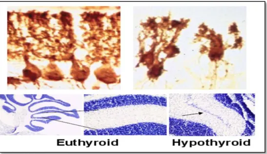
FIGURE 2 - Postnatal morphological changes in the rodent cerebellum after neonatal hypothyroidism. Bernal J. Thyroid hormones in brain development and function. Nat Clin
Pract Endocrinol Metab, 2007; 3:249-259 (modified) [10].
Upper panel: Purkinje cells in a normal (left) and hypothyroid rat (right). Lower panel: persistence of the external granular layer (arrow) in a hypothyroid mouse cerebellum.
FIGURE 3 - Myelination in the anterior commissure of euthyroid and hypothyroid rats.
Bernal J. Thyroid hormones in brain development and function. Nat Clin Pract Endocrinol
Metab, 2007; 3:249-259 (modified) [10].
Upper panels: transversal section of the anterior commissure stained for myelin. The lower panels: electromicroscopy analysis. The number of myelinated axons is reduced in the hypothyroid rats
16
1.3.2.
EFFECTS ON OXYGEN CONSUMPTION, HEAT
PRODUCTION AND FREE RADICAL FORMATION
Triiodothyronine increases oxygen consumption and heat production in part by stimulation of Na+-K+ ATPase in all tissues except the brain, spleen and testis. This contributes to the increased basal metabolic rate (total somatic oxygen consumption at rest) and the increase sensitivity to heat in hyperthyroidism and decrease sensitivity to heat in hypothyroidism. Thyroid hormones stimulate mitochondria, augmenting the cell’s oxidative capacity. They also induce changes in the mitochondrial inner membrane protein and lipid composition that increase oxidative metabolism by both genomic and nongenomic effects. The reduced efficiency of oxidative metabolism caused by thyroid hormone is also reflected in the increased futile cycling of intermediary carbohydrate metabolites [1].
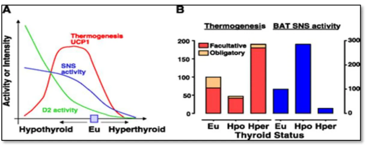
FIGURE 4 - Relationship between brown adipose tissue facultative thermogenesis and obligatory thermogenesis, sympathetic BAT stimulation and type II iodothyronine 5′-deiodinase activity as a function of thyroid status in rodents acclimated at room temperature. Silva J. Thermogenic mechanisms and their hormonal regulation. Physiol
Reviews, 2006; 86: 435-464 (modified) [11].
A: BAT thermogenesis and UCP1 (red line), SNS activity (blue line), and D2 activity (green line)
expressed as continued function of hypothyroid and hyperthyroid state departing from the euthyroid condition B: obligatory thermogenesis and BAT facultative thermogenesis (left axis) and BAT SNS activity (right axis) in the euthyroid status compared with hypothyroidism and hyperthyroidism.
Abbreviations: BAT-brown adipose tissue, D2-5′-deiodinase, Eu-euthyroid, Hper-hyper, Hpo-hypo
17
1.3.3.
EFFECTS ON THE FAT TISSUE AND
CARBOHYDRATE METABOLISM
Hyperthyroidism increases hepatic gluconeogenesis and glycogenolysis, as well as intestinal glucose absorption, and they may also be thyroid hormone-mediated decreases in insulin sensitivity. Thus, hyperthyroidism can worsen glycemic control in patients with diabetes mellitus. On the other hand, reduced glucose absorption from gastrointestinal tract accompanied by prolonged peripheral glucose accumulation, gluconeogenesis, diminished hepatic glucose output and reduced disposal of glucose are hallmarks of hypothyroidism. In overt or subclinical hypothyroidism, insulin resistance leads to glucose-stimulated insulin secretion. In subclinical hypothyroidism, diminished rate of insulin stimulated glucose transport rate caused by perturbed expression of glucose transporter type 2 gene (GLUT 2) translocation may lead to insulin resistance. Cholesterol synthesis and degradation are both increased by thyroid hormones. Lipolysis is increased, releasing fatty acids and glycerol into circulating plasma. Thyroid hormones also play important roles in the development and function of brown and white adipose tissue. Thyroid hormones can induce white adipose tissue differentiation from preadipocytes in young rats [12]. Triiodothyronine not only induces intracellular lipid accumulation and various adipocyte-specific markers such as malic enzyme and glycerophosphate dehydrogenase, but also stimulates adipocyte cell proliferation and fat cell cluster formation [12, 13]. Several human studies have shown that chronic hypothyroidism or hyperthyroidism, as well as acute T3 treatment did not affect serum leptin levels [14, 15]. However, one study showed that hypothyroid patients had increased leptin levels, but the increase correlated with adiposity [16]. Another study showed that hyperthyroid patients treated with thiamazole increased their leptin level [17].
18
FIGURE 5 - Effects of thyroid hormones on carbohydrate metabolism. Sinha R, Singh
B, Yen P. Thyroid hormone regulation of hepatic lipid and carbohydrate metabolism.
19
1.3.4.
EFFECTS ON THE LIVER
Thyroid hormones have multiple effects on liver function including stimulation of enzymes regulating lipogenesis and lipolysis as well as oxidative processes [19, 20]. It has been well known for many years that hypothyroidism is associated with hypercholesterolemia with elevated serum intermediate and low-density lipoprotein (LDL) cholesterol concentrations [21]. The major mechanism for these effects maybe lower cholesterol clearance resulting from decreased LDL receptors. Furthermore, the genotype of the LDL receptor gene may influence the elevation of serum LDL cholesterol concentrations in hypothyroid patients and their response to thyroxine treatment [22]. An additional mechanism may be decreased hepatic lipase activity in hypothyroidism which decreases conversion of intermediate-density lipoproteins to LDL and high-density lipoprotein (HDL) metabolism [23, 24]. Thyroid hormones also have been shown to regulate the expression of several important proteins and enzymes involved in cholesterol metabolism and synthesis such as the LDL receptor, cholesterol ester hydrolase, and cholesterol acyltransferase [25, 26].
20
1.3.5.
EFFECTS ON THE CARDIOVASCULAR SYSTEM
Thyroid hormones lower systemic vascular resistance, increase blood volume, and have inotropic and chronotropic effects on cardiac function. The combination of these effects on both, the circulation and the heart itself, results in increased cardiac output. Hyperthyroid patients have a high output circulation state, whereas hypothyroid patients have low cardiac output, decreased stroke volume, decreased vascular volume, and increased systemic vascular resistance [27]. These changes in cardiac function ultimately depend on the regulation of target genes within the heart and indirect effects due to hemodynamic changes.
Thyroid hormones increase expression of the more rapidly contractile isoforms of myosin heavy chain, the α isoforms, which contributes to enhanced systolic function. In myocardium, T3 also alters expression of different isoforms of the Na+/K+- ATPase genes, increases expression of β-adrenergic receptors, and decreases the concentration of the inhibitory G protein Gi α. The rate of diastolic relaxation of the heart is related to intracellular Ca2+ concentration and sarcoplasmic reticulum Ca2+-ATPase activity. The ATPase is an ion pump that removes calcium from the cytosol and stores in the sarcoplasmic reticulum during diastole. This decrease in the intracellular Ca2+ generated during systole then leads to muscle relaxation. Hypothyroid rats had decreased level of Ca2+-ATPase mRNA that could be markedly stimulated by T3 administration [28]. T3 also has been shown to regulate expression of several ion channels in heart such as the voltage-gated potassium channel, Na+/K+- ATPase, and the hyperpolarization activated cyclic nucleotide-gated channel [29, 30]. Additionally, thyroid hormones can regulate β-adrenergic receptor number in the heart and may thereby enhance sensitivity to catecholamines [31]. Finally, a novel and potentially exciting therapeutic use of T3 as an inotropic agent has been in cardiac surgery [32]. Novitsky showed improved cardiac function and hemodynamics when brain-dead organ donors were pretreated with T3 postoperatively [32]. A small group of patients that underwent cardiac bypass surgery and were treated with postoperatively with T3 also showed some benefit [33]. However, a large randomized study showed that although T3
21 increased cardiac output and decreased systemic vascular resistance in patients who underwent coronary-artery bypass surgery, there was no improvement in outcome or changes in postoperative therapy [34].
T3 also increases the rates of both depolarization and repolarization of the sinoatrial (SA) node, increasing heart rate. Consequently, thyroid hormones have positive inotropic and chronotropic effects on the heart, which along with the heightened adrenergic sensitivity, accounts for the increased heart rate and contractility in hyperthyroidism and the reverse in hypothyroidism.
Thyroid hormones also lower peripheral vascular resistance, and increase intravascular volume, which contributes further to the increase in cardiac output associated with thyroid hormone action [3].
FIGURE 6. Effects of thyroid hormone on cardiovascular hemodynamics. Klein I,
Danzi S. Cardiovascular Involvement in General Medical Conditions, Circulation, 2007; 15:1725-1735 (modified) [35].
T3 affects tissue thermogenesis, systemic vascular resistance, blood volume, cardiac contractility, heart rate, and cardiac output as indicated by the arrows. Abbreviations: Hyper- hyperthyroidism; hypo- hypothyroidism
22
1.3.6.
EFFECTS ON THE SKELETAL SYSTEM
Thyroid hormones are critical for normal bone growth and development. In children, hypothyroidism can cause short structure and delayed closure of the epiphyses. Thyroid hormones stimulate bone turnover, increasing bone resorption and, to a lesser degree, bone formation. Thyroid hormones may act on bone via stimulation of growth hormone (GH) and insulin-like growth factor I (IGF-I) or by direct effects on target genes. Biochemical studies have shown that thyroid hormones can affect the expression of various bone markers in serum, reflecting changes in both bone formation and resorption [36, 37, 38]. There is enhanced calcification and bone formation coupled to increased bone resorption in hyperthyroid patients [37, 39]. Additionally, the time interval between formation and subsequent mineralization of osteoid is shortened. The net effect on these bone cells is bone resorption and loss of trabecular bone thickness in hyperthyroidism. There also is marked increase in porosity and decreased cortical thickness in cortical bone in hyperthyroid patients [40, 41]. These effects can lead to osteoporosis and increased fractures. Little is known about direct thyroid hormones effects on osteoclasts. Recently, thyroid receptor protein was detected in a human osteoclastoma and in human bone samples by immunostaining, suggesting that TH might have direct effects on osteoclasts. However, two groups have used a bone slice resorption assay to show that functionally isolated osteoclasts were unable to respond directly to T3 by increasing bone resorption, and could only do so if other bone cells were present [42, 43]. These results would suggest that thyroid hormones may not have a direct effect on bone resorption but may mediate its effects via paracrine factors secreted by osteoblast cells.
23
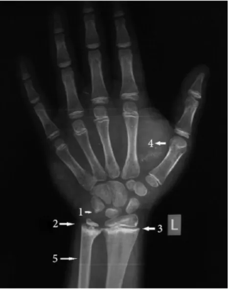
Figure A. X-ray skull: enlarged sella, Figure B. X-ray wrist showing bone age hypoplastic maxillary and frontal sinuses of 10 years (chronological age 24)
1. Epiphysis of pisiform just appearing
2. Irregular ossification of growth plate
3. Sclerotic band at radial metaphysis
4. Soft tissue thickening
5. Pencil thin cortex
Figure C: X-ray pelvis 1. Unfused Figure D: X-ray spine 1. Bullet shaped vertebral head epiphysis 2. Unfused L1 vertebra. 2. Osteoporosis 3. Increased apophysis 3. Pencil thin cortex. inter vertebral spaces
4. Persistent tri radiate cartilage
FIGURE 7 - Radiological manifestations of juvenile hypothyroidism. Patidar P, Philip R,
24
1.3.7.
EFFECTS ON PULMONARY AND
GASTROINTESTINAL SYSTEM
Thyroid hormones maintain ventilator responses to hypoxia and hypercapnia in the brain stem respiratory center. Consequently, in patients with severe hypothyroidism, hypoventilation can occur. Thyroid hormone also regulates respiratory muscle functions, and they can be weakened in hypothyroidism, leading to a sense of breathlessness.
Thyroid hormones promote gut motility, which can result in increased motility and hyperdefecation (increased frequency of formed bowel movements) in hyperthyroidism. Slowed bowel transit and constipation occur in hypothyroidism.
25
1.3.8.
EFFECTS ON THE ENDOCRINE SYSTEM
In hypothyroid children, impaired growth hormone release slows longitudinal growth. Hypothyroidism can cause delayed puberty by impairing gonadotropin-releasing hormone (GnRH) and gonadotropin secretion. Conversely, primary hypothyroidism can also cause precocious puberty. Perhaps as an effect of very high TSH levels on gonadotropin receptors. In adults, hypothyroidism causes hyperprolactinemia in a minority of affected women. Menorrhagia and anovulation are common in hypothyroid women, the latter resulting in infertility. The responsiveness of the hypothalamic-pituitary-adrenal axis to stress is blunted in hypothyroid patients. A slowing of the cortisol metabolic clearance rate compensates for this in the hypothyroid state. Conversely, restoration of euthyroidism can rarely provoke adrenal insufficiency as cortisol metabolism is accelerated in patients with diminished cortisol reserve due to concomitant disease affecting the adrenal axis. In hyperthyroidism, accelerated aromatization of androgens to estrogens and increased sex hormone-binding globulin levels contribute to the gynecomastia and elevated total testosterone levels seen in affected men. Hyperthyroidism can also impair normal GnRH and gonadotropin regulation of ovulation and menses, causing infertility and amenorrhea, respectively [3].
26
1.4. HYPOTHYROIDISM AND THE CARDIOVASCULAR
SYSTEM
Thyroid hormone receptors are rich in the myocardium, so the heart is very sensitive to the thyroid hormones [45]. The cardiovascular signs and symptoms of thyroid disease are some of the most profound and clinically relevant findings that accompany both hypothyroidism and hyperthyroidism. Based on the understanding of the cellular mechanisms of thyroid hormone action on the heart and cardiovascular system, it is possible to explain the changes in cardiac output, cardiac contractility, blood pressure, vascular resistance, and rhythm disturbances that result from thyroid dysfunction. There are many regulatory effects of thyroid hormones, such as cardiac protein transcription and gene expression; these are effective, especially in cardiovascular endothelial and smooth muscle cells [46]. Therefore, thyroid hormone deficiency could result in significant changes in the cardiovascular system.
Hypothyroidism has various cardiovascular manifestations including:
Impaired diastolic function
Impaired myocardial contractility
Decreased cardiac output and decreased heart rate
Increased systemic vascular resistance [47]
Endothelial dysfunction [48, 49]
Pericardial effusion
Heart failure [50, 51]
Cardiac arrhythmias
27
1.4.1.
PATOPHYSIOLOGY
1.4.1.1.
VASCULAR RESISTANCE
Thyroid hormones relax vascular smooth muscle cells, thereby reducing peripheral vascular resistance [45]. Conversely, hypothyroidism causes a decrease in the release of endothelial-derived relaxation factor (EDRF), which in turn promotes contraction of these cells thereby increasing peripheral vascular resistance. This change results in reduction in cardiac output (in part because the heart cannot increase contractility to compensate) and tissue perfusion. Tissue oxygen utilization is also decreased; thus, arteriovenous (A-V) oxygen extraction is similar to the normal subjects.
1.4.1.2.
CARDIAC CONTRACTILITY
All measures of left ventricular performance are impaired in both short and long-term hypothyroidism, leading to a reduction in cardiac output. There is also a decrease in the rate of ventricular diastolic relaxation; thus, compliance and diastolic filling are impaired [52, 53, 54]. The reduced ventricular performance is probably multifactorial. Possible mechanisms include increases in afterload and changes in expression of the genes for myocardial calcium regulatory proteins [54, 55]. Several enzymes involved in regulating calcium fluxes in the heart are controlled by thyroid hormone, including the calcium-dependent adenosine triphosphatase and phospholamban [45]. Hypothyroid-dependent decreases in the expression and activity of these enzymes could potentially impaired systolic performance and diastolic relaxation. β-adrenergic receptor expression is also decreased, resulting in a blunted response to catecholamine mediated increase in inotropy.
28
FIGURE 8 - Effect of L-thyroxine replacement in patients with hypothyroidism on serum T4, TSH, and cardiac contractility as measured by the PEP as a function of LVET.
Contractility increased (as demonstrated by a fall in PEP/LVET) in a dose-dependent fashion. Crowley W, Ridgway E, Bough E, et al. Noninvasive evaluation of cardiac function in hypothyroidism. Response to gradual thyroxine replacement. N Engl J Med 1977; 296:301 (modified) [56].
Abbreviations: L-thyroxine: levothyroxine; LVET: left ventricular ejection time; PEP:
29
1.4.2.
CLINICAL MANIFESTATION
It has been long recognized that some of the most characteristic and common signs and symptoms of thyroid disease are those that result from the effects of thyroid hormone on the heart and cardiovascular system. Those include:
Exertional dyspnea and exercise intolerance, although these symptoms are probably due to skeletal muscle dysfunction
Bradycardia
Hypertension resulting from the increase in vascular resistance and the fall in endothelial-derived relaxing factor (EDRF)
Cardiac dysfunction with poor contractility and/or dilatation
Edema, often nonpitting
Cardiac arrhythmias
Pericardial effusion, which occur in approximately 25 percent of patients and may be quite large
30
1.4.2.1.
BLOOD PRESSURE
Thyroid hormones play a role in blood pressure homeostasis. In patients who had undergone total thyroidectomy for thyroid cancer, withdrawal of thyroxine for 6 weeks results in an increase in serum norepinephrine and aldosterone concentration, and an increase in blood pressure with a greater rise in diastolic pressure (126/85 compared with 120/76 mmHg at baseline) [57]. Diastolic blood pressure varies directly with serum thyroid-stimulating hormone levels and may vary the entire spectrum of thyroid disease.
Approximately 20 to 40 percent of hypothyroid patients have hypertension, even though cardiac output is reduced. The hypertension is primary diastolic and the pulse pressure is diminished. In hypertensive hypothyroid patients, the serum levels of renin are low and there is an increased prevalence of salt sensitivity confirming the importance of the increase in systemic vascular resistance [55, 58]. Among large group of patients with hypertension, however, hypothyroidism is a contributory factor in only a small percentage.
31
1.4.2.2.
CARDIAC DYSFUNCTION
In hypothyroidism, the upstroke of the pulse can be slow and the left ventricular apical impulse weak. In some patients on physical exam the heart sounds can be distant and the heart may be enlarged. These findings, plus dyspnea, exercise intolerance, and edema, may make it seem as if the patient has congestive heart failure. However, heart failure due solely to hypothyroidism is rare [45].
Electrocardiograms may show low voltage and nonspecific ST segment and Q wave changes. Occasionally, large pericardial effusion can occur characterized by a high protein and cholesterol content. They are rarely hemodynamically important and should be managed with thyroid hormone replacement, not by needle or surgical drainage. The latter can lead to hemodynamic worsening.
FIGURE 9 - Large pericardial effusion in severe hypothyroid woman with no underlying heart disease.
32
1.4.2.3.
CORONARY ARTERY DISEASE
Hypothyroidism may result in accelerated atherosclerosis and coronary artery disease, presumably because of associated hypercholesterolemia, hypertension, and elevated homocysteine levels [53, 55]. Potential mechanism in addition to lipid abnormalities and diastolic hypertension include elevated concentrations of C-reactive protein (CRP) and endothelial dysfunction [59, 60]. Direct evidence of such an effect of overt hypothyroidism is lacking. However, in a study of 1149 postmenopausal women in the Netherlands, those with subclinical hypothyroidism were more likely to have a history of myocardial infarction and had a higher frequency of calcification of the aorta [68]. Thyroxine therapy may reverse the cardiovascular changes associated with hypothyroidism. In a large study of patients with hypothyroidism who were evaluated for clinical evidence of ischemic heart disease after the initiation of thyroid hormone therapy, new or worsening angina or acute myocardial infarction were rare, and more patients had improvement in angina symptoms [50]. These findings reinforce the important and potentially beneficial effects of thyroid hormone in improving the efficiency of myocardial oxygen consumption and simultaneously lowering systemic vascular resistance [50]. Whether patients with subclinical hypothyroidism should be treated is still the subject of disagreement, but from a cardiac perspective, treatment offers benefit with minimal risk.
33
1.4.2.4.
EDEMA
Periorbital edema and nonpitting edema of the hands and feet are characteristic features of hypothyroidism, albeit rare today. Nonpitting edema is due to interstitial accumulation of glycosaminoglycans (hyaluronic acid and chondroitin sulfate), with associated extravascular water retention while plasma volume is decreased [45, 52]. Some patients have pitting edema of the feet and legs, probably secondary to an increase in albumin content of the interstitial fluid [61]. Ascites, pleural and scrotal effusions may also be present.
A) B)
FIGURE 10 - Edema in patients with hypothyroidism A) Periorbital edema; B)
34
1.4.3.
LABORATORY TESTS
1.4.3.1.
LIPIDS
Dyslipidemia is common in hypothyroidism. The usual findings are high serum total and low-density lipoprotein (LDL) cholesterol concentration. Some patients have high serum very-low-density lipoprotein (VLDL) cholesterol concentration, and a few have hypertriglyceridemia.
The spectrum and frequency of lipid abnormalities that can occur was illustrated in a report from the Mayo Clinic which evaluated 295 patients with hypothyroidism. Hypercholesterolemia was present in 56 percent of the patients, hypercholesterolemia and hypertriglyceridemia in 34 percent, and hypertriglyceridemia in 1.5 percent; only 8.5 percent had a normal lipid profile [62]. The changes in serum LDL cholesterol have been related to reduced expression of LDL receptors and decreased hepatic and biliary LDL cholesterol clearance. Another study evaluated the prevalence of hypothyroidism in patients referred for hyperlipidemia. Among 1509 consecutive patients, hypothyroidism was present in 4.2 percent, approximately twice the incidence than in the general population [63]. In view of the increased prevalence of hypercholesterolemia, especially in patients with overt hypothyroidism (TSH>20 μIU/mL), the question of optimum therapy often arises. If hypothyroidism is present, the patient should be treated for 3 to 4 months with thyroid hormone to normalized serum TSH. If the serum lipid concentrations are not then normal, specific lipid-lowering therapy may be indicated. It should be noted that hypothyroidism may predispose to the development of statin-associated myopathy and that use of statins may unmask hypothyroid myopathy.
35
1.4.3.2.
HOMOCYSTEINE
Some patients with hypothyroidism have high serum homocysteine concentrations, which fall toward if not to normal with LT4 therapy.
1.4.3.3.
CREATINE KINASE
Many hypothyroidism patients have high serum creatine kinase (CK) concentrations. The isoenzyme distribution is almost completely CK-MM, with less than 4 percent constituting CK-MB, indicating skeletal muscle, not myocardial, origin [64]. However, as many as 14 percent of patients with hypothyroidism have a raised serum concentration of CK-MB, which can be confusing in the evaluation of chest pain. The problem is obviated by measurement of serum troponin I, which is normal in hypothyroidism [65].
36
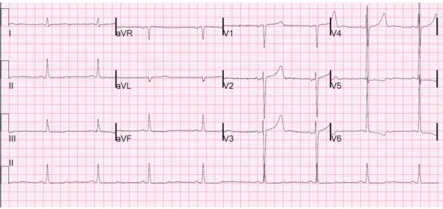
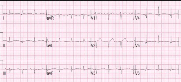
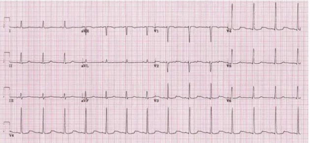
1.4.4.
RHYTHM DISTURBANCES
In addition to slow pulse rate, hypothyroid patients may have ventricular premature beats and rarely ventricular tachycardia with a long QT interval (torsades de pointes). This can be especially problematic in patients with underlying ischemic heart disease or known ventricular arrhythmias.
EKG changes in hypothyroidism involve:
1) Bradycardia
2) Right bundle branch block (RBBB) 3) Flat or inverted T wave
4) QRS prolongation 5) QT prolongation 6) Torsades de pointes
37
FIGURE 11 - Sinus bradycardia.
38
FIGURE 13 - Flat T waves.
39
FIGURE 15 - QT prolongation.
40
2. THESIS AND AIMS OF THE STUDY
Thyroid hormones affect various functions of the heart including contractility and chronotropic functions of the heart. The association of hyperthyroidism with atrial tachyarrhythmias is well established but little is known about the association of hypothyroidism and cardiac arrhythmias. Though EKG changes in hypothyroidism, including prolongation of QT interval, QRS interval and bradycardias, are well known, still little is known about the clinical significance of these EKG changes including the incidence and prevalence of cardiac arrhythmias and the need for periodic surveillance in these patients for arrhythmias.
Increased QT has been found to be associated with an increased incidence of malignant ventricular arrhythmias and sudden death, however a few clinical observations showed that sudden death is uncommon in hypothyroidism despite the marked lengthening of the QT interval [75, 76, 77]. There were also studies which showed that QT dispersions improved after the L-thyroxine treatment in patients with primary hypothyroidism [78]. This suggests that the L-thyroxine replacement therapy may reduce malignant ventricular arrhythmia and sudden cardiac death in patients with hypothyroidism.
Patients with thyroid dysfunction are as such at increased risk for cardiovascular morbidity and mortality through an increased prevalence of ischemic heart disease and congestive heart failure (CHF), both of which predispose to the development of cardiac arrhythmias. Independent of these conditions it is unsure if hypothyroid status per se predisposes to cardiac arrhythmias.
41
2.1. THESIS OF THE STUDY
Despite these facts about cardiac arrhythmias in hypothyroid patients, very few clinical studies have addressed the prevalence of cardiac arrhythmias in hypothyroidism. This relatively large a retrospective age-, gender- and ethnicity-matched case control study was conducted to analyze differences, if any, in the prevalence of cardiac arrhythmias between hypothyroid patients and euthyroid control group. One of the aims of this study is to throw light on the prevalence of arrhythmias in hypothyroidism and detect necessitates for future large scale prospective studies to better define the risk of such ventricular arrhythmias and the effects of thyroid supplementation on this risk. Identifying the life-threatening arrhythmias in patients with hypothyroidism can help to detect disease earlier, prolong survival, and decrease overall mortality in this group of patients. These patients may need more intensive preventive care for arrhythmias than general population.
42
2.2. AIMS OF THE STUDY
1) Detect the prevalence of cardiac arrhythmias in patients with hypothyroidism.
2) Analyze differences in the prevalence of cardiac arrhythmias between hypothyroid patients and patients with normal thyroid function.
3) Establish clinical significance and the need for periodic surveillance for arrhythmias in hypothyroid patients.
43
3. PATIENTS, MATERIALS, AND METHODS
3.1. STUDY DESIGN AND PATIENTS ENROLLMENT
Approval for the study was obtained from the Albert Einstein Medical Center Ethics Committee (IRB ID: 4601 EXE). Retrospective chart analysis was performed. Demographic data including age, gender, and ethnicity were collected. Age, gender, and ethnicity matched controls were selected from the euthyroid group. Each hypothyroid patient was matched against the euthyroid subject by random allocation in chronological order of hospitalization. Data regarding specific arrhythmias were obtained from the EKG, chart documentation of known past medical history and from telemetry recordings during the index hospitalization. History of cardiac arrhythmias included:
1) Tachyarrhythmias
atrial fibrillation (AFib)
atrial flutter (AF)
atrial tachycardia
atrioventricular reentrant tachycardia (AVRT)
atrioventricular node reentrant tachycardia (AVNRT)
ventricular tachycardia (VT)
nonsustained ventricular tachycardia
44 2) Bradyarrhythmias
sinus bradycardia
atrioventricular block (AVB)
junctional rhythms
idioventricular rhythm
Left ventricular systolic and diastolic function were evaluated based on transthoracic echocardiogram result, chest x-ray, physical exam and elevated B-type natriuretic peptide (BNP) level. Patients were classified into three categories:
1. Any congestive heart failure if either systolic or diastolic function was compromised. 2. Systolic CHF if ejection fraction (EF) was below 50% (Cardiologist at Albert Einstein
Medical Center, Philadelphia use criteria of EF<50% to diagnose systolic CHF). 3. Only diastolic dysfunction per Echocardiography report (based on E/A ratio).
Typical chest x-ray changes in patients with congestive heart failure included:
pulmonary venous congestion, interstitial edema, pleural effusion, and cardiomegaly. Only patients who based on Framingham diagnostic criteria met two major criteria (acute pulmonary edema, cardiomegaly, hepatojugular reflex, neck vein distention, paroxysmal nocturnal dyspnea or orthopnea, rales, third heart sound gallop) or one major and two minor criteria (ankle edema, dyspnea on exertion, hepatomegaly, nocturnal cough, pleural effusion, and tachycardia >120 beats per minute) were included as congestive heart failure patients.
45 From 5642 patients who were admitted to the Cardiology Floor at Albert Einstein Medical Center in Philadelphia, Pennsylvania, United States of America between 1st of June 2011 and 31st of May 2012, there were 214 patients who met criteria for potential subjects for the study based on the level of thyroid stimulating hormone >10 μIU/mL followed by confirmation with low level of free thyroxine. After applying exclusion criteria described below, 152 patients were selected as a study group. 152 subjects who were euthyroid based on TSH level were age-, gender- and ethnic- matched and selected as a control group. The main reasons for admissions to Cardiology Floor were: chest pain, myocardial infarct, unstable angina, syncope, congestive heart failure exacerbation, atrial fibrillation with rapid ventricular response and cardiomyopathy.
46
3.1.1.
SUBJECTS
Patients with TSH level >10 μIU/mL followed by confirmation with low level of circulating free thyroxine were included in the hypothyroid group and considered eligible for the subject group after applying specific cardiac exclusion criteria as listed in Table 2.
TABLE 2 - Exclusion cardiac criteria for hypothyroid group.
TABLE 2. EXCLUSION CRITERIA FOR HYPOTHYROID GROUP
1. Patients with a pacemaker implanted
2. Patients with an Implantable Cardioverter Defibrillator (ICD) 3. Patients with congenital long QT syndrome
4. Patients within 30 days post myocardial infarction (MI)
5. Patients with arrhythmias due to documented electrolyte abnormalities-hypokalemia, hyperkalemia, hypomagnesemia, hypocalcemia, hypercalcemia
47 All patients who were taking medications that might interfere with the accuracy of TSH measurement were also excluded from the study.
Drugs that can increase TSH include the following:
Dopamine antagonists
Chlorpromazine
Haloperidol
Iodine-containing drugs
Amiodarone (amiodarone-induced hypothyroidism)
Drugs that can decrease TSH include the following:
Metformin
Dopamine
Levodopa
Bromocriptine
Glucocorticoids (>0.5 mg/day dexamethasone, 100 mg/day hydrocortisone)
Somatostatin analogs (octreotide, lanrerotide)
48
3.1.2.
CONTROL GROUP
Subjects who were euthyroid based on physical examination and TSH level were chosen as a control group, with the TSH cut-off defined as within the lab reference range for the assay performed. Such patients were included in the euthyroid group after applying specific cardiac exclusion criteria as listed in Table 3.
TABLE 3 - Exclusion cardiac criteria for euthyroid group.
TABLE 3. EXCLUSION CRITERIA FOR EUTHYROID GROUP
1. Patients with a diagnosis of hypothyroidism and on levothyroxine supplementation
2 Patients with a pacemaker implanted
3 Patients with an Implantable Cardioverter Defibrillator (ICD) 4 Patients with congenital long QT syndrome
5 Patients within 30 days post myocardial infarction (MI)
6 Patients with arrhythmias due to documented electrolyte abnormalities-hypokalemia, hyperkalemia, hypomagnesemia, hypocalcemia, hypercalcemia
49
3.2. STATISTICAL ANALYSIS
Statistical analysis was performed using Statistical Package for the Social Sciences (SPSS) Platform, Stata/IC 13 for Windows 10 (version 11.0, SPSS Inc, Chicago, Ill, USA) and Microsoft Excel. Group differences were analyzed using Student t-test for parametric data and Mann-Whitney U test for nonparametric data. Group differences in the prevalence of individual cardiac arrhythmias were analyzed using Chi-square test. Continues variables were presented as mean +/- standard deviation and categorical variables were presented as the percentage. A value of p < 0.05 was considered statistically significant.
50
3.3. METHODS
3.3.1.
PLASMA THYROID STIMULATING HORMONE
LEVEL
Thyroid stimulating hormone stimulates the production and secretion of the metabolically active thyroid hormones, thyroxine and triiodothyronine, by interacting with a specific receptor on the thyroid cell surface. TSH is composed of two non-covalently linked subunits, designated α and β. Although the α subunit of TSH is common to luteinizing hormone (LH), follicle stimulating hormone (FSH), and human chorionic gonadotropin (hCG), the β subunits of these glycoproteins are hormone specific and confer biological as well as immunological specificity. The synthesis and secretion of TSH is stimulated by the thyrotropin releasing hormone (TRH), the hypothalamic tripeptide, in response to low levels of circulating thyroid hormones. Elevated levels of T3 and T4 suppress the production of TSH via a classic negative feedback mechanism. Recent evidence also indicates that somatostatin and dopamine exert inhibitory control over TSH release, suggesting that the hypothalamus may provide both an inhibitory and stimulatory influence on pituitary TSH production [1].
The measurement of plasma TSH is the commonly accepted and most sensitive screening test for primary thyroid disorder, because the pituitary gland responds with great changes in its secretion, even to slight changes in the levels of free thyroid hormones. The sensitivity of its measurement in the diagnostics of tissue hormone excess is estimated to be higher than 95%, and the specificity-approximately 90%, while its daily fluctuations are very small and are of no importance for the interpretation of results [83, 84, 85].
The determination of TSH serves not only as a preliminary test in the differentiation of the thyreologic state of the population, but also plays the role of a predictive factor of the
51 occurrence of malignant changes within the nodules [86]. In the past, the TSH test was the only test performed in the diagnostics of thyroid function; however, it seems that for a genuine and objective assessment of the thyreologic state, the level of TSH, together with fT4 level, should be determined, which allows the identification of patients with pathology of the hypothalamo-pituitary system with respect to thyroid axis. At the same time, an abnormal TSH level makes it necessary to determine the peripheral hormone levels, and form of free thyroid hormones fT4 and fT3, which enables evaluation of the intensity of thyroid function disorders and foresee its consequences [87, 88].
The currently applied methods for the determination of TSH are characterized by a much higher sensitivity and specificity, due to the use of two monoclonal antibodies identifying two different epitopes of the TSH, which allowed the elimination of cross-reactivity. A National Academy of Clinical Biochemistry guideline specifies that sensitivity, or lower limit of detection, for TSH assays should be less than 0.02 μIU/mL (third-generation methods). This permits patients with nonthyroidal illness to be distinguished from those with primary hyperthyroidism. This is particularly important in patients hospitalized with nonthyroidal illness where TSH concentration as low as 0.02 μIU/mL may be encountered [89].
52
TABLE 4 - Typical TSH findings.
TSH Free T4 Free or total T3
Probable interpretation
High Normal Normal Subclinical hypothyroidism High Low Low or
normal
Primary hypothyroidism Low Normal Normal Subclinical hyperthyroidism Low High or normal High or normal Primary hyperthyroidism Low Low or normal Low or normal
Non-thyroidal illness; pituitary (secondary) hypothyroidism
Normal High High Thyroid hormone resistance syndrome
FIGURE 17 - Serum TSH in various states of thyroid function. Dunlap D. Clinical
53
3.3.1.1.
TSH TEST PROCEDURE
All patients included in the study had TSH level checked with Roche Elecsys System using Cobas E 411 analyzer provided by LabCorp Laboratory. Roche Elecsys system is intended to be an immunoassay for the quantitative determination of thyrotropin in human serum and plasma and is standardized to the World Health Organization (WHO) Second International Standard for Human TSH (IRP 80/588). It is a third-generation assay for TSH and has a functional sensitivity as low as 0.014 μIU/mL. In this study the reference TSH interval was 0.450−4.500 μIU/mL.
Only patients with TSH level >10 μIU/mL followed by confirmation with low levels of circulating thyroid hormone levels (fT4) were selected as a study group (hypothyroid group). Patients with the TSH cut-off defined as within the lab reference range for the assay performed were chosen as a control group (euthyroid group).
TEST PROCEDURE
Entire procedure took about 15-20 minutes.
1) First, the written consent for the blood work was taken from every patient and patients were informed about possible complications which included:
Excessive bleeding
Fainting or feeling light-headed
54 2) The area on the arm was cleaned with an antiseptic solution
3) An elastic band was tied around the arm to increase blood flow within the vessel
4) A needle was inserted into the vein to draw blood. The blood was collected in a small tube attached to the needle (red-top tube or gel-barrier tube)
5) The tube labeled with patient’s name and medical record number (MRN) was sent to the lab for analysis
6) LabCorp Laboratory measured TSH level using Roche Elecsys system with Cobas E 411 analyzer
7) All abnormal results were confirmed with fT4 test and uploaded in the computer system
55
TABLE 5 - TSH test characteristics.
Feature Specification
Laboratory LabCorp
Test name Thyroid-stimulating Hormone (TSH) Test Number 004250
Methodology Electrochemiluminescence immunoassay (ECLIA) Specimen Serum
Volume 0.8 mL Minimum volume 0.3 mL
Container Red-top tube or gel-barrier tube
Collection If a red-top tube was used, separated serum was transported to a plastic transport tube
Storage Room temperature
Stability Room temperature 14 days
Refrigerated 14 days Frozen 14 days Freeze/thaw cycles Stable x3
56
TABLE 6 - Reference interval of TSH test (units μIU/mL).
Age Range (μIU/mL)
0 to 6 days 0.700−15.200 7 days to 3 months 0.720−11.000 > 3 months to 12 months 0.730−8.350 1 to 5 years 0.700−5.970 6 to 10 years 0.600−4.840 >10 years 0.450−4.500
57
3.3.1.2.
ELECTROCHEMILUMINESCENCE (ECLIA)
Roche Elecsys System uses Electrochemiluminescence (ECLIA) method to measure TSH level. Based on this technology and combined with well-designed, specific and sensitive immunoassays, Elecsys delivers reliable results. ECLIA is a kind of luminescence produced during electrochemical reactions in solution. It combines the analytical advantages of chemiluminescent analysis (absence of background optical signal) with ease of reaction control by applying electrode potential. Enhanced selectivity of ECLIA analysis is reached by variation of electrode potential thus controlling groups that are oxidized/reduced at the electrode and take part in ECLIA reaction.
In electrogenerated chemiluminescence, electrochemically generated intermediates undergo a highly exergonic reaction to produce an electronically excited state that emits light. ECLIA excitation is caused by energetic electron transfer (redox) reactions of electrogenerated species. Such luminescence excitation is a form of chemiluminescence where all reactants are produced electrochemically on the electrodes. ECLIA is usually observed during the application of potential (voltage) to electrodes of electrochemical cell that contains solution of luminescent species (polycyclic aromatic hydrocarbons, metal complexes) in aprotic organic solvent (ECLIA composition) [90]. The development of ECLIA immunoassays is based on the use of a ruthenium-complex and tripropylamine (TPA). The chemiluminescence reaction for the detection of the reaction complex is initiated by applying a voltage to the sample solution resulting in a precisely controlled reaction [91].
58
FIGURE 18 - Elecsys TSH. Electrochemiluminescence (ECLIA). Roche Diagnostics
(modified).
Elecsys TSH test (ECLIA method) was done by LabCorp laboratory technician in 3 steps:
1) 1st incubation (9 minutes)
50 μL of sample, a biotinylated monoclonal specific antibody and a monoclonal TSH-specific antibody labeled with a ruthenium complex were incubated. A sandwich-complex was formed with TSH carrying a biotinylated and a ruthenylated anti-body against different epitopes on human TSH.
2) 2nd incubation (9 minutes)
After addition of streptavidin-coated microparticles the complex became bound to the solid phase via interaction of biotin and streptavidin.
3) Measurement
The reaction mixture was aspirated into the measuring cell where the microparticles were magnetically captured onto the surface of the electrode. Unbound substances were then removed. Application of a voltage to the electrode then induced chemiluminescent emission
59 which was measured by a photomultiplier. The signal yield was roughly proportional to the TSH concentration in the sample.
TABLE 7 - Sets used in Elecsys TSH test.
Device name Feature Description Catalog number
Reagent Elecsys TSH 200 tests 11731459 Calibrator TSH CalSet 4x1.3 mL 04738551 Control PreciControl Universal 2x3 mL each 11731416 Analyzer PreciControl TSH 4x2 mL 11776479 Assay Diluent MultiAssay 2x16 mL 03609987
TABLE 8 - Elecsys TSH test characteristics.
Feature Specification
Testing time 18 min
Test principle One-step sandwich assay Detection/Operating
Principle
Chemiluminescence Solid Phase Micro-particle Sample type Serum and plasma
Antibody Monoclonal anti-TSH mouse antibody
Solid phase binding principle
Biotin and streptavidin Analyzer Reagents On-board Storage
60
calibrator & control
Cap/septum for increased reagen stability and evaporation control Reagent, Calibrator and Control Liquid Calibration and Control Stability Unopened
•At 2-8°C up to the stated expiration date
Opened
•28 days / 4 weeks at 2-8°C Calibration 2 point
Sample material Serum, Li-, Na-, NH4+-heparin plasma, K3-EDTA, Na-citrate, NaF, K-oxalate plasma
Sample volume 50 μL
Detection limit 0.005 μIU/mL Functional sensitivity 0.014 μIU/mL Measuring range 0.005 - 100 μIU/mL
Traceability 2nd IRP WHO Reference Standard 80/558 Total imprecision
(NCCLS)
cobas e 411 analyzer, E2010: 1.8 - 8.7% cobas e 601 / e 602 modules, E170: 3.2 - 7.2%
Expected values 0.27 - 4.2 μIU/mL (95th percentile) Analyzer Sample
Detection
Liquid Level Detection Clot Detection
Calibrator Matrix Horse serum with added recombinant TSH Analyzer Host
Interface
61
3.3.2.
ELECTROCARDIOGRAPHY (EKG)
All subjects underwent standard 12-lead EKG, acquired using the MAC 5500 electrocardiograph (GE Healthcare, Milian, Italy) at a paper speed of 25 mm/s and 10 mm/Mv. Patients were informed of the procedure to be performed, emphasizing that it is painless and harmless but they must lie still, breathe normally and refrain from talking. Proper skin preparation, with shaving if necessary, was required to reduce impedance and ensure adhesion of the electrode. This greatly helped to minimize the appearance of artifacts that can sometimes cause significant diagnostic errors. All recordings were performed in a controlled environment through spontaneous breathing, the subsequent 15 minutes of adjustment in the supine position. EKG interpretations were performed by certified cardiologist from Department of Cardiology at Albert Einstein Medical Center in Philadelphia, United States of America.
62
3.3.2.1.
EKG LEADS PLACEMENT
1. Positioning patient
The standard position of the patient was used- supine with head flat or at no more than a 45-degree angle, with arms and legs free. If the patient could not lie with their head flat or at no more than 45-degrees, patient was positioned for comfort. Altered position was documented, so the physician could consider the position when interpreting the EKG.
2. Skin preparation
By protocol each area where electrodes are to be placed must be cleaned properly. Alcohol pad was used to de-fat the skin and a dry cloth or 2 by 2 gauze pad were used to gently abrade the top layer of cells of the skin. Not only does this ensure full contact of the electrode with the skin for best conduction, but also reduced noise and improved the quality of the recorded EKG. A special consideration was made for hairy chests; shaving was done only when it was necessary.
63
TABLE 9 - Skin preparation.
Normal Oily Diaphoretic
If skin was very oily or
soiled, it was washed with soap and water; dry
Briskly rub skin with dry cloth to dry
Skin was cleaned with alcohol pad and allowed to dry
Briskly rub skin with alcohol pad- allow to dry
Clean skin with alcohol pad- allow to dry
Briskly rub skin with dry 2x2 pad
Briskly rub skin with dry 2x2 pad
Briskly rub skin with dry 2x2 pad
64
3. Lead electrode placement
Total of 10 electrodes were used. There were four electrodes for limb leads named RA (right arm), LA (left arm), RL (right leg) and LL (left leg). There were six electrodes for chest leads named V1 through V6. EKG technician checked if electrodes adhered to the skin and laid flat against the skin. Electrodes could point in any direction as long as they maintained full contact with the skin when connected and there was no stress or pulling on the lead wires. Before performing the EKG, double check was made that the leads clips/wires were securely attached and going to the correct electrode.
65
FIGURE 19 - Placement of the limb electrode. Electrocardiogram leads, Electrocar
Diogram, June 2012 [135].
FIGURE 20 - Placement of the precordial electrodes. Clores L, ECG leads placement,