Uniwersytet Medyczny
Im. Karola Marcinkowskiego
w Poznaniu
mgr Klaus Lücke
Isolation of Fetus Cells from
Maternal Blood
Pre-clinical Study
PRACA NAUKOWA NA STOPIEŃ
DOKTORA NAUK BIOLOGICZNYCH
PROMOTOR
Prof. dr hab. n. med. Grzegorz H. Bręborowicz
Katedra i Klinika Perinatologii I Ginekologii
CONTENTS
1 Abbreviations ... 5 2 Introduction ... 8 2.1 Prenatal diagnostics ... 8 2.2 Fetal cells ...10 2.3 Isolation techniques ...132.4 The technology of the nanodetector ...15
2.5 Nanotechnology ...16
3 Aims of this work ...19
4 Material ...20
4.1 HLA-G antigen ...20
4.2 Antibodies ...20
4.2.1 Human antibodies – phage display ...21
4.2.2 Purification of the antibody ...21
4.2.3 Structure of the used antibodies ...21
4.3 Cell Lines ...22
4.4 Animals ...23
4.4.1 Rats ...23
4.4.2 Overview: Studied groups ...23
4.5 Animal study outline ...25
5 Methods ...28
5.1 Surface of the wire ...28
5.1.1 Nanostructure (horizontal approach) ...29
5.1.2 Nanostructure (vertical approach) ...33
5.2 Binding of the antibody to the surface of the wire ...34
5.2.1 Activation of the hydrogel and coupling of antibodies ...35
5.5 Surface plasmon resonance (SPR) ...38
5.6 Flow system ...38
5.7 Visualization of cells ...39
5.8 Safety testing including animal trials (EN ISO 10993) ...40
5.8.1 Description of the device including any materials that will be in contact with tissues or body fluids ...40
5.8.2 Relevant inspections ...41
5.8.3 Cytotoxicity test ...41
5.8.4 Acute systemic toxicity ...43
5.8.5 Autopsy and organ weight ...44
5.8.6 Hemocompatibility ...45
5.9 Intended medical application of the nanodetector including any necessary storage and handling requirements ...54
5.9.1 Sterilization and packaging ...54
5.9.2 Application and usage ...55
5.10 Statistical analysis ...57
6 Results ...58
6.1 Horizontal approach with nanoisland ...58
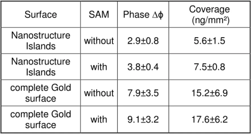
6.1.1 Amount of antibodies bound to nanostructured and complete gold surfaces ...58
6.1.2 Calculation of the number of antibodies per nanospot ...59
6.2 Vertical approach with different hydrogels adsorbed onto gold surface ...62
6.2.1 Hydrogels based on agarose and polyacrylic acid (PAA) ...62
6.2.2 Hydrogels based on polyethylene imine (PEI) and polyacrylic acid (PAA) ...63
6.2.3 Concentration of carboxyl groups ...64
6.3 Preclinical in vitro testing ...70
6.3.1 Flow cytometry with the antibody ...70
6.3.2 Immunohistochemistry (IHC) ...71
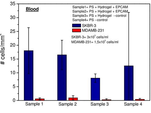
6.3.3 Cell binding experiments in spiked blood on chips ...72
6.3.4 Cell binding experiments in peripheral blood of pregnant women .73 6.4 Cytotoxicity tests ...74
6.4.2 Investigation of the cytotoxicity of antibody solutions ...76
6.4.3 Study of the cytotoxicity of the nanodetector with an elution test .77 6.5 Animal tests ...78
6.6 Hemocompatibility test ...90
6.6.1 Thrombosis ...90
6.6.2 Coagulation ...94
6.6.3 Specific coagulation factors ...95
6.6.4 Hemolysis ...96
6.7 Abrasion tests on the nanodetector ...96
7 Discussion ...97
7.1 Functionality of the nanodetector ...97
7.2 Amount of antibody on the nanodetector ...98
7.3 Device risk analysis and risk assessment (EN ISO 14971) ...99
7.3.1 Introduction ...99
7.3.2 Wire, gold and hydrogel ...99
7.3.3 Application time ... 100
7.3.4 Safety ... 100
7.4 Evaluation of the potential effects which might originate from nanodetector-bound antibodies ... 100
7.4.1 The use of fully human antibodies is preferred to murine, humanized or chimeric antibodies ... 102
7.5 Therapeutic monoclonal antibodies ... 102
7.6 Risk analysis and assessment including a description of the risk graph .. 104
7.7 Claims and intended performance of the device that are to be verified in a clinical study ... 105
7.8 Possible other applications ... 105
7.9 Can enough cells be caught ? ... 106
8 Conclusions ... 107
9 Summary ... 108
10 References ... 109
1
Abbreviations
AFM Atomic force microscopy ALAT Alanine aminotransferase ALARP as low as reasonable possible ALP Alkaline phosphatase
ANCA Anti-neutrophil cytoplasmic antibody ASAT Aspartate aminotransferase
AT anucleated trophoblasts Bil Bilirubin
bp Base pairs
BUN Blood urine nitrogen BW Body weight
CCP Cyclic citrullinated peptide CD Cluster of differentiation CIP Clinical investigation plan CK Cytokeratin
CMV Cytomegalovirus CRP C-reactive protein CT Cytotrophoblasts
CVS Chorionic villus sampling DC Density gradient centrifugation Df Dilution factor
DLE Discoid lupus erythematosus DNA Deoxyribonucleic acid
EDC (N-(3-Dimethylamino-propyl)-N`-ethylcarbodiimide hydrochloride) EGF(-R) Epidermal growth factor(-receptor)
ELISA Enzyme-linked immunosorbent assay EMEA European Medicines Agency
Ery Erythrocytes
FACS Fluorescence activated cell sorting FDA Food and Drug Administration FISH Fluorescence in situ- hybridization FPA Fibrinopeptide A
Glu Glucose
HGB Hemoglobin
HLA Human leukocyte antigen HRP Horse radish peroxidase HTC Hematocrit
TAT Thrombin-antithrombin III complex ICC Immunocytochemistry
IDT Interdigital transducers
INR International Normalized Ratio ket Ketones
kDa kilo Dalton leu Leukocytes Ig Immunoglobulin IL Interleukin
MACS Magnetic activated cell sorting MCH Mean corpuscular hemoglobin
MCHC Mean corpuscular hemoglobin concentration MCV Mean corpuscular volume
MPV Mean platelet volume
MRI Magnetic resonance imaging MSAFP Maternal serum alpha-fetoprotein N/A Not applicable
NHL Non-Hodgkin lymphoma NHS N-Hydroxysuccinimide nit Nitrite
NK Natural killer cells NSL Nanosphere lithography NT Nuchal translucency NTC No template control NTD Neural tube defect
PALS Periarteriolar Lymphoid Sheaths PBS Phosphate buffered saline PCR Polymerase chain reaction PCT Platelet Hematocrit
PGD Preimplantation genetic diagnostics pI Isoelectric point
PPP Platelet-poor plasma PRP Platelet-rich plasma PRT Plasma recalcification time RA Rheumatoid arthritis RBC Red blood cell
RDW Red blood cell distribution width RISH RNA in situ-hybridization
RU Resonance units
SAM Self assembled monolayer
SCID Severe combined immunodeficiency SEM Scanning electron microscopy SG Specific gravity
SOP Standard operating procedure SPR Surface plasmon resonance ST Syncytiotrophoblast
SPR Surface Plasmon resonance TMB Tetramethylbenzidin
TNF Tumor necrosis factor Ubg Urobilinogen
UV Ultra-violet
VNTR-PCR Variable number of tandem repeats-PCR WBC White blood cell
2
Introduction
2.1 Prenatal diagnostics
At present there are a number of different methods which are applied to perform prenatal diagnostics according to the time of gestation and other factors. The most common methods used for prenatal diagnostics are amniocentesis, cordocentesis and chorionic villus sampling (CVS). The obtained fetal cells can be analysed for chromosomal aberrations.
Transabdominal amniocentesis is the most commonly used procedure to obtain fetal cells for cytogenetic analysis. During amniocentesis a sample of amniotic fluid is taken. The procedure is performed between weeks 15 to 18 of gestational age [1]. An accurate sample for analysis is provided in more than 99 % of the cases. Results are usually available within one to two weeks. Risks of this method include fetal loss, chorioamnionitis, fetal injury and maternal Rh sensitization, each of which is very uncommon. The highest risk is fetal loss at less than 0.5 %. The rate of miscarriages after 15 weeks of gestation varies and is most commonly quoted to be 1 % [2]. This risk increases in twin pregnancies up to 2.73 %. Early amniocentesis, before week 15 of gestation increases the risk of fetal loss to between 3 and 5 %. This also increases the risk of congenital foot deformities (mainly talipes equinovarus). A possible explanation for this could be the leakage of amniotic fluid after the procedure, with a subsequent reduction of the space and pressure within the amniotic sac. Early amniocentesis is not commonly used in prenatal genetic diagnosis, because of mentioned problems and first-trimester Chorionic villus sampling (CVS) has replaced it in everyday practice.
CVS has some disadvantages compared with amniocentesis. First, the procedure related pregnancy loss rate is somewhat higher than that of amniocentesis. Second, the incidence of limb deficiencies is greater in cases of CVS performed before the completed ninth week of pregnancy. Third, there is a 2 % false-mosaicism rate during the laboratory evaluation of the chorionic tissue [2]. The risks of CVS greatly depend on the time in pregnancy and on the procedure (transabdominal or transcervical) how it is carried out.
Evans and Wapner [3] conclude in comparision to [2], that the procedure induced miscarriage rate following mid trimester (16-20 weeks) amniocentesis in experienced hands appears to be 1/300. In addition to this, “early amniocentesis” (≤ 13 completed weeks) even with very experienced operators has a risk of fetal loss approaching 1/50 with a risk of talipes
equinovarus between 1 and 2 %. For operators experienced in both first trimester CVS and mid-trimester amniocentesis the two procedures have comparable safety outcomes. For parents desiring a prenatal diagnosis before week 13 of gestation, CVS is currently the safest procedure.
Further methods for prenatal diagnoses include the sampling of fetal blood and preimplantation genetic diagnosis (PGD) [1]. Fetal blood sampling is applied only under special circumstances and carries a risk of spontaneous abortion of between 1 and 2 %, i.e. higher than amniocentesis or CVS. PGD is performed in cases where the family carries a known genetic disorder. In this procedure a single cell is taken from the early embryo and analysed using polymerase chain reaction (PCR).
Non-invasive prenatal diagnostic techniques include those using visualization techniques and maternal serum screening. Of the visualization techniques ultrasonography is most commonly used. With the sonography one can screen for fetal trisomy 21 by measuring the thickness of a fluid-filled space behind the fetal neck, otherwise known as the nuchal
population with the highest estimated risk included 77% of trisomy-21 cases [4]. In a different study, ultrasound detected an absence of the nasal bone in approximately 65 % of fetuses with trisomy 21 and in 1% of fetuses with a normal karyotype [5]. Combining absence of the nasal bone with first-trimester NT and screening of maternal serum free-ß-human chorionic gonadotropin (hCG) and pregnancy-associated plasma protein-A (PAPP-A) resulted in the detection of 90 % of fetuses with Down syndrome with a false-positive rate of 5 % [5]. However, while these approaches are useful in screening for Down syndrome, they find limited application in diagnosing fetal aneuploidy.
Cordocentesis is a method to obtain a sample of fetal blood to assess fetal disorders, fetal infections, or isoimmunization or may be used for rapid fetal karyotyping. It also has been used to supply fetal treatment such as transfusions. Fetal loss or spontaneous abortion is reported in approximately 1% to 2% of cases, making the risk associated with this procedure higher than that with amniocentesis or CVS.[6]
Other less common techniques are MRI (magnetic resonance imaging) and fetal echocardiography. All these techniques are not strictly considered to be diagnostic. They only give a strong suggestion of possible fetal developmental problems that are indicative for further tests such as amniocentesis, cordocentesis or CVS.
Maternal serum screening gives another indicator for fetal diagnosis called maternal serum α-fetoprotein (MSAFP) which is elevated in approximately 90% of cases of anencephaly and in approximately 80% of cases of open spina bifida. Maternal serum markers indicative of the Down syndrome in the second trimester are MSAFP in combination with human chorionic gonadotropin and unconjugated estriol concentrations (these constitute the “triple screen” for fetal aneuploidy). Decreased concentrations of all 3 analytes may also be an indicator for fetal trisomy 18. MSAFP screening for the Down syndrome, however, only has a positive screening rate of approximately 5 % and a positive predictive value of approximately between 3 % and 5 % so that the great majority of those who have a positive screening result have a normal outcome [1].
Fetal RhD blood type in Rh-positive pregnant women can be reliably determined by analyzing maternal plasma. Furthermore, genetic diseases where the mother does not have the genetic alteration can be diagnosed by analyzing maternal plasma. However, plasma analysis is not meaningful about the health of the fetus and therefore cannot be used for prenatal diagnosis of maternally inherited genetic diseases and aneuploidies. This could only be done at the moment with amniocentesis or CVS.
In other investigation fetal DNA is in the centre of interest. They isolate and concentrate the fetal DNA circulating in the peripheral blood of the mother instead of fetal cells. Like in a recently published paper from Fan et al. [7] fetal DNA was statistically examined with shotgun sequencing or experimental digital PCR [8]. It is also shown, that pregnancies with Down syndrome fetuses exhibit 1.7 fold higher levels of cell-free fetal DNA in the circulation of the mother than in normal pregnancies [9]. Lo the pioneer for fetal DNA showed in a clinical trial that fetal trisomy 21 could be detected in high risk pregnancies with high sensitivity and specificity by a multiplexed sequencing technique of maternal plasma DNA [10]. The study compared a large number of trisomy 21 and unaffected pregnancies results against full karyotyping. This study reports the use of massively very expensive parallel genomic sequencing (so called next generation sequencing) for medical diagnostics and can only determine trisomy 21. But it seems still difficult to declare a sample positive for a chromosomal trisomy or monosomy based on statistical data. Therefore, it would be more recommendable to apply a quantitative real-time PCR or a FISH-analysis with whole fetal cells. In conclusion it appears to be necessary to isolate and analyse cells and not only free DNA. Alternative approaches use fetal cellular material circulating in the maternal blood for screening and evaluating the health of a fetus. The bases for such approaches are fetal cells released into the maternal circulation at the interface between mother and fetus in the uterus. These cells – trophoblasts or nucleated erythroid cells – survive for a limited time and are
then destroyed setting free fetal DNA. There are several reports in which the free fetal DNA was employed for the detection of a trisomy 21. The problem is that free and unprotected fetal DNA is broken down in the mother´s blood to small fragments which are reliable sources for diagnostic purposes only in the case of a trisomy when statistical methods are used for analysis. The method described by Papageorgiou et al., e.g.,[11] is based on the methylated DNA immunoprecipitation (MeDiP) methodology in combination with real-time quantitative PCR (qPCR) to achieve fetal chromosome dosage assessment, this is compared to normal and trisomy fetal DNA. Its final statistical analysis can only be used for the detection of some trisomies, not for the detection of other fetal genetic defects. In addition these small fragments of fetal DNA are hidden and veiled by a tremendous amount of
maternal DNA fragments also circulating in the blood. It seems to be much more convenient and promising to capture the complete fetal
cells with the entire genome of the fetus instead of searching for fetal DNA breakdown products in the blood. The cells may even contain several nuclei and they protect the fetal DNA from being broken down so that they represent the target of choice for fetal material isolation methods and they constitute the appropriate basis for comprehensive analyses of the genetic status of a fetus.
Since only a limited number of fetal cells circulate in maternal blood, procedures to enrich the cells and enable single cell analysis with high sensitivity are required, which is the focus of this work.
2.2 Fetal cells
There are three different fetal cell types floating in the maternal blood: fetal leukocytes, trophoblast cells and fetal erythroid/nucleated red blood cells. All these cells have advantages and disadvantages in relation to isolation, detection and the prenatal diagnostics.
Fetal leukocytes could be more easily enriched and are able to grow in culture, but they are very persistent in the maternal blood. Erythrocytes are only short-dated in the blood, but it is not easy to distinguish between fetal and maternal erythrocytes. Nucleated erythrocytes are abundant in first trimester fetal blood, during the yolk sac and liver phases of hematopoeisis. If fetomaternal transfusion is reflective of fetal blood, a 1000 to 1 red-cell-to-white-cell ratio is present. The advantage of trophoblasts over erythroblasts is the fact that they are of fetal origin only. It is possible for erythroblast cells to be of maternal origin, as they appear during physiological morphogenesis of erythrocytes.
Analyses of trophoblast cells have some advantages. First of all the trophoblast cell is involved in the implantation of the blastocyst. The trophoblasts cover the outer part of the blastocyst as an epithelial layer [12] and arrange the anchorage of the blastocyst with the endometrium [13]. The trophoblast cells surrounding the embryoblast infiltrate the endometrium and start to dissolve the decidual cells which release necessary nutrients for the proliferating trophoblasts. The outer trophoblastic layer is called syncytium consisting of syncytiotrophoblasts (ST). Cytotrophoblasts (CT) from the inner layer differentiate and move to the outer part as syncytiotrophoblasts. The syncytium breaks through the endometrium and enables a connection to the maternal capillaries. Parts of the syncytium are filled with blood for the maintenance of the placenta and the fetus.
During early pregnancy fetal cells circulate in maternal blood. Schmorl first observed fetal-maternal traffic of trophoblasts into maternal circulation in 1893 [14],[15]. Focusing the isolation of trophoblast cells from the maternal circulation during early pregnancy, however, different cell types have to be distinguished. These cells are syncytiotrophoblasts (STs),
trophoblasts are regularly found in maternal peripheral blood. Those originate from endovascular trophoblasts that migrate into the lumen of the spiral arteries to replace the endothelial cells during early pregnancy.
In the following two images the differentiation and aging of trophoblasts in discriminative stages is mapped. Figure 1 presents the trophoblastic pathway of differentiation. In the uterus the villous trophoblast stem cells (with dark nuclei) differentiate, fuse, and maintain the multinucleated ST. Within the ST, a second differentiation pathway takes place resulting in the accumulation of aged nuclei in syncytial knots (black arrows in villi on the lower left).
The extravillous trophoblast stem cells (with dark nuclei) are localized at the basement membrane of anchoring villi in the most proximal part of the cell columns. Their postproliferative daughter cells invade maternal tissues as interstitial trophoblasts, penetrating the endometrium and the first third of the myometrium (light grey arrow). A subset of the interstitial trophoblasts reaches the walls of the spiral arteries and becomes endovascular trophoblasts (dark grey arrow to the right) [12].
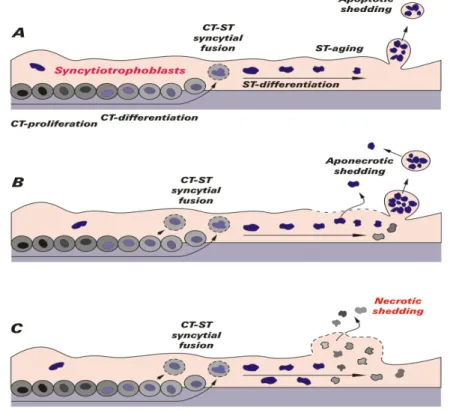
Figure 2 Modes of releasing syncytial material into the maternal circulation
Different steps of trophoblast-differentiation and the release of apoptotic syncytial knots are illustrated in Figure 2. In image A the normal apoptotic shedding is shown. The life cycle of a villous trophoblast comprises the following stages: CT-proliferation and differentiation, syncytial fusion of CT with the ST, further differentiation inside the ST and packing of old material into apoptotic syncytial knots. These corpuscular structures are surrounded by a sealed plasma membrane when they are extruded into maternal circulation and do not induce any inflammatory response of the mother. In part B the aponecrotic shedding is explained. Finalization of the apoptotic cascade may fail because of energetic or other problems. At these sites necrotic breakdown of the plasma membrane may take place. Thus, the already apoptotically cleaved material is released by necrosis (aponecrosis) and may induce inflammation. In part C the necrotic breakdown of syncytial sites is presented which releases cell-free trophoblast material into maternal circulation. Thus an inflammation is caused.
Because the ST fragments may be trapped in the capillary bed of the lungs as a result of their large size, and because the anucleate microvillous fragments are not useful for prenatal diagnosis, one has to focus on the isolation of extravillous CT circulating in maternal blood. Extravillous trophoblast invasion into maternal tissue is a process that starts at implantation, rises to a peak in the mid second trimester, and declines rapidly thereafter. Trophoblast cells vary in size from 20 to 200 µm and contain differing numbers of nuclei, depending on the trophoblast type.
The class I major histocompatibility complex (MHC-I) encodes the classical (class Ia) and nonclassical (class Ib) molecules. HLA-G is a class nonclassical Ib molecule and the genes show limited allelic variation compared with the class Ia genes. HLA-G has commanded centre stage among reproduction immunologists because its expression is restricted to extravillous trophoblasts [16]. It has not been convincingly demonstrated in any other fetal or normal adult tissue with the exception of fetal thymus where HLA-G is found in
able to suppress the cellular and humoral immune response being triggered by the classical HLA class I (HLA-A, B, C) or class II (HLA-DR, DQ, DP) molecules. HLA-G molecules [20]:
(1) inhibit the proliferation and cytotoxic functions of T cells and natural killer (NK) cells [21]
(2) drive T cells into an immunosuppressive profile or provoke the generation of regulatory T cells
(3) induce the apoptosis of endothelial cells
(4) inhibit the differentiation of antigen-presenting cells (5) alter the cytokine profile towards a Th2 polarization
(6) up-regulate inhibitory receptors on all kinds of effectors cells.
As secreted/shed HLA-G (sHLA-G) and membrane-anchored molecules exhibit the same receptor specificity, both HLA-G and its soluble counterparts are potent regulator molecules for the innate and acquired cellular immune response. In line with its strong immune tolerogenic functionality, HLA-G is strongly expressed during pregnancy but an ectopic expression is also found in thymus, cornea, and erythroid cells as well as in blood cells [22] and in nonphysiological situations such as transplantation, cancer, infections, and autoimmunity [23].
Indeed, HLA-G was the first HLA class I molecule being identified on trophoblast cells. So far, no pregnancy in which all of the proteins derived from the HLA-G gene are absent has been reported. Thus, HLA-G seems largely to be responsible for the reprogramming of local maternal immune response towards protecting the fetus.
Concerning the tasks of HLA-G it was already mentioned that it has an anti-viral function between the maternal and fetal interface. HLA-G is expressed during the first month of pregnancy in certain cytotrophoblasts and seems responsible for a normal pregnancy without complications. Soluble HLA-G was also found in amniotic fluid and cord blood [24].
Trophoblasts are epithelial cells, which are shed into the maternal blood stream as early as the sixth week of gestation [25] [26], but unlike lymphoid and myeloid cells of fetal origin, they do not persist for years after delivery [27]. Detection of trophoblast cells were found to be optimal within the time window between 9 and 13 weeks [28]. A trend was seen by Knight et. al. [29] towards increasing Trophoblast levels with increasing gestational age. They did a comparision between pre-eclampsia and normal pregant women and could see clear increase in trophoblasts in the case of pre-eclampsia. Their number regresses after delivery [30]. Trophoblasts do not persist after delivery.
2.3 Isolation techniques
To avoid the relatively complication-loaded invasive methods to obtain fetal cells, a method is needed to enrich fetal cells in sufficient numbers from peripheral blood of the mother. It is known that already from the 6th week of pregnancy fetal cells are in the maternal circulation. These include blood cells of the fetus (such as CD71 positive fetal erythroid progenitor cells) and fetal cells of placental origin (such as HLA-G positive trophoblasts). The cell number of these fetal cells in relation to the maternal cells is so small - 2 to 25 cells / ml maternal blood - that maternal blood is not readily suitable for molecular genetic studies. There are different techniques used for the sorting and the enrichment of fetal cells. The fluorescent-activated cell sorter (FACS) is one method for isolating cells from complex cell mixtures, the other one is the magnetic-activated cell separation system (MACS). Table 1 lists several works that have been tried to isolate trophoblasts from maternal blood. The method of enrichment is the biggest problem, as the subsequent investigations are able to identify or characterize individual cells.
Hawes et al. [31] used antibodies specific for ST to identify large multinucleated fragments of trophoblasts in maternal blood and used these cells for prenatal diagnosis of β-thalassemia. Mueller et al. [32] used the same antibodies to detect STs and subsequently applied the polymerase chain reaction (PCR).
By means of flow cytometry cell sorting, Cacheux et al. [33] identified extravillous CTs in a woman known to carry a fetus with a 47, XYY karyotype. Sbracia et al. [34] used HLA-G antibodies in flow cytometry cell sorting followed by PCR and correctly identified the fetal sex in 9 patients. Durrant et al. [35] identified CT cells with a monoclonal antibody and correctly predicted the fetal gender in 5 out of 6 male gestations, with no false-positive results in 7 female gestations.
According to Bianchi [27] in 90 samples of blood from women mainly in the second trimester of pregnancy a mean number of 19 male fetal cells were detected (1.2 cells per ml maternal blood). In contrast, in 109 samples (16 ml, maternal blood, 2nd trimester of pregnancy) the mean number of previous pregnancy fetal male cells obtained from female fetuses was 2 (0.1 cells per ml maternal blood are false positives). The cell number was determined using Y-chromosomal PCR without cell separation. For the detection of the fetal sex also the FISH-method was used.
One result of the identification of fetal cells in a maternal peripheral blood sample is shown in Figure 3, in which one single HLA-G positive cell was stained with a 7-amino-4-methylcoumarin-3-acetic acid labeled anti-HLA-G antibody (A) [36]. The same cells are visualized through the X-chromosomal specific signal (blue) in part B. HLA-G positive cells show a green staining for the Y-chromosome. In conclusion the analysis of the HLA-G positive cell revealed that the cell in the peripheral blood of the mother was of male fetal origin.
and fluorescence in situ hybridization [36]. A: Viewed with a triple filter visualizing blue 7-amino-4-methylcoumarin-3-acetic acid stained HLA-G. B: Single X chromosome–specific signal (blue dot) viewed with Spectrum Aqua filter visualizing Spectrum Aqua probe. C: Y-chromosome–specific signal (yellow-green dot).
Table 1 Overview of the characteristics of studies focused on the use of circulating trophoblasts for prenatal diagnosis (examples for positive cell selection taken from Oudejans, et al. [37].
Study Stage Sample Select. Antibody Method Identification Target
Author Year wks (ml) method
Hawes [31] 1994 10-18 10-50 + FDO MACS Morphology
Durrant [35] 1996 10-14 15-20 + 340 MACS PCR Y
Lim [38] 1999 14-20 25 + and - CD45/
340 MACS FISH XY
Hviid [39] 1999 1st trim 20 + and - CD45/
LK26
DC +
MACS VNTR-PCR TH01
Schueler [40] 2001 6-14 30-40 + Cocktail MACS ICC + RISH /+
FISH Cockt.+ XY Lim [41] 2001 9-35 20 + and - CD45/ 340 MACS FISH XY Koumantaki [42] 2001 8-12 16-18 + and - multiple DC/ MACS VNTR-PCR Multi.
Van Wijk [36] 2001 8-18 25-30 DC FISH XY
Vona [43] 2002 11-12 5 ISET + MD PCR VNTR-PCR ICC XY Multi. CK Guetta [44] 2005 17-20 20 + MEM-G/9 DC + MACS PCR XY
Zhang [45] 2008 35-40 6 DC ICC, PCR HLA-G,
Y
DC, density gradient centrifugation; ICC, immunocytochemistry; MACS, magnetic activated cell sorting; RISH, RNA in situ - hybridization; VNTR-PCR, variable number of tandem repeats-PCR; + positive selection; - negative selection.
2.4 The technology of the nanodetector
The goal in this work is to isolate trophoblast cells by catching them in vivo in the vein of a pregnant woman. For isolating trophoblast cells the recognition of the HLA-G antigen by an antibody is essential. Various types of noncovalent interactions may contribute to antibody binding of antigen, including electrostatic forces, hydrogen bonds, van der Waals forces and
medical device, which is making use of different technologies to bind specific antibodies on a novel nanodetector device. The main issue is generating biocompatible surfaces that enable new diagnostic devices for intracorporeal in vivo application.
Although it is generally possible to isolate fetal cells from maternal blood in a complex combination of magnetic separation and density gradient centrifugation practical, widely applicable and standardized methods for the enrichment of fetal cells from maternal blood are not available. Therefore, to secure a 100% prenatal diagnosis in clinical practice the invasive amniocentesis or CVS has to be used as before, with the associated risks to the fetus.
All the existing techniques are applied to blood samples in-vitro. In this work a new nanodetector will be introduced, which is based on a biofunctionalized wire and enables the capture of fetal cells (trophoblasts) from the maternal blood stream in-vivo. An antibody against a special surface marker antigen of fetal trophoblasts is covalently bound to a hydrogel. By targeting these specific antigens, trophoblasts can attach to the catheter. Trophoblasts are the target, because there are have known antigens on the cell surface and commercial antibodies exist are available.
The catheter is intented to be inserted into a peripheral vein of a pregnant woman for about 30 min and specifically captures fetal trophoblasts from the blood stream. Afterwards cytogenetic methods are applied for the detailed analysis of chromosomal aberrations (e. g. the Down syndrome). The device is intended to be used from the 10th week of pregnancy on.
Due to the fact that the amount of trophoblasts is extremely low, a routine blood sample is unable to offer the required sensitivity. Furthermore, it is generally regarded as desirable to avoid taking any more blood than absolutely necessary from pregnant women.
The nanodetector should be used in prenatal screening for pregnant women to determine if their fetus has a particular disorder, such as the Down syndrome. Clinical indications for a high risk of a fetus disorder are the age of the women as well as prenatal biochemical and biophysical tests, family history, physical examination and results from ultrasound examination.
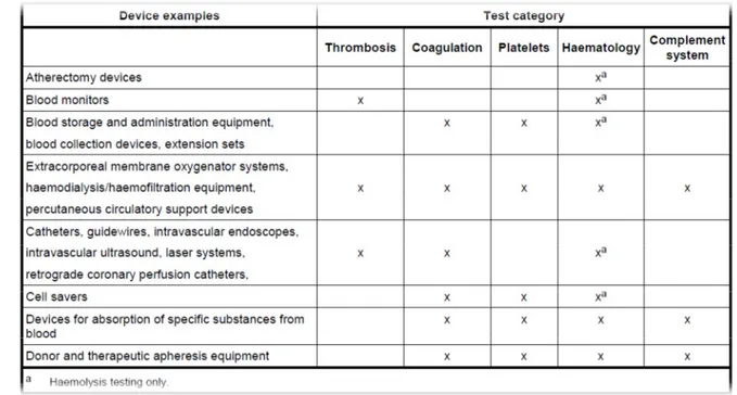
For the intended use of the nanodetector in the arm vein a contact with the circulating blood is necessary over a period of less than 1 h. According to European Norm ISO 10993-1 Table 1 the following safety tests are therefore to be considered: Cytotoxicity test according to EN ISO 10993-5, Hemocompatibility test according to EN ISO 10993-4, Irritation test according to EN ISO 10993-10 or Intracutane reactivity according to EN ISO 10993-10, Sensibility tests according to EN ISO 10993-10 and Acute systemic toxicity according to EN ISO 10993-11. Special emphasis will be laid on the Acute systemic toxicity test with a large animal trial.
2.5 Nanotechnology
Nanotechnology offers new opportunities for the development of a new diagnostic devices for isolating rare cells. Nanotechnology is defined by small feature sizes, a clear definition does not exist. In general nanomaterials have structural features between isolated atoms and bulk materials in the range of about one to 100 nanometers, physical attributes substantially different from those displayed by either atoms or bulk materials [46]. This changes characteristic properties of nanoparticles [47] and because of the small feature sizes applications like electron tunneling are enabled. Surface modifications change the chemical characteristic for binding reactions, like antibodies [46].
• for drug delivery systems including microchips, microneedle-based transdermal therapeutic systems, and assembled systems, [56], [57], [58], or
• nano/micro devices are in development for both diagnosis and therapy (theragnosis) which can improve the personalized medicine [59].
In many biosensors, biological materials are applied to nanostructures or a plain gold surface covering a non-binding surface, like a medical wire, to generate a horizontal or a vertical nanostructure. In this work it should be analysed which surface is the most effective for isolating cells in vivo.
Two principal factors determine the properties of nanomaterials to differ significantly from other materials: increased relative surface area and quantum effects. These factors can change or enhance properties such as reactivity, strength, and electrical characteristics. As a particle decreases in size, a greater proportion of atoms are found at the surface compared to those inside. For example, a particle with a diameter of 30 nm has 5 % of its atoms on its surface, at 10 nm 20 % of its atoms, and at 3 nm 50 % of its atoms. Thus nanoparticles have a much greater surface area per mass unit compared to larger particles. As catalytic chemical reactions occur at surfaces, this means that a given mass of material in nanoparticulate form will be much more reactive than the same mass of material made up of larger particles. This can affect the optical, electrical and magnetic behaviour of materials, particularly as the structure or particle size approaches the smaller end of the nanoscale.[60] An effective diagnostic nanodetector depends on at least two components: a specific target binding structure, the sensor, and the appropriate presentation of this sensor to the target. While sensors are readily available nowadays, the presentation of the sensor to the target limits nanodetector application and sensitivity. Two approaches were developed to present sufficient sensors to target molecules by first coupling to a large-area, two-dimensional highly ordered metallic nanostructures using nanosphere-lithography (NSL) and second by applying a vertical hydrogel to a gold surface. Self-assembly of the hexagonal closed-packed monolayer of latex spheres (LS) is a basis of NSL. This technique is used for the creation of masks for deposition of various materials, typically by evaporation or sputtering. It is known that NSL can be used to make honeycomb lattices of triangularly shaped islands on various substrates. Using LS with different diameters, one can change the spacing and size of the periodically arranged islands. By annealing the samples at a temperature of about 70 % of the melting point of the bulk material and adjusting the time of the thermal treatment, spherical particles can be obtained. Nanodots and nanorods can be obtained by multiple metal depositions at different deposition angles. A variety of complex morphologies, ranging from cup-like structures to rods and wires, are possible using this technique.[61] Applying this technique to a medical wire allows to produce diagnostic nanodetectors with large area, two-dimensional gold nanostructures of 50 to 100 nm size. A typical example of these structures is shown in Figure 4.
Figure 4 Typical atomic force microscopy images of a guide wire coating. a) Two-dimensional mask on a guide wire created by using latex particles with a diameter of 500 nm. b) Two-dimensional Au–triangle nanostructures. c) Samples presented in b) after annealing show spherical Au-nanoparticles with a diameter of 50 nm.
3
Aims of this work
The first goal of this study was to develop a nanodetector to isolate trophoblast cells to catch them in vivo in the peripheral blood stream in a pregnant woman. The second goal was the pre-clinical analysis for a clinical study with pregnant woman. In order to achieve the goal a common medical guide wire was used as a base for developing a nanodetector. Different technologies were applied to bind specific antibodies on the nanodetector against surface antigens of trophoblast cells which were targeted.
• Development of a nanodetector
a. finding out an optimized nanostructure surface (horizontal or vertical approach),
b. characterisation of the antibody specificity and avidity as well as determination of the antibody amount on the nanodetector surface,
• Proof of the functionality of the nanodetector to isolate trophoblast cells:
a. determination of nanodetector cell binding efficiency in
in vitro settings,
b. evaluation of the ability to detect trophoblast cells in a perpiherial blood stream of pregnant woman
• Assessment of the safety of the nanodetector for the clinical use in pregnant women:
a. determination of the nanodetector cytotoxicity,
b. evaluation of the risk for triggering thrombosis – hemocompatibility,
c. perform an animal trial to rule out an acute systemic toxicity,
d. evaluation of the immunogenicity of a nanodetector components.
4
Material
4.1 HLA-G antigen
Since HLA-G has been demonstrated to be expressed only in extravillous trophoblasts [16] and not in any other fetal or normal adult cells or tissues it was chosen as the prime target for antibodies. In this work a commercial available antibody and a F(ab) fragment, which were to be developed, for functionalizing the nanodetector. HLA-G is a nonclassical major histocompatibility complex class I (MHC-I) molecule consisting of 3 domains. MHC-I complexes are localized in plasma membranes and are involved in immunological defense and tolerance mechanisms differentiating between self and foreign molecules and cells. The overall structure of HLA-G is similar to that of other members of the MHC-I family - like HLA-A2, HLA-B44, HLA-C3 and HLA-E. They share a significant sequence similarity or even identity - HLA-G, e.g. 82 % with HLA-A2. [62]. In order to develop specific antibodies against HLA-G which do not cross-react with the other structurally related MHC-I molecules an amino acid sequence alignment was used to determine sequences unique for HLA-G and therefore suitable as potential antigens in the panning process of the development of recombinant human antibodies. Three such sequences were found. These sequences are located in the three different domains of HLA-G and they were the basis for synthesizing 4 peptides as antigens for the page display.
Alpha 1-Domain: Peptide 1 : E61 E E T R N T K A H A Q T D R M N L Q T L R G83 Alpha 2-Domain: Peptide 2: I142 S K R K C E A A N V A E Q R R A Y L E G T163 Peptide 3: E148 A A N V A E Q R R A Y L E G T C V E W L H R Y L E N174
Alpha 3-Domain, ILT-2 binding site:
Peptide 4:
H193 P V F D Y E A T L R C W A206
4.2 Antibodies
Polyclonal antibodies are widely used as detection reagents in research and diagnostics but they are a batch-dependent limited resource and also contain antibodies with unknown specificity. A big milestone in antibody generation was the generation of monoclonal antibodies (mAbs) by hybridoma technology which is based on the fusion of antibody producing spleen cells from immunized mice or rats with immortal myeloma cells [63]. Although the development of the hybridoma technology was a landmark event in
rejected by the human immune system. Exbio provides a mouse antibody called MEM-G/9 which reacts with native form of human HLA-G1 on the cell surface as well as with soluble HLA-G5 isoform in its beta2-microglobulin associated form.
To reduce the risk of immunogenicity today most of the novel therapeutic antibodies are of human origin or at least partially humanized. Many approaches to overcome these limitations of antibody production have been tried, such as to generate either chimeric antibodies or humanized antibodies or using mice that are genetically engineered to produce antibodies with human sequences or the recombinant human antibody technology – a fully in
vitro method for developing highly specific and high-affinity antibodies. 4.2.1 Human antibodies – phage display
Today suitable recombinant human antibodies are identified with the help of very large libraries of antibody genes. These libraries typically contain human antibody gene sequences, as they were originally used to produce human antibodies for therapeutic purposes. In this work the Human Combinatorial Antibody Library (HuCAL®) technology of MorphoSys was used. The suitable antibodies that bind to a given antigen must be identified by selection rather than by screening. There are numbers of selection techniques like phage display, ribosome display and yeast display. Phage display is widespread and the most popular and best established system for selection over the last decade.
Phage display is based on the fusion of antibody fragments on a surface protein of the phage M13. In that way the antibody is presented on the surface and can be selected by “phage panning”, which is somewhat similar to solid-phase immunoassays [64]. In this process the antigen of interest is immobilized on microplate wells, on magnetic beads or on a column. The phages are then added. After washing to remove all non-specific materials, the bound phages are eluted and amplified by replication in new host cells. The selection procedure is repeated several times, resulting in a population that consists almost entirely of phages that express the desired antibodies. After the selection steps, the antibody genes are isolated and inserted into an expression vector.
4.2.2 Purification of the antibody
After transfection into new host cells and growing the cells, they were lysed and the antibody lysate was tested by ELISA for the presence of antigen-specific antibody material. This soluble antibody fragments produced by Escherichia coli are purified by one step affinity chromatography using the His-6-tag, which has been fused to the C-terminus of the antibody and is completed by the metal chelate Ni-NTA. To remove endotoxins from the antibody solution the antibodies were additionally purified with Endo-Trap® columns.
4.2.3 Structure of the used antibodies
The antibody used for the functionalization of the nanodetector is a murine IgG1 (see Figure 5) and a human F(ab). Therefore the antibody lacks completely the Fc part and consists only of the Fab-fragment (see Figure 6). Additionally, the antibody possesses two different tags: a His-Tag and a Cys-Tag. The Cys-Tag could be responsible for the dimerisation of two antibodies resulting in a Fab-Dimer.
Figure 5 Schematic diagram of the basic symmetrical structure of immunoglobulins (antibodies) composed of two heavy and two light chains.1. Fab fragment, 2. Fc part, 3. heavy chain (consisting of the variable region VH with the hypervariable CDR regions, the constant regions CH1, CH2 and CH3 and the hinge region); 4. light chain (consisting of VL and CL regions), 5. antigen binding site.
Figure 6 Schematic drawing of the antibody format.
4.3 Cell Lines
Used cell lines:Name Description source Number
Jeg-3 human choriocarcinoma DSMZ ACC 463
K-562-HLA-G
Transinfected cell line
HLA-G LMU Munich
K-562 WT Wild type LMU Munich
MG-63
Human osteoblastic cell
line ATCC CRL-1427
NHDF-c adult humane Fibroblasten PromoCell C-12302
Jar human choriocarcinoma DSMZ ACC 462
4.4 Animals
4.4.1 RatsFemale Wistar rats, both pregnant and non-pregnant ones, were used in the trial. The trial was carried out in accordance with ISO 10993-2. The animals were bred at the Department of Toxicology, University of Medical Sciences, Poznan, Poland.
All the tested rats were four months old at the start of the study. The pregnant animals were 4 months ± 20 days old at the end of the experiment.
The minimum number of animals included in each studied group was 5. The control groups of non-pregnant and pregnant rats consisted of 12 animals each.
All animals were kept under standardised husbandry at the Department of Toxicology. The rats were housed in Tecniplast (1291H001) stainless-steel cages, which were maintained at the temperature of 22±2oC, and a relative humidity of 50±10%. The 12/12 hours light/dark cycle was maintained throughout the study.
The animals were fed with standardised normal LABOFEED H (PN ISO 9001) produced by the Feeds and Concentrates Production Plant, Certificate of Quality System No 181/1/98, Kcynia, Poland. The animals were fed every day at the same time, in the morning with a 10 % surplus added to the amount consumed on the previous day. Water was available ad libitum.
To adapt the animals to the new environment, the rats were kept in husbandry for 3-5 days in groups of two to three animals before the start of the tests.
During the observation, no changes in the behaviour and motoricity of animals were observed. The condition of skin, mucosa and fur was not altered.
4.4.2 Overview: Studied groups
The animals were divided into several experimental groups [65].
Two negative control groups (Control, Pregnant-Control) without any surgical procedures were used. Another control group comprised animals in which the surgical procedures were performed in the same way as in the case of animals belonging to the experimental groups, however, the catheters were not introduced (Control/Catheter/Catheter Gold, Pregnant-Control/Catheter/Catheter Gold). In the case of rats from the other control groups, the catheters, which were not covered with an antibody were introduced (Catheter, Pregnant/Catheter, Catheter Gold, Pregnant/Catheter Gold).
The experimental groups (Table 2) of non-pregnant and pregnant animals were established in order to determine the systemic toxicity to small amounts of antibody, which the nanodetector is coated with. For this purpose, the antibody was injected into the peritoneal cavity in three different doses and autopsy was done at different times after administration of the antibody (Table 2).
Table 2 Outline of the animal trial
Group symbol Group description
Non-pregnant rats Pregnant rats
Control
Control group; autopsy; Control group; autopsy
Catheter Catheter introduction; autopsy after 30 min.
Catheter Gold Catheter gold introduction; autopsy after 30 min.
Control/Catheter/ Catheter Gold
Control group; incision, preparation of carotid vein; autopsy after 30 min.
Control group; incision, preparation of carotid vein; autopsy after 30 min.
Catheter + Anti-HLA-G Catheter covered with Anti-HLA-G introduction; autopsy after 30 min.;
Catheter covered with Anti-HLA-G introduction; autopsy after 30 min.
Catheter Gold + Anti-HLA-G
Catheter gold covered with Anti-HLA-G introduction; autopsy after 30 min.
Catheter gold covered with Anti-HLA-G introduction; autopsy after 30 min. Anti-HLA-G 0.05 ng/2 hours Antibody administration 0.05 ng/250 g; autopsy after 2 h; Antibody administration 0.05 ng/250g; autopsy after 2 h Anti-HLA-G 0.10 ng/2 hours Antibody administration 0.10 ng/250 g; autopsy after 2 h Antibody administration 0.10 ng/250 g; autopsy after 2 h Anti-HLA-G 0.25 ng/2 hours Antibody administration 0.25 ng/250 g; autopsy after 2 h Antibody administration 0.25 ng/250 g; autopsy after 2 h Anti-HLA-G 0.05 ng/24 hours Antibody administration 0.05 ng/250 g; autopsy after 24 h Antibody administration 0.05 ng/250 g;autopsy after 24 h Anti-HLA-G 0.10 ng/24 hours Antibody administration 0.10 ng/250 g; autopsy after 24 h Antibody administration 0.10 ng/250 g; autopsy after 24 h Anti-HLA-G 0.25 ng/24 hours Antibody administration 0.25 ng/250 g; autopsy after 24 h Antibody administration 0.25 ng/250 g; autopsy after 24 h Anti-HLA-G 0.05 ng/6 or 3 weeks Antibody administration 0.05 ng/250 g; autopsy after 6 weeks
Antibody administration 0.05 ng/250 g; autopsy after 3 weeks
Anti-HLA-G
0.10 ng/6 or 3 weeks
Antibody administration 0.10 ng/250 g; autopsy after 6 weeks
Antibody administration 0.10 ng/250 g; autopsy after 3 weeks
Anti-HLA-G
0.25 ng/6 or 3 weeks
Antibody administration 0.25 ng/250 g; autopsy after 6 weeks
Antibody administration 0.25 ng/250 g; autopsy after 3 weeks
a period of 30 minutes. After this time, the autopsy was performed. The used nanodetector contained about 1.5 ng of the antibody, with and without gold nanostructures.
The animal trial was performed in accordance with the guidelines provided in the Ministry of High Education Report from 1959, and the UNESCO Declaration of Animal Rights from 1978 (Paris).
An approval was released from the Local Committee of Bio-Ethics for Animal Experiments (Wielkopolska District) (agreement – 16/2007) on February 19th, 2007.
The pregnancy was induced in 4-month-old rats and the experiments were performed (or terminated) on day 20 of the pregnancy.
4.5 Animal study outline
The study groups A to R, i.e. PA to PR for pregnant animals (in alphabetical order, see table 4), were established in order to determine the systemic toxicity to small amounts of antibody which the nanodetector is coated with. For this purpose the antibody was injected into the peritoneum in three different doses/ study groups (0.05 ng, 0.1 ng and 0.25 ng). The antibody used is a mouse monoclonal antibody against human HLA-G. The reason behind using an antibody from a rodent in another rodent is the fact that the final product contains a fully human antibody to be inserted into humans, the results obtained should thus be comparable to the real situation. A rat antibody was not used as this is not available. In addition, three different time spans for observation were included in the trial (2 hours, 24 hours and 6 weeks i.e. 3 weeks for pregnant rats).
Control group: Control groups L and PL were performed as negative controls
without insertion of the nanodetector but with performing the same minimal invasive procedure to be used before nanodetector insertion. Additional controls included nanodetectors without the special gold nanometer sized structuring (group R and PR) and nanodetectors without antibody coating (groups M and N). The nanodetector used contained about 1 ng of antibody, with and without gold nanostructures.
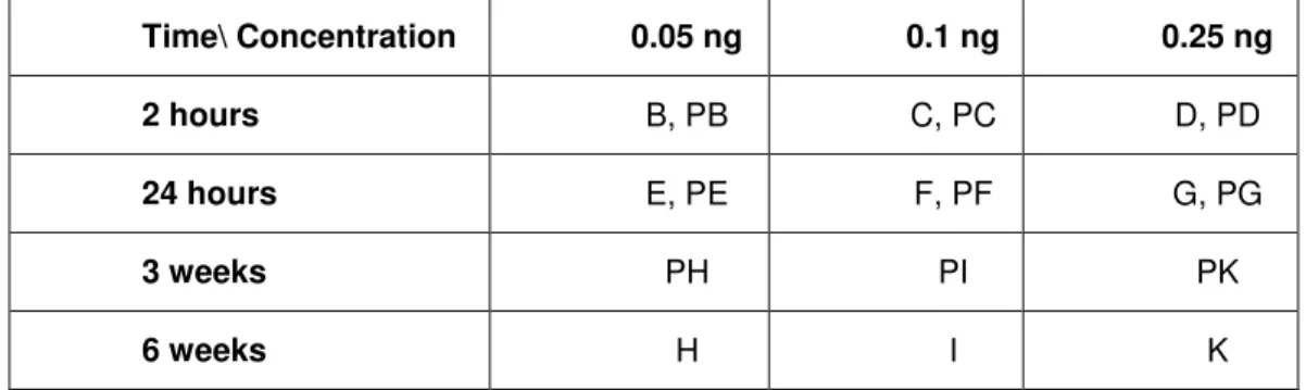
Study Groups – Antibody Injection: Groups B to K (respectively PB to PK for pregnant animals) were injected with increasing concentrations of the antibody into the peritoneum. The solutions of the anti HLA-G monoclonal antibody in concentrations of 0.05; 0.1 and 0.25 ng in 0.5 ml were prepared using 0.9 % NaCl. The finished dilution was injected into the peritoneum of the rats from selected groups. Afterwards, the rats were observed for different time spans (from 2 hours to 6 weeks). See Table 3.
Table 3 Outline of study groups for injection of the test substance (monoclonal mouse anti-human HLA-G antibody).
Time\ Concentration 0.05 ng 0.1 ng 0.25 ng 2 hours B, PB C, PC D, PD
24 hours E, PE F, PF G, PG
3 weeks PH PI PK
6 weeks H I K
For each group hematological tests and clinical biochemistry tests were performed. The urinalysis as well as the tests for gross pathology (analysis of different organs) were only done for certain study groups. Body weight and body weight changes were also recorded where applicable. The tests were performed in accordance with ISO 10993.
The different study groups with dates of experiments are listed in Table 4 (below)
Table 4 Outline of the animal trial including a description of the groups included in the study, the dates the experiments were performed, the numbering of animals included in each group and the numbering of documentation for each experiment.
GROUP NUMBER DATE / Number
A Control 1-20 20.03.2007 1-12
PA Pregnant – Control 501-520 26.06.2007 501-512
B Anti-HLA-G 0,05 ng/2 hours 61-80 4.05.2007 61-66
PB Pregnant Anti-HLA-G 0,05 ng/2 hours 581-600 28.06.2007 581-586
C Anti-HLA-G 0,10 ng/2 hours 81-100 4.05.2007 81-86
PC Pregnant Anti-HLA-G 0,10 ng/2 hours 601-620 28.06.2007 601-606
D Anti-HLA-G 0,25 ng/2 hours 101-120 4.05.2007 101-106
PD Pregnant Anti-HLA-G 0,25 ng/2 hours 621-640 28.06 .2007 621 -626
E Anti-HLA-G 0,05 ng/24 hours 181-200 10.05.2007
11.05.2007
181-186 section PE Pregnant Anti-HLA-G 0,05 ng/24 hours 641-660 28.06.2007
29.06.2007 641-646 section F Anti-HLA-G 0,10 ng/24 hours 201-220 10.05.2007 11.05.2007 201-206 section PF Pregnant Anti-HLA-G 0,10 ng/24 hours 661-680 28.06.2007
29.06.2007 661-666 section G Anti-HLA-G 0,25 ng/24 hours 221-240 10.05.2007 11.05.2007 221-226 section PG Pregnant Anti-HLA-G 0,25 ng/24 hours 681-700 28.06.2007
29.06.2007 681-686 section H Anti-HLA-G 0,05 ng/6 weeks 121-140 4.05.2007 21.06.2007 121-126 section PH Pregnant Anti-HLA-G 0,05 ng/3 weeks 701-720 20.06.2007
10.07.2007
701-706 section
I Anti-HLA-G 0,10 ng/6 weeks 141-160 4.05.2007 21.06.2007
141-146 section PI Pregnant Anti-HLA-G 0,10 ng/3 weeks 721-740 20.06.2007
10.07.2007 721-726 section K Anti-HLA-G 0,25 ng/6 weeks 161-180 4.05.2007 21.06.2007 161-166 section PK Pregnant Anti-HLA-G 0,25 ng/3 weeks 741-760 20.06.2007
10.07.2007
741-746 section
L Control/Catheter/ Catheter Gold 241-260 18.05.2007 241-246
PL Pregnant Control/Catheter/ Catheter Gold 561-580 28.06.2007 561-566 M Catheter 21-40 4.04.2007 26.04.2007 17.07.2007 21-23 24-25 26-31 N Catheter Gold 41-60 4.04.2007 26.04.2007 17.07.2007 41-43 44-49 50-55
O Catheter Gold + Anti-HLA-G 261-280 20.06.2007
21.06.2007
261-266 267 PO Pregnant – Catheter Gold + Anti-HLA-G 521-540 27.06.2007 521-526
R Catheter + Anti-HLA-G 281-300 21.06.2007 281-285
5
Methods
5.1 Surface of the wire
In pre-clinical studies, nanodetectors were produced and assembled for use in minimal invasive diagnostic procedures by nanostructuring the surface of spring wires that are used in hospitals for patients. Modification of a nanostructured commercially available, health-board approved spring wire with biofunctional components is presented in Figure 7. Regarding the nanodetector device: to the gold-coated surface of the guide wire human antibodies to human cell surface target molecules are attached through a linker molecule. The tendency of proteins or cells to physically adsorb onto a substrate without specific receptor recognition is known as nonspecific adsorption. This type of contamination reduces the efficacy of nanodetctor. Undesirable features are high background noise and “false positives” results. Poly(ethylene glycol) (PEG) and PEG based polymeric materials have been used for many biological applications because of PEG’s capacity to resist protein and cell adhesion, as well as its nontoxicity and its non-immunogenicity. This modification allows specific detection of circulating rare cells present in the blood stream of a pregnant woman (fetal cells).
Figure 7 Detection of target cells (fetal cells, tumor cells) by binding to Au-modified nanoparticles, conjugated with target-cell-specific antibodies. Au-nanoparticles decorate the nanostructured surface of a guide wire. Antibodies are covalently bound to the gold nanoparticle by a linker-system. This technique is the first nano-sandwich approach for the production of an in vivo- nanodetector device.
Different methods of linker systems were tried to attach antibodies to the nanostructured gold surface.
The second approach is a nanodetector consisting of a wire which is covered with a hydrogel. Most commonly used surfaces in current nanobiotechnology applications are hydrophilic polymer or oligomer layers covalently attached to a substrate like gold due to the simplicity of the surface chemistry and the formation of ordered assemblies shown in Figure 8.
Figure 8 Nanodetector covered with hydrogel and antibodies
5.1.1 Nanostructure (horizontal approach)
As the basis of a covalent binding surface mostly gold nanoparticles are used. Despite the described advantages of the nanotechnology, fabrication of the surface modification is still a problem . Therefore, urface modifications in the nanostructure range are still investigated to address theirr sophisticated and challenging fabrication because little differences in size can make a difference in the performance. . Györvary et all [66] build a closedly packed monolayer of 4 nm amino-functionalized CdSe nanoparticles on a protein surface layer lattice. Heddle et all [67], captured and attached gold nanodots with a prepared protein surface. He was able to build gold nanodots onto the SiO2 layer of a metal oxide semiconductor. Other techniques are sputtering [68]. These are different approaches to generate the similar patterns of defined nanostructured islands on a surface.. For the horizontal nanostructured surfaces, small regions of gold are formed through deposition to another material. Self-assembled nanodots range from 10 to a few 100 nm in diameter. Nanosphere-lithography is an established method to generate with structured lithographic masks, nanoislands with dimensions exceeding 100 nm.
Nanoparticles or nanostructures can be modified by established surface chemistry protocols to functionalize the nanostructure or the small sized gold islands by adsorbing untagged antibodies. But the molecules are attached randomly. Alternatively, the antibodies can be bound via an interface of a self assembling monolayer (SAM), creating a surface bearing carboxylic end groups enabling subsequent coupling chemistries. Particle size characterization is important, because of the size similiarity to antigen distances on cells. Nanoparticles are not detectable by normal optical microscopy, so electron microscopy or scanning electron microscopy have be used to obtain the information, but electron microscopy cannot recognize antibodies or PEG structures. Other techniques are elavuated by Hall et all. [69]. By too tight packing these antibodies to the nanostructure surface the risk of steric hinderance in binding the respective antigen is increased.. This would prevent binding of antibodies to the respective antigen on the target cells. This effect can by reduced or avoided by the introduction of non-binding materials surrounding the islands of the nanostructure surfaces. In this work the nanosphere-lithography was used to generate small nano-islands.
5.1.1.1 Necessary steps for nanosphere lithography (NSL)
1. Preparation of a 2D-mask made from polystyrene particles with a mean diameter of 0.44 µm.
2. Coating of the wire surface with this mask.
3. Application of a gold coating by evaporation of titanium (for better surface contact) and gold.
4. Removal of the mask by various cleaning steps.
5. Structural characterization of the nanostructured wire surface by SEM. 6. Passivation against non-specific cell binding.
7. Sterilization of the nanodetector.
Step 1: Preparation of a 2D-mask made from polystyrene particles with a mean diameter of 0.44 µm.
This method is well known and already published by the group of Prof. Giersig [61, 70-72] A schematic diagram of this method is shown in Figure 9.
Figure 9 Schematic diagram of the preparation of a 2D-mask consisting of polystyrene particles
Application of a dispersion of polystyrene particles to the water surface in a beaker by using a Pasteur pipette.
Centralizing the particle layer by using a tenside in order to achieve a monolayer.
Transfer of the mask to the medical wire substrate.
Figure 10 SEM Image of the 2D maske from latex Particel with 500 nm diameter The mask will cover 2 cm of the tip of wire
Step 2: Coating of the wire surface with this mask Figure 10.
The coating mask consists of polystyrene particles which applied to the tip of the wire see Figure 11
Figure 11 The image of a Seldinger wire wrapped with the mask under a light microscope showing the characteristic light refraction for the mask
Step 3: Application of the gold coating by evaporation of titanium (to encourage adhesion) and gold.
The next step is the coating with gold by evaporation. In order to encourage the adhesion of a gold layer on a substrate a layer of titanium is initially applied to the surface. After coating with titanium a layer of gold is applied by evaporation in the same evaporation chamber.
Step 4: Removal of the mask by various cleaning steps.
The removal of the mask is accomplished by using adhesive tape and several washing steps in an ultrasound bath.
Step 5: Structural characterization of the nanostructured wire surface by SEM.
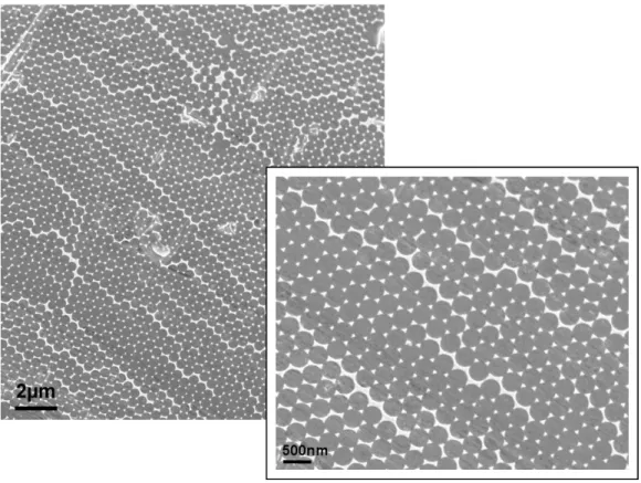
On the medical wire an array of nanometer-sized gold islands has now been created. This array of gold islands is now analysed by SEM as seen in Figure 12.
Figure 12 SEM images showing at different magnifications the arrays of gold islands produced by NSL.
Step 6: Passivation against non-specific cell binding
In order to avoid non-specific cell binding in the regions between the gold nano-structures, the medical wires are passivated by means of a PEG2000-Urea-silane solution (Figure 13). The passivation steps are as following:
b. Then the wires are placed inside a Schlenk flask filled with 10 ml of a dry solution of toluene.
c. One drop of distilled, degassed triethylamine (Et3N) is added to this
solution.
d. 10 mg of PEG2000-Urea-silane are dissolved in 5 ml of a dry solution of toluene employing a separate container. The solution is then added to the Schlenk flask.
e. The Schlenk flask is sealed up and the nanostructured wires are kept in this passivation solution for 4-24 h at 80°C.
f. Afterwards, the wires are rinsed with ethyl acetate and then in methanol and finally dried in a stream of nitrogen.
Figure 13 Chemical formula of the PEG2000-Urea-silane employed for the passivation
Step 7: Sterilization
The nanodetector is thoroughly cleaned and sterilized by steam before being transferred to a laminar flow hood to be coated with the antibody. All materials used during production of the nanodetector are cleaned and sterilized accordingly.
5.1.2 Nanostructure (vertical approach)
With galvanization techniques the first 2 cm of the medical wire were covered with a complete gold surface. The thickness of the gold surface is between 0,5 – 1 µm and the galvanization was done by a company (OTEK; Briselang).
There are two principal strategies to attach a polymer to a surface: grafting from and grafting onto. Grafting from means the in-situ polymerization from a surface, while grafting onto involves the covalent attachment of previously formed polymer chains to a substrate. Grafting from is believed to possess an inherent advantage: Theoretically, it is supposed to result in higher densities of polymer chains covering the surface. When the polymer chains are growing at the interface, only monomers of small to moderate size have to access the ends of the growing chains, a process which takes place with high diffusion rates and which does not imply steric hinderance. On the contrary, in the grafting onto scenario, entire polymer chains need to access a surface and react there. Diffusion of polymers is generally slower, and especially when some polymer chains are already attached, the access of further polymer chains to a reaction site on the surface will be sterically limited.
In 1990, Löfås and Johnsson [73] introduced a method to modify noble metal