Epidemiology of sport-related injuries
S
port-related injuries may occur during training or competition [1]. They usually incur damage to musculoskeletal structures such as muscles, ligaments, and bones. Due to their complexity spinal injuries should be treated as hazardous to a patient’s life and health [2]. On the average, 30-45 injuries occur in one million athletes, 5-14% of them being spinal injuries [3, 4]. Injuries are more often experienced by sub-elite and less experienced athletes [5]. Some authors claim the group most exposed to injuries are adult men since their strength, muscle mass, and level of aggression are higher than women’s or male adolescents’ [6, 7].The intersex differences with regard to sport-related injuries appear to be far less significant. Female athletes experience a higher scale of indirect hostility, while male athletes a higher scale of assault [8]. An interesting research question is whether more impulsive and aggressive individuals sustain injuries of the lumbar spine more often.
Epidemiology of lumbar spine injuries
Spinal injuries affect from 1 to 30% athletes and constitute one of the basic injuries eliminating athletes from training sports [9]. The precise etiology of spinal injuries is rather difficult. Bulging, extruding, protruding, or sequestered intervertebral discs can result in lumbar disk disease – a frequent source of low back pain (LBP). The most frequent cause of spinal injuries is excessive muscle tone affecting the nerve roots. Other types of injuries are associated with transverse and compressive vertebra fractures, and often with injuries to soft tissue
TRENDS
in
Sport Sciences
2017; 1(24): 5-11 ISSN 2299-9590
Genetic and environmental risk factors for lumbar disc
disease in Olympic grappling athletes: a review
ILDUS AHMETOV1, PRZEMYSŁAW LUTOMSKI2, BARBARA POSPIESZNA3, MACIEJ JURASZ2,
DARIUSZ WIELIŃSKI4, AGNIESZKA DOBERSKA5
Abstract
The authors examine genetic and environmental risk factors for lumbar disc disease in Olympic grappling athletes. The spine is the primary site of injuries in combat sports involving kicking, striking, throwing and joint locking. The execution of such techniques involves multiple repetitions of rapid movements, short maximal muscle contractions, usually with heavy external loads, and frequent training bouts with a partner. This type of training is associated with a significantly increased risk of injuries and overloads of the lumbar spine. The consequences of musculoskeletal overloads are often caused by the insufficient elasticity of the soft tissues.
KEYWORDS: grappling athletes, lumbar spine injuries, genetic/ /environmental factors.
Received: 15 August 2016 Accepted: 21 January 2017
Corresponding author: bpospieszna@gmail.com
1 Kazan State Medical University, Laboratory of Molecular
Genetics, Kazan, Russia
2 University School of Physical Education, Department of
Sport Medicine, Poznań, Poland
3 Adam Mickiewicz University in Poznań, Department of
Tourism and Recreation, Poznań, Poland
4 University School of Physical Education, Department of
Anthropology and Biometrics, Poznań, Poland
5 University School of Physical Education, Department of
in the lumbar region. The compression of nerve roots in athletes is often caused by herniation of the nucleus pulposus [10].
Physiological characteristics of combat sports
Combat sports are characterized by short-lasting, mostly dynamic, acyclical physical efforts of very high intensity, with short recovery breaks during a single bout or round, and – in the best combat sports practitioners – ability to continue fighting after a few-minute breaks [11, 12, 13].
Detailed physiological characteristics of particular combat sports depend on the ratio between dynamic and static efforts, high- and low-intensity exercises, number of short explosive efforts, as well as the competitor’s age category, body weight class, and competitive level [14]. Combat sport performance involves a vast array of physical activities with a variable contribution of aerobic and anaerobic energy metabolism [15]. The performance of short and rapid movement techniques such as blows, holds, throws, chokes, locks or kicks – which require a quick release of high power – is dominated by non-lactic anaerobic energy metabolism, while the muscle contraction energy derives usually from phosphate substrates [13, 16]. However, during efforts of longer duration and higher intensity the muscle contraction energy comes from anaerobic glycolysis, as it is unquestionably indicated by high lactate levels (up to 20 mmol/L) in athletes immediately after a bout or a round of combat [12, 13, 17, 20].
Although a single bout lasts usually a few minutes and the efforts involved are dominated by anaerobic energy metabolism, depending on a combat sport and its characteristics it may also entail periods of lower intensity as well as breaks featuring aerobic energy metabolism [14]. Moreover, the requirements to fight several bouts during a single day make the competitors’ aerobic capacity crucial [13]. High aerobic capacity makes the work of the fatigued body more effective, and first of all, accelerates the process of recovery before each consecutive effort. This is confirmed by high maximal oxygen uptake values (VO2max) in combat sports athletes, ranging from 50 to 65 ml O2·kg-1·min-1
[12, 13, 17, 20].
The above observations show that in combat sports – especially in disciplines involving a significant number of short, explosive, anaerobic efforts (e.g. grappling) – physical training should concentrate on the development of significant muscle force, high tolerance to lactic acid build up, and high aerobic capacity [15, 18]. Performance of such motor tasks during training sessions involves
multiple repetitions of rapid movements, short maximal muscle contractions – usually with heavy external loads – and frequent training bouts with a partner. Therefore this type of training is ultimately associated with an elevated risk of injuries and overloads of the lumbar spine. Paradoxically, the risk of injury-related low back pain prevalence is high in both: athletes, who feature the maximal levels of physical activity and physically non-active individuals (Figure 1).
The low level of physical fitness has previously been shown to be associated with the increased risk of low back disorders [21, 22]. Elfering et al. [23] found a higher risk of lumbar disc degeneration among those who did not participate in sports activities at all. The role of physical fitness (VO2max) as a risk factor for
herniated lumbar disc disease (HLDD) remains unclear. No association has been observed between the level of physical fitness and the risk of hospitalization due to HLDD during a 30-year continuation [24].
Nature vs nurture
The major focus of this part of the study is to evaluate the relative contributions of nature (inherited abilities) vs nurture (environmental effects) to the lumbar disc disease. According to some authors the main question is not whether there is a genetic component associated with sport performance, endurance/power trainability, anaerobic power, sports-related injuries, etc. – it is after all well known that acute exercise may even induce muscle injury [25] – but which genetic profiles
challenge elite performance in certain sports, and what is the level of susceptibility to disease [26].
Researchers have been assessing the impact of these factors in terms of their power and frequency. This issue is, however, complicated as it involves not only sex and anthropometric traits (body height, spinal mobility) as products of the genetic and environmental impact studied separately, physical and psychosocial characteristics, but also interactions between the genotype and the environment [27]. Lifting heavy loads, torsional stress, and driving motor vehicles are among the best-identified environmental risk factors.
An important question is which genes are involved in the mentioned problem? Another key research issue is whether there are any anthropological differences between athletes, for example, whether Asian athletes may suffer more often from LDD? If so, does it result from differences in body build or other genetic predispositions, or perhaps some environmental habits such as resting squats common among the population of South Korea, Vietnam and Laos. It would be interesting to examine how beneficial a resting squat is during training, which definitely causes stretching of low back muscles and fasciae.
Due to the specificity of judo, its practitioners constitute a highly interesting study sample. Min et al. [28] in their study of genetic predispositions towards lumbar disc degeneration (LDD) among judo practitioners showed that LDD is frequently experienced by athletes, and that it involves a spectrum of symptoms. They argued that a key role in the process of lumbar disc degeneration is played by the cartilage intermediate layer protein (CILP). This protein was later cloned and Lorenzo not only deduced its amino-acid sequence [29], but also showed that CILP was present in the mid-zone of human articular cartilage [30]. CILP plays a role in cartilage scaffolding and it antagonizes TGF-beta1 (TGFB1) and IGF1 functions. Overexpression of the CILP gene may lead to impaired chondrocyte growth and matrix repairs, and indirectly promote inorganic pyrophosphate (PPi) supersaturation in aging and osteoarthritis cartilage. Min et al. [28] carried out an analysis of association for the functional single-nucleotide polymorphism (SNP; 1184T/C, rs 2073711) of the CILP gene in Japanese collegiate judo athletes. By using logistic regression analysis and associations of lumbar disc degeneration with the CILP C allele (odds ratio = 4.1) and body weight (odds ratio = 1.06), they concluded that the CILP gene 1184T/C polymorphism is a significant risk factor for LDD occurrence in Japanese judokas. Its possible impact on other ethnic groups remains, however, unknown.
Min et al. [28] indicate that CILP gene genotyping could also yield important information about genetic predispositions towards LDD. The question remains whether there are other genes that play a similar role in LDD incidence. Such genes could be two collagen IX alleles associated with sciatica and lumbar disc herniation. Intervertebral disc degeneration has been shown to be related to an aggrecan gene polymorphism, a vitamin D receptor, and matrix metalloproteinase-3 gene alleles [31].
Considering the genetics of LDD in combat sports it seems important that COL1A1 may be associated with an increased risk of tendinopathy, tendon ruptures, and shoulder dislocations. Posthumus et al. [32] investigated a rare TT genotype of the Sp1 polymorphism in COL1A1 (G/T; rs 1800012) and observed a significant underrepresentation of participants with ACL ruptures [32]. It cannot be excluded that a T allele of the Sp1 binding site polymorphism enhances the binding of the Sp1 factor, causing an increased expression of the COL1A1 gene and production rate of alpha-1 chains. Genetic studies have been increasingly concerned with the search for potential major genes affecting LDD.
Classification of spinal injuries with particular consideration of the lumbar – sacral region
The causes underlying low back pain or spinal mobility restraints can be found in at least one of the following three complex spinal subsystems [33]:
1. The neural subsystem (nerves of ligaments, tendons and muscles, central nervous system);
2. The passive subsystem (vertebrae, facet joints, ligaments, synovial capsules and passive mechanical properties of skeletal muscles);
3. The active subsystem (muscles and tendons surrounding the spinal column).
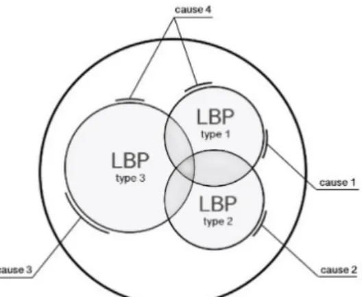
Figure 2. Non-specific LBP may consist of LBP with different
Figure 2 illustrates challenges faced by orthopedists or physical therapists in classifying the causes of non-specific mechanical low back pain.
On the other hand, according to Bayfield [34], there is no non-specific mechanical low back pain, but only “non-specific doctors”, which is an express reference to various diagnostic shortcomings of the condition. For physicians and physical therapists finding at least approximate causes of low back pain is of crucial significance, as only then can any conservative or operative treatment be successful. Otherwise the pain will not subside, and the recovery will be only temporary (risk of recurrence) or will never take place. A detailed attempt to classify the causes of low back pain, crucial for differential diagnosis, was made by Maitland [5].
Intervertebral disc degeneration
The intervertebral disc is not fully adapted to absorb all the loads of the standing body posture. The degeneration of the nucleus pulposus of the disc is, in fact, caused by the imperfect adaptation of the spine in evolution [36]. The degeneration is caused by ischemia and the resultant insufficient hydration of the intervertebral discs. In normal conditions, during development and at birth, intervertebral discs have some vascular supply to the cartilage endplates and the annulus fibrosus.
Under impingement the capillary bed deteriorates and the direct nutrition of the discs becomes inhibited. The metabolism of the fibroblasts of intervertebral discs becomes merely osmosis-dependent. Such circumstances lead to the production of deficient fibers and ground substance, which, in turn, may cause involutional lesions of the disc. The healing of the intervertebral disc occurs only in growing persons, and involves the filling of the damaged site with deficient fibrocartilage, which may lead to calcification and ossification by the penetration of osteoblasts from adjacent vertebral bodies [37]. In some cases a disc may deteriorate completely and becomes replaced with connective tissue resulting in the formation of osseous connections between the neighboring vertebrae.
Three stages of lumbar disc degeneration can be distinguished:
• degeneration of the nucleus pulposus, • herniation of the nucleus pulposus, • fibrosis of the nucleus pulposus.
The cause of degeneration of the nucleus pulposus is the decreasing content of mucopolysaccharides and their molecular mass. It results in dehydration of the nucleus pulposus and lowering of oncotic pressure in the nucleus pulposus. Dehydration makes the disc more
vulnerable to compressive forces. The consequences of deteriorated properties of the collagen fibers and ground substance are fissures in the outer fibrous ring of the disc known as anulus fibrosus disci intervertebralis, which impair the disc elasticity and lead to herniation of the nucleus pulposus [38].
Herniation of the nucleus pulposus is slow and gradual. The nucleus pulposus impinges against the anulus fibrosus owing to the positive pressure inside the disc. When the outer fibrous ring is ruptured, the nucleus pulposus bulges into the fissure. Further protrusion constrains the posterior longitudinal ligament which under pressure moves away from the vertebral body causing hernia [30].
Almost all cases of disc herniation are always postero-lateral in nature owing to the presence of the posterior longitudinal ligament in the spinal canal, which supports the central component of the fibrous ring. Parts of the nucleus pulposus penetrating the ligament as loose bodies in the vertebral canal may slide on the nerve root and get stuck in the intervertebral space. In general, however, a loose part moves under the posterior longitudinal ligament along the ligament fibers, or laterally along the nerve root towards the intervertebral space [36, 40]. A massive disc protrusion may also ensue, i.e. the bulging of the fibrous ring and shifting of the nucleus pulposus backwards. When the degenerative lesions of the ring are large also parts of the ring would bulge into the vertebral canal together with the nucleus pulposus.
A large displacement of the nucleus pulposus is irreversible due to the high pressure inside the intervertebral disc [36]. This pressure rises during athlete’s muscle contractions activated by coughing, sneezing, excessive spinal flexion and extension, or lifting heavy loads. The consequence of the sequestration is releasing fragments of the nucleus pulposus which can get stuck between the edges of the vertebral bodies. In such case every move of the spine may result in displacing these loose parts into the spinal canal [39]. The fibrosis of the intervertebral disc is scarring resulting from the process wound self-repair. It constrains the intervertebral space and results in pathological proliferative changes in the endplates.
Most often, a single loose disc fragment in the mucoprotein gel is more mobile and shifts backwards. The remaining part of the intervertebral disc is subject to degenerative changes, fibrosis, and immobilization. Degenerative lesions can also be observed on the displaced parts of the nucleus pulposus. They give rise to osteophytes due to the loss of tissue elasticity, dehydration, and damage by granulation
tissue, and thus to hardening. Ossification may appear at the site of the posterior longitudinal ligament rupture [37]. Degenerative changes may also affect the fibrous ring. Its posterior part, which is under constant pressure and irritation, becomes fibered, contracted and hardened. These lesions result in constrained mobility of the vertebral bodies until total immobilization of a given spinal region. Excessive impingement can also cause lesions in the vertebral bodies, manifested by the sclerotization of the tissue under the endplates. In the last stage of the degenerative disc disease the intervertebral spaces become narrowed. In the process of fibrosis the space may contract to below one-fourth of its original height. This may induce changes in the facet joints and articular surface incongruence and overloading of the joints, which are then affected by degenerative and deformative changes.
The last stage of the disease may also involve the fibrous ankylosis of the intervertebral joints. If the discs are not damaged at other levels, the pain subsides [41]
Pathogenesis and evolution of lumbar pain related to disc herniation
The main complaint of athletes is pain caused by irritation during protrusion of the innervated structures by the meningeal branches of the spinal nerve. Its sensory fibers are located in the posterior part of the fibrous ring, posterior longitudinal ligaments, vertebral periosteum and synovial capsules of the facet joints. The most acute pain sensation is caused by the primary pains related to disc degeneration, which include irritation of nerve roots and the impingement by the osteophytes. The most sensitive to pain are the peripheral nerve roots, that, when impinged, cause acute localized pain [36]. Similar pain sensations are caused by the impinging bone spurs around the intervertebral space. Secondary disc-related pains are caused by the narrowing of the height of intervertebral discs, and they radiate from facet joints and muscles. Articular surface incongruence occurs then in the facet joints.
It is also assumed that nerve root pain can be caused by biochemical factors. The impingement on a nerve root causes changes in its microcirculation and an inflammatory reaction manifested by an edema and nerve fiber atrophy. Additionally, the direct contact with the acidic metabolites of the nucleus pulposus and the capsule of the nerve root induces an inflammatory reaction of the adjacent nerve fibers [37].
The role and necessity of spinal elasticity
Soft tissue plays a crucial role in the stabilizing of the spine [42]. Its elasticity is an important determinant
of maintaining good fitness and of effective injury prevention. There is, however, contradictory clinical evidence regarding the role of the spinal soft tissue elasticity in injury prevention or treatment. For example, grapplers and dancers, whose spinal soft tissue is sufficiently elastic thanks to effective training, also experience injuries of the intervertebral disc [43]. Grappling places very specific demands on the muscular system, and they must be accounted for during preventive exercises aimed at lowering the risk of injuries. The consequences of musculoskeletal system overloading are often caused by the insufficient elasticity of the soft tissue.
Combat sport training involves sparring and strength and speed exercises. Particularly important are exercises with a partner. It may happen that the cause of the overloads is the large disproportion of body mass between the competitors. In their strength training combat sport practitioners focus primarily on the development of maximal force and explosive strength. This all demands maximal engagement with relatively heavy external loads, which may overload structures stabilizing the intervertebral discs.
Excessive training of this type decreases the elasticity of musculoskeletal structures and leads to injuries.
References
1. Gabbett TJ. The training-injury prevention paradox: should athletes be training smarter and harder? Br J Sports Med. 2016; 50(5): 273-280. doi: 10.1136/ bjsports-2015-095788.
2. Segan RD, Cassidy C, Bentkowski J. A discussion of the issue of football helmet removal in suspected cervical spine injuries. J Athl Train. 1993; 28: 294-305. 3. Armsey TD, Hosey RG. Medical aspects of sports:
epidemiology of injuries, preparation physical examination, and drugs in sports. Clinics Sports Med. 2004; 23: 255.
4. Bhopal AM, Richardson JB. Articular cartilage: structure, injuries and review of management. Brit Med Bull. 2008; 87: 77-95.
5. Birrer R. Trauma epidemiology in the martial arts: the results of eighteen year international survey. Am J Sports Med. 1996; 24: 72-79.
6. Touminen R. Injuries in national karate competitions in Finland. Scand J Med Sci Sports. 1995; 5: 44-48.
7. Zetaruk M, Violan M, Zurakowski D, et al. Safety recommendations in Shotokan Karate. Clin J Sports Med. 2000; 32: 421-425.
8. Keeler LA. The differences in sport aggression, life aggression, and life assertion among adult male and
female collision, contact, and non-contact sport athletes. J Sport Behav. 2007; 30: 57-76.
9. Mortazavi J, Zebardast J, Mirzashahi B. Low back pain in athletes. Asian J Sports Med. 2015; 6: e24718. 10. Nadler SF, Malanga GA, DePrince M, et al. The
relationship between lower extremity injury, low back pain, and hip muscle strength in male and female collegiate athletes. Clin J Sport Med. 2000; 10: 89-97. 11. Belotti P, Benzi G, Dal Monte A, et al. Classificazione
degli sport e determinazione dei mezzi di allenamento. Atleticastudi (Athletic studies). 1978; 3-4: 29-46 [in Italian].
12. Chaabène H, Hachana Y, Franchini E, et al. Physical and physiological profile of elite karate athletes. Sports Med. 2012; 42: 829-843. doi: 10.2165/11633050-000000000- -00000.
13. Franchini E, Del Vecchio FB, Matsushigue KA, Artioli GG. Physiological profiles of elite judo athletes. Sports Med. 2011; 41: 147-166.
14. Tabben M, Chaouachi A, Mahfoudhi HM, et al. Physical and physiological characteristics of high-level combat sport athletes. J Combat Sports Mart Arts. 2014; 5: 1-5. doi: 10.5604/20815735.1127445.
15. Todorov I, Bratić M, Nurkić M, Radovanović D. The influence of physiological characteristics on the competitive success of judo athletes. Facta Univ Series: Phys Edu Sport. 2013; 11: 317-323.
16. Karnincic H, Tocilj Z, Uljevic O, et al. Lactate profile during Greco-Roman wrestling match. Sports Sci Med. 2009; 8: 17-19.
17. Smith MS. Physiological profile of senior and junior England international amateur boxers. J Sports Sci Med Combat Sports Special Issue. 2006; 74-89.
18. Davis P, Wittekind A, Beneke R. Amateur boxing: activity profile of winners and losers. Int J Sports Physiol Perform. 2013; 8: 84-91.
19. Kraemer WJ, Fry AC, Rubin MR, et al. Physiological and performance responses to tournament wrestling. Med Sci Sports Ex. 2001; 33: 1367-1378.
20. Franchini E, Takito MY, Kiss MA, et al. Physical fitness and anthropometrical differences between elite and non-elite judo players. Biol Sport. 2005; 22: 315-328. 21. Stevenson JM, Weber CL, Smith JT, et al. A longitudinal
study of the development of low back pain in an industrial population. Spine (Phila Pa 1976). 2001; 26: 1370-1377. doi: 10.1097/00007632-200106150-00022.
22. Heneweer H, Picavet HS, Staes F, et al. Physical fitness, rather than self-reported physical activities, is more strongly associated with low back pain: evidence from a working population. Eur Spine J. 2011; 21: 1265-1272.
23. Elfering A, Semmer N, Birkhofer D, et al. Risk factors for lumbar disc degeneration: a 5-year prospective MRI study in asymptomatic individuals. Spine (Phila Pa 1976). 2002; 27: 125-134. doi: 10.1097/00007632- -200201150-00002.
24. Jørgensen MB, Holtermann A, Finn Gyntelberg, et al. Physical fitness as a predictor of herniated lumbar disc disease – a 33-year follow-up in the Copenhagen male study. Musculoskelet Disord. 2013; 14: 86.
25. McKune AJ, Semple SJ, Peters-Futre EM. Acute exercise-induced muscle injury. Biol Sport. 2012; 29: 3-10. 26. Gronek P, Wieliński D, Gronek J. Genetic and
non-genetic determinants of aggression in combat sports. Open Life Sci. 2015; 10: 7-8.
27. Gagneur J, Stegle O, Zhu C, et al. Genotype-environment interactions reveal causal pathways that mediate genetic effects on phenotype. PLoS Genet. 2013; 9(9): e1003803. doi: 10.1371/journal.pgen.1003803. 28. Min SK, Nakazato K, Okada T, et al. The cartilage
intermediate layer protein gene is associated with lumbar disc degeneration in collegiate judokas. Int J Sports Med. 2009; 30: 691-694. doi: 10.1055/s-0029-1214380. 29. Lorenzo P, Neame P, Sommarin Y, et al. Cloning and
deduced amino acid sequence of a novel cartilage protein (CILP) identifies a proform including a nucleotide pyrophosphohydrolase. J Biol Chem. 1998a; 273: 23469-23475.
30. Lorenzo P, Bayliss MT, Heinegard D. A novel cartilage protein (CILP) present in the mid-zone of human articular cartilage increases with age. J Biol Chem. 1998b; 273: 23463-23468.
31. Williams AG, Wackerhage H. Genetic testing of athletes. Med Sport Sci. 2009; 54: 176-186. doi: 10.1159/000235704.
32. Posthumus M, September AV, Keegan M, et al. Genetic risk factors for anterior cruciate ligament ruptures: COL1A1 gene variant. Br J Sports Med. 2009; 43: 352-356. doi: 10.1136/bjsm.2008.056150.
33. Panjabi MM. The stabilizing system of the spine. Part I. Function, dysfunction, adaptation, and enhancement. J Spinal Dis. 1992; 5: 383-389.
34. Bayfield D. Chiropractic manipulative skills, II ed., 2009, Elsevier.
35. Maitland G, Hengeveld E, Banks K, English K. Maitland’s vertebral manipulation, VII ed., 2005, Elsevier.
36. Brown S, Rodrigues S, Sharp C, et al. Staying connected: structural integration at the intervertebral disc-vertebra interface of human lumbar spines. Eur Spine J. 2016 Apr 15 [Epub ahead of print]. doi 10.1007/s00586-016-4560-y. 37. Steele J, Bruce-Low S, Smith, et al. Can specific loading
intervertebral disc? Spine J. 2015; 15: 2117-2121. doi: 10.1016/j.spinee.2014.08.446.
38. Li X, Dou Q, Kong Q. Repair and regenerative therapies of the annulus fibrosus of the intervertebral disc. J Coll Physicians Surg Pak. 2016; 26: 138-144. doi: 02.2016/ JCPSP.138144.
39. Lee SH, Jeong YJ, Kim NH, et al. The factors associated with the successful outcomes of percutaneous disc decompression in patients with lumbar herniated nucleus pulposus. Ann Rehabil Med. 2015; 39: 735-744. doi: 10.5535/arm.2015.39.5.735.
40. Bonetti M, Zambello A, Leonardi M, Princiotta C. Herniated disks unchanged over time: Size reduced after oxygen-ozone therapy. Interv Neuroradiol. 2016; pii: 1591019916637356. [Epub ahead of print]
41. Miller TT. Imaging of disk disease and degenerative spondylosis of the lumbar spine. Semin Ultrasound CT MR. 2004; 25: 506-522.
42. George SZ, Delitto A. Management of the athlete with low back pain. Clin Sports Med. 2002; 21: 105-132. 43. Gleim GW, McHugh MP. Flexibility and its effects on