Corresponding author: mgebska@pum.edu.pl
1 Pomeranian University of Medical Science, Department of Musculoskeletal System Rehabilitation, Szczecin, Poland 2 High School of Education and Therapy them. prof. Kazimiera Malinowska in Poznań, Poland
3 State Vocational College them. S. Staszica in Piła, Poland 4 Specialist Hospital in Piła, Department of Gynecology and Obstetrics, Piła, Poland
MAGDALENA GĘBSKA1, KRYSTYNA OPALKO2,3, PIOTR RYNIO4,
KATARZYNA WEBER-NOWAKOWSKA1, EWELINA ŻYŻNIEWSKA-BANASZAK1,
WOJCIECH GARCZYŃSKI2, ŁUKASZ KOŁODZIEJ1
Evaluation of bioelectrical activity of masseter muscles in women
with myogenic disorders of the stomatognathic system
TRENDS
in
Sport Sciences
2018; 1(25): 43-48 ISSN 2299-9590 DOI: 10.23829/TSS.2018.25.1-6 BackgroundT
he percentage of patients suffering from stomatognathic system disorders quite differs among the authors.According to McNeil, about 75% of population might be diagnosed with one of the dysfunction symptoms, but only about 33% of them will feel pain [18].
According to Kleinrok about 40%-90% of population complains about symptoms deriving from the SS [13]. The authors agree that the symptoms begin to appear more often between 20 a 40 year of life, and affect women 4 times more often than men [1, 15, 16].
One of the etiological factors contributing do development of the SS disorders are excess tone and muscular activity connected to long-lasting and not physiological load to the SS and difficulty of adapting to the excess of load [19].
Trigger points (TrPs) might often appear in the masticatory muscles that don’t function properly [11].
Abstract
Background. One of the etiological factors of the stomatognathic system (SS) disorders is excessive tension and muscle activity. These factors are associated with long-term non-physiological overload of SS tissues. Palpation of masticatory muscles allows for subjective assessment of muscle tension and tenderness. One of the available tools which are an important part of the examination is non-invasive global electromyography − sEMG
(ang. surface electromyography). Objective. The aim of the study
is to evaluate the bioelectrical activity of masseter muscles in patients with stomatognathic disorders. All disorders are myogenic origin. In addition reference standards for the masseter muscle
in healthy people will be definied. Material and Methods. The
study was conducted in a group of 104 women (Group I) with a myogenic pain disorders. All women were examined sEMG masseter muscles at rest and during maximal contraction. To defined standarized vaues for the masseter muscle authors
formed a control group (Group II). Results. Comparison of
the two groups revealed statistically significant differences in amplitude sEMG masseter muscle. In Group I, the average value of the amplitude of the EMG masseter muscle at rest and during maximal contraction is higher than in Group II.
Conclusions. The results testify to generating greater static and dynamic loads on the surfaces of the temporomandibular joints. The higher amplitude values in female group with myogenic disorders of the SS confirms that in patients with myogenic pain disorders sEMG test is a valuable addition to the diagnosis. KEY WORDS: surface electromyography, stomatognathic system, masseter muscle.
Received: 12 July 2017 Accepted: 06 February 2018
The authors have stated that the existence of active TrPs in a muscle is a signal of pain of myogenic origin and has connection with the neuromuscular system disorders and the existence of the active electric potentials [8]. The rate of the SS disorders successfully treated mostly relies on the correct diagnosis [7, 17]. Palpation of stomatognathic system gives only a subjective assessment of excess muscle tone and tenderness. So the detection of differences, measurement of the changes of muscular tone, as well as the rate of success of the treatment the right and left side is difficult.
Because of this, diagnosing the SS dysfunctions is done by a specialist relying on signs and symptoms. Nowadays, because of advances in physics and biomedical engineering, additional tests bring significant support to clinical management.
Instrumental diagnosis helps to measure both the rate of dysfunction and the rate of success in treatment [8]. Surface electromyography (sEMG) is one of the measuring tools that significantly contribute to muscle function assessment [3, 4, 22].
There are two kinds of EMG used in neurophysiology. The first kind, nEMG – elementary, relies of analyzing the function of individual motoric units [12]. It is conducted by putting needle electrodes into a muscle. The second one − sEMG (surface electromyography) uses surface electrodes. It assesses the fuction of all motoric units in an examined muscle [12]. In dentistry, the noninvasive eEMG is used more often. The sEMG test allows for obtaining objective data about muscular bioelectric activity (masseter muscle, temporal muscle, suprahyoid muscles, infrahyoid muscles and others), testing symmetry of their function, obtaining graphical representation and data analysis on both sides of the face. Advocates of this neurophysiological diagnostic test state that it should be used to measuring of increased or decreased muscular tone, measuring mandibular rest position, treating of parafunctional activity, assessing the therapeutic effect after using occlusal splint therapy, after orthodontic and physiotherapeutic treatment [5, 6, 9, 10, 21].
Aim of Study
Authors attempted to establish reference values of the masseter muscle in workload and in relax.
Material and Methods
We have tested 104 women (group I) aged 20 to 45 (average age: 33).
Occurrence of pain and dysfunction of the myogenic stomatognathic system was found using the Boumann
Functional Manual Diagnostics of the Stomatognathic System. The patients have undergone surface electromyography (sEMG) testing of the masseter muscles during relax and workload.
The control group (group II) consists of 100 women aged 20 to 45 (average age: 33) who have not been diagnosed with disorders of the stomatognathic system or pain of the craniofacial area.
The sEMG testing done in the group II was used to define reference values for the masseter muscle, because there is no such data in the available literature.
Recording of sEMG signals from the masseter muscles in group I and group II was done using dual-channel device NeuroTrac MyoPlus 2 with NeuroTrac software. During the EMG testing the Clinical Mode was used. In order to obtain precise sEMG measurements, we used a band-pass filter, which guarantees that 50Hz and 60Hz frequencies (coming from power gird lines) would not interfere with muscle activity recordings (measured in microvolts).
The filtering allows for sEMG measurement made with an accuracy of 0.1 µV.
In order to avoid magnetic interference, during sEMG testing, the measuring device was not located close to mobile phones (<4m) or any other sources that could interfere with data obtained. The testing was done using unipolar electrodes, which were located in a distance of 20 millimeters from each other. The key principle used during all the tests was placing the electrodes at the center of the belly of the masseter muscle, in parallel to its fibers. In order to precisely locate the electrodes, each time a thorough palpation of the muscle was done. The testing of the master muscles was performed while the patient was sitting in an upright position, with a neutral position of the head and hands resting at the knees and feet resting on the ground. Before the electrodes were fixed, the skin was cleaned with a disinfection agent (Skinsept). The passive electrode was placed in a cervical region – C7 (cervical spine).
During the sEMG testing of the masseter muscles, the Neuro Track MyoPlus 2 device was put on a small table on a side of the patient. The sEMG testing procedure consists of two parts:
1. The sEMG testing of the masseter muscle at rest: The testing was done when a patient was relaxed. The alveolar arches were slightly open during the test. In order to avoid recording of signals from the orbicularis oculi muscle, the patients had their eyes closed during the test. The patients were asked not to swallow during the test and to keep their tongue at resting position.
2. The testing of bioelectrical activity of the masseter
muscle during maximal contraction (grinding of teeth in a position of maximal intercuspation).
Recording of sEMG signal was done with a patient sitting, grinding her teeth with maximal possible strength for duration of 5 seconds.
Device-compatible computer software records minimal and maximal potential values and then calculates average potential values.
The results underwent statistical analysis using computer software (Statistica 2010). Statistical analysis was done using the following tests: Shapiro Wilk test, Wilcoxon signed-rank test, Student’s t-test, Mann–Whitney U test; 95% confidence interval (CI) has been assumed.
Results
First, results of sEMG testing of the masseter muscle at rest and during workload on the right and left side in group II (control group) are presented:
During statistical analysis of sEMG values of the masseter muscle at rest and during workload on both sides of the face in group II (control), no statistically significant difference has been found (p=0.29).
Median of sEMG amplitude for the right masseter muscle was 2.0 µV (= 1.9 µV), and 1.9 µV (= 1.9 µV) for the left one (Table1).
Statistical analysis of sEMG measurment results for the masseter muscle during workload for the right and left side showed no significant difference (p = 0.30).
Table 1. Profile of distribution of sEMG values [µV] of the masseter muscle at rest and during workload on the right and left
side in group II
Profile of distribution
sEMG [Vµ] of the masseter muscle at rest (n = 100)
sEMG [µV] of the masseter muscle – workload (n = 100)
Side of face Side of face
Right Left Right Left
min-max 1.0–2.5 1.0–2.5 52.5–79.3 51.5–79.9 Q1-Q3 1.4–2.3 1.5–2.2 60–73.6 60.5–73.1 me 95% CI 2,0 1.75–2.1 1.9 1.65–2.05 67.5 65.5–69.2 66.3 64.2–70 X– (SD) 1.9(0.5) 1.9(0.4) 67.2(7.6) 66.9(7.7) Materiality level Shapiro-Wilk test < 0.001 < 0.001 < 0.001 < 0.001
Wilcoxon signed-rank test 0.29 0.30
Key: n – group size, min – minimal value, max – maximal value, Q1 – first quartile, Q3 – third quartile, me − median, X– − arithmetic mean, SD – standard deviation, CI − confidence interval
Table 2. Profile of distribution of sEMG values [µV] of the masseter muscle at rest and during workload on the right and left
side in group I (with pain of the stomatognathic system)
Profile of distribution
sEMG [µV] of the masseter muscle at rest (n=100)
sEMG [µV] of the masseter muscle – workload (n=100)
Side of face Side of face
Right Left Right Left
min-max 13.6–92.9 11.9–95.9 120–397 142–522 Q1-Q3 22.2–36.5 31.4–55.8 219–344 226–319.5 me 95% CI 28.1 26.4–32.1 43.4 37.3–48.9 277.5 255–302 275.5 259–298 X– (SD) 31.7(13) 45.6(18.9) 273(76) 278(65) Materiality level Shapiro-Wilk test < 0.001 < 0.001 < 0.002 < 0.051
Wilcoxon signed-rank test < 0.001 0.52
Key: n – group size, min – minimal value, max – maximal value, Q1 – first quartile, Q3 – third quartile, me − median, X– − arithmetic mean, SD – standard deviation, CI − confidence interval
Median of sEMG amplitude for the right masseter muscle was 67,5 µV (= 67.2 µV), and 66.3 µV (= 66.9 µV) for the left one (Table 1).
Lack of statistically significant difference for sEMG measurments in group II between the right and the left muscle at rest and during workload proves homogeneity of the group.
Table 2 shows results form the masseter muscle measurements in group I.
Statistical evaluation of sEMG measurements of the masseter muscle at rest on the right and left side in group I, showed statistically significant difference (p < 0.001).
The median of amplitude for the right masseter muscle was 28.1 µV (= 31.7 µV), and 43.4 µV (= 45.6 µV) for the left one.
Statistical evaluation of sEMG measurements of the masseter muscle during workload on both sides of the face, in the group with pain, showed no statistically significant difference (p = 0.52).
The median of sEMG amplitude for the right masseter muscle was 277.5 µV (= 273 µV) and 275.5 µV (= 278 µV) fir the left one (Table 2).
Table 3 shows sEMG amplitude values [µV] of the masseter muscles at rest and during workload.
Analysis of the results shown in Table 3 suggests existence of statistically significant differences (p = < 0.0001) in values of sEMG amplitudes of the masseter muscle (at rest and during workload) between group I and II, which is precisely shown in Figure 1 and 2
Discussion of results
Electromyographic equipment allows for good visualization of the testing, recording of session and its results, as well as advanced automated analysis. The advantage of its use is possibility to precisely define average values of the potentials transmitted to muscles through motoric neurons.
The literature available is lacking any reference norms for the superficial part of the masseter muscle, so it was the reason why we decided to analyze muscular amplitude values in the first step.
The authors have observed that during analysis of bioelectrical muscular activity of the masseter muscles in group of 104 women with SS dysfunction and pain in craniofacial area, sEMG amplitude is higher.
This applies both for muscles at rest (right side 31.7 µV, left side 45.6 µV) and for workload (right side 277.5 µV, left side 275.5µV). In the group with pain,
Table 3. Average values of the sEMG amplitudes [µV] of the masseter muscle at rest and during workload in group I and II
Group analyzed Profile of
distribution Control group (n = 100)
Group with SS dysfunction and
pain (n = 104) Materiality level (Mann-Whitney U test)
Side of face Right Left Right Left
sEMG [µV] of the masseter muscle Workload me X–(SD) 2.0 1.9(0.5) 1.9 1.9(0.4) 28.1 31.7(13) 43.4 45.6(18.9) < 0.0001 Rest me X–(SD) 67.5 67.2(7.6) 66.3 66.9(7.7) 277.5 273(76) 275.5 278(65) < 0.0001 Key: n – group size, me − median, X– − arithmetic mean, SD – standard deviation
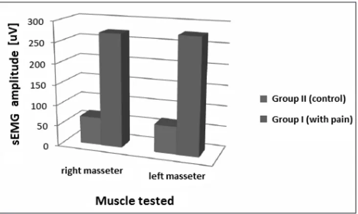
Figure 1. Average sEMG amplitude values of the masseter
muscle at rest and during workload in group I and II
Figure 2. Average sEMG amplitude values of the masseter
there is statistically significant asymmetry of muscular bioelectric activity at rest between right and left side of face (p < 0.001).
During workload, the amplitude values difference between right and left muscles were not statistically significant (p = 0.52).
The results in group I prove that there is larger load generated on the surfaces of the temporomandibular joints. This may lead to disorders of the articular disk and generate mirco- and macrotraumas in adjacent tissues of the stomatognathic system.
There are no standardized reference values of the masseter muscles in literature, which would be based on large study groups.
Testing 100 healthy people, who constituted the control group, enabled us to set a reference point for the results of the patients with SS dysfunction and craniofacial pain. In the control group the sEMG amplitude values at rest and during workload on the right and on the left side were almost equal averaged at 1.9 µV.
During workload the average amplitude value for the right muscle was 67.2 µV and 66.9 µV for the left one. In this group there were no differences in the amplitude values of the masseter muscle on the right and on the left side, both at rest and during workload.
Keeping balance of tone of the masticatory muscles allows the SS to work physiologically without any dysfunctions appearing.
Bioelectrical activity of the masseter muscle showed statistically significant difference between groups with and without pain, both at rest and during workload (p < 0.0001). Higher amplitude values in group I prove that sEMG is a valuable diagnostic instrument for patients with myogenic pain of the SS.
When comparing sEMG values of the masseter muscle with publications of other authors, it can be noticed the they are not consistent [14, 20, 23].
The cause of this inconsistency could be used of different electromyographic equipment, which differs in signal reception sensitivity, software and electrodes used.
Conclusions
The results of the study prove that there are larger static and dynamic loads on surfaces of temporomandibular joints among women with pain located within the SS. Higher amplitude values found in a population of women with myogenic SS dysfunctions confirm that sEMG testing a valuable diagnostic instrument for patients with myogenic pain of the SS.
References
1. Aidi N, Piotrowski P, Mehr K, Głowacka A. Częstość występowania zaburzeń czynnościowych narządu żu- cia – przegląd piśmiennictwa. Twój Prz Stomatol. 2012; 12: 56.
2. Akamatsu FE, Ayres BR, Saleh SO, et al. Trigger points: an anatomical substratum. BioMed Res Int. 2015; 623287.
3. Botelho AL, Melchior de O, da Silva AM, da Silva MA. Electromyographic evaluation of neuromuscular coordination of subject after orthodontic intervention. Cranio. 2009; 27(3): 152-158.
4. Botelho AL, Silva BC, Gentil FH, Sforza C, da Silva MA. Immediate effect of the resilient splint evaluated using surface electromyography in patients with TMD. Cranio. 2010; 28(4): 266-273.
5. Choi KH, Kwon OS, Jerng UM, Lee SM., Kim LH, Jung J. Development of electromyographic indicators for the diagnosis of temporomandibulardisorders: a protocol for an assessor-blinded cross-sectional study. Integr Med Res. 2017; 6(1): 97-104.
6. Dalewski B, Chruściel-Nogalska M, Frączak B. Occlusal splint versus modified nociceptive trigeminal inhibition splint in bruxism therapy: a randomized, controlled trial using surface electromyography. Aust Dent J. 2015; 60(4): 445-454.
7. Drobek W. Diagnostyka układu stomatognatycznego. Konieczność diagnostyki układu ruchowego narządu żucia przed leczeniem ortodontycznym i rehabilitacją protetyczną. Twój Prz Stomatol. 2009; 5: 14-17.
8. Gębska M, Opalko K. Pionierska analiza stawu skronio- wo-żuchwowego. Med Trib Stomatol. 2016; 2: 34-35. 9. Gębska M, Opalko K. Leczenie dysfunkcji układu stoma-
tognatycznego. Med Trib Stomatol. 2016; 1: 26-29. 10. Grossi GB, Garagiola U, Santoro F. Measuring
effectiveness of orthognathic surgery by electromyography: a restrospective clinical study. Minerva Stomatol. 2017; 66(3): 98-106.
11. Halmova K, Holly D, Stanko P. The influence of cranio-cervical rehabilitation in patients with myofascial temporomandibular pain disorders. Bratisl Lek Listy. 2017; 118(11): 710-713.
12. Huber J, Kulczyk A, Lisiński P, Lipiec J. The use of surface electromyography for diagnosis of muscle dysfunction with pain symptoms. Trends Sport Sci. 2013; 3(20): 135-139.
13. Kleinrok M. Zaburzenia czynnościowe układu ruchowego narządu żucia. Tom I. Wyd. V poprawione, Czelej, Lublin, 2012: 46-48.
14. Lauriti L, et al. Influence of temporomandibular disorder on temporal and masseter muscles and occlusal contats
in adolescents: an electromyographic study. BMC Musculosceleted Disorders. 2014; 15: 123.
15. Liu F, Steinkeler A. Epidemiology, diagnosis, and treatment of temporomandibular disorders. Dental Clinics of North America. 2013; 57(3): 465-479.
16. Łapuć M, Gołębiewska M, Kondrat W. Częstość występowania i diagnostyka dysfunkcji narządu żucia u pacjentów w wieku 20-30 lat. Mag Stomatol. 2011; 2: 12-17.
17. Łapuć M, Gołębiewska M, Sierpińska T. Zastosowanie badań EMG i T-Scan w diagnostyce pacjentów z dysfunkcją układu ruchowego narządu żucia. Mag Stomatol. 2008; 5: 24-28.
18. McNeill C. Management of temporomandibular disorders: concepts and controversies. J Prosthet Dent. 1997; 77: 510-522.
19. Pietruski JK, Pietruska MD. Dysfunkcja narządu żucia – przyczyny, diagnostyka, leczenie. Mag Stomatol. 2013; 12: 42-48.
20. Sójka A, Huber J, Hędzelek W, Wiertel-Krawczuk A, Szymankiewicz-Szukała A, Seraszek-Jaros A, et al.
Relations between the results of complex clinical and neurophysiological examinations in patients with temporomandibular disorders symptoms. Cranio. 2018; 36(1): 44-52.
21. Szyszka-Sommerfeld L, Woźniak K, Matthews-Brzo- zowska T, Kawala B, Mikulewicz M. Electromyographic analysis of superior orbicularis oris muscle function in children surgically treated for unilateral complete cleft lip and palate. J Craniomaxillofac Surg. 2017; 45(9): 1547-1551.
22. Vedana L, Landulpho AB, Andrade E, Silva F, Buarque E, Silva WA. Electromyographic evaluation during masticatory function in patients with temporomandibular disorders following interocclusal appliance treatment. Electromyogr. Clin. Neurophysiol. 2010; 50(1): 33-38.
23. Woźniak K, Lipski M, Lichota D, Szyszka- -Sommerfeld L. Muscle fatigue in the temporal and masseter muscles in patients with temporomandibular dysfunction. BioMed Res Int. 2015; 269734.